

Loading, please wait...
Loading, please wait...

Understanding phenotypic variations in craniocervical junction disorders remains an essential clinical objective for neurologists and pediatricians. For instance, recent cohort investigations into familial Chiari malformation type 1 have provided critical clarity regarding how inherited traits govern clinical expression. Clinicians often encounter substantial variability in tonsillar herniation, symptom chronicity, and anatomical disruption across different patients. Consequently, examining multiplex pedigrees allows physicians to determine whether distinct clinical presentations follow shared familial trajectories or sporadic anatomical variations.
Chiari malformation type 1 fundamentally involves the downward herniation of cerebellar tonsils through the foramen magnum. Typically, tonsillar ectopia exceeds five millimeters, although smaller descents become clinically meaningful when cerebrospinal fluid pathways face obstruction. This mechanical crowding disrupts normal pulsatile fluid movement between the intracranial vault and the upper spinal canal. As a result, patients experience sustained hydrodynamic pressure shifts and localized neural compression along the craniocervical junction. Furthermore, chronic tonsillar descent impinges upon the lower brainstem and cervicomedullary junction. Secondary neurological complications frequently develop from this altered biomechanical environment. For example, hydrocephalus emerges when posterior fossa crowding impedes fourth ventricular outflow. In addition, disturbed CSF flow dynamics promote fluid accumulation within the spinal cord parenchyma, driving syrinx development. Therefore, clinicians should look beyond simple anatomical measurements on static magnetic resonance imaging. Instead, practitioners must evaluate how CSF circulatory disruptions cause sensory deficits, motor weakness, and neuropathic pain. Understanding these mechanical processes helps neurologists anticipate neurological decline before permanent damage occurs. Early physiological assessment enables timely surgical decompression that effectively halts disease progression.
Recent investigations analyzing multiplex families demonstrate that clinical phenotypes cluster significantly across generations. Specifically, researchers assessed twenty-four new families containing fifty-seven affected individuals to quantify intrafamilial concordances. Consequently, statistical analysis revealed that familial Chiari malformation type 1 exhibits strong concordance for age of symptom onset. This correlation suggests that shared genetic loci govern the temporal presentation of tonsillar ectopia across relatives. Moreover, prominent symptoms such as occipital headaches demonstrated significant intrafamilial similarity. Similarly, posterior neck pain displayed substantial concordance among affected family members. These observations indicate that inherited structural traits shape individual pain pathways and symptom severity. In contrast, unrelated cohorts display highly divergent clinical presentations and unpredictable progression rates. Furthermore, family members frequently exhibit shared sensory disturbances, vestibular abnormalities, and autonomic dysfunction. Thus, clinicians should systematically construct three-generation pedigree charts whenever they diagnose an index case. Recognizing shared phenotypic clustering helps medical teams identify affected relatives early. Ultimately, familial screening prevents prolonged diagnostic delays and optimizes timely conservative or neurosurgical management.
Beyond isolated headaches, central nervous system comorbidities exhibit striking familial aggregation. In particular, syringomyelia demonstrated statistically significant intrafamilial concordance among affected kindreds. This finding means that an individual whose relative has syringomyelia carries elevated vulnerability to developing similar spinal fluid cavities. Syringomyelia typically arises from turbulent CSF motion that drives fluid accumulation into the central spinal canal. Consequently, expanding syrinx cavities cause cape-like sensory dissociation, muscular atrophy, and chronic extremity weakness. Similarly, hydrocephalus showed pronounced familial concordance across the evaluated cohorts. Tonsillar herniation obstructs fourth ventricular egress, thereby precipitating progressive ventricular enlargement and raised intracranial pressure. Moreover, underlying genetic variants that regulate neuroependymal integrity and cranial vault ossification likely explain this joint vulnerability. Therefore, identifying hydrocephalus or syringomyelia in an index patient warrants comprehensive neuraxis MRI evaluation for all affected relatives. Clinicians must actively monitor these patients for subtle signs of myelopathy or cognitive slowing. In addition, proactive surveillance ensures prompt neurosurgical decompression before permanent spinal cord parenchymal damage ensues. Early structural interventions reliably stabilize neurological function and promote syrinx cavity regression.
Intriguingly, phenotypic clustering in familial cohorts extends beyond structural neural deformities to systemic and cognitive conditions. Specifically, cohort data demonstrated significant intrafamilial aggregation of neurodevelopmental conditions, most notably dyslexia. This association suggests that genetic mutations disrupting craniocervical bone formation may simultaneously affect early neurodevelopment and cortical organization. Moreover, researchers documented remarkable concordance for hereditary connective tissue disorders, especially hypermobile Ehlers-Danlos syndrome. Generalized ligamentous laxity compromises stability at the atlantoaxial and craniocervical junctions. Consequently, dynamic hypermobility permits excess downward brainstem translation, aggravating tonsillar impaction through the foramen magnum. In addition, abnormal extracellular matrix composition alters dural compliance, diminishing normal dampening of CSF pressure pulses. Therefore, pediatricians and neurologists must routinely evaluate patients with tonsillar ectopia for hypermobility and neurocognitive challenges. Identifying connective tissue laxity avoids improper therapies, including forceful spinal manipulation, which can trigger catastrophic neurological injury. Furthermore, recognizing neurodevelopmental overlap allows educators and developmental pediatricians to initiate supportive interventions promptly. A comprehensive diagnostic perspective ensures tailored holistic care that addresses both systemic collagen integrity and craniocervical biomechanics.
Surgical management of tonsillar ectopia requires individualized evaluation of symptom severity, syringomyelia presence, and craniocervical stability. Posterior fossa decompression remains the gold standard operative intervention to restore unobstructed cerebrospinal fluid pathways. However, neurosurgeons must adapt surgical techniques when managing familial cases with systemic hypermobility or spinal fluid cavities. For example, patients with extensive syrinx formation generally require duraplasty to guarantee adequate volume expansion. Conversely, patients with connective tissue laxity face elevated risks of postoperative cranial settling, cerebrospinal fluid leaks, and pseudomeningoceles. In such vulnerable individuals, neurosurgeons should avoid aggressive bone resection or consider primary occipitocervical fusion. For Indian clinicians, these familial findings provide vital practical guidance. In India, where joint and extended families frequently share clinical histories, physicians can readily elicit familial patterns. When evaluating a patient with cough-induced occipital headaches, clinicians should actively query sibling symptoms. Furthermore, expanding magnetic resonance imaging availability across Indian cities facilitates prompt diagnostic workups for at-risk relatives. Multidisciplinary teams uniting neurologists, neurosurgeons, and pediatricians optimize surgical timing and prevent irreversible myelopathy. Ultimately, adopting family-centered screening models improves long-term outcomes and reduces neurological disability across generations.
Familial Chiari malformation type 1 arises from inherited polygenic factors and structural variants that perturb embryonic posterior fossa development. These genetic alterations disrupt normal craniocervical bone formation, creating an undersized occipital compartment. Consequently, the overcrowded posterior fossa forces cerebellar tonsils to herniate caudally through the foramen magnum into the spinal canal.
Neurosurgical teams utilize familial phenotypic information to anticipate surgical risks and operative complexities. Relatives of patients with syringomyelia frequently require duraplasty to restore physiological subarachnoid pathways. Conversely, kindreds exhibiting hereditary connective tissue disorders demand conservative bone resection or spinal stabilization to prevent postoperative cervical instability, persistent cerebrospinal fluid leaks, and pseudomeningocele formation.
Routine magnetic resonance imaging for entirely asymptomatic relatives remains controversial and is rarely warranted. Instead, clinicians recommend comprehensive clinical examinations and detailed pedigree assessments. However, if first-degree relatives develop exertion-induced occipital headaches, neck pain, paresthesias, or progressive clumsiness, physicians should promptly order craniocervical and spinal magnetic resonance imaging to detect early tonsillar herniation or syringomyelia.
Disclaimer: This content is for informational and educational purposes only and does not constitute medical advice. Refer to the latest local and national guidelines for clinical practice.
References

Read summarized clinical updates, watch expert medical content, and earn CME certifications right from your smartphone.
Open your smartphone camera to scan and install.





A seminal cohort study reveals significant intrafamilial phenotypic concordance in familial Chiari malformation type 1, showing strong aggregation for age of onset, occipital headaches, neck pain, syringomyelia, hydrocephalus, and connective tissue disorders, guiding tailored neurosurgical decision-making.
Today

A multicenter study introduces BAT-AKI, a biomarker-aware transformer that achieves superior acute kidney injury prediction and post-onset prognostic modeling across diverse hospital electronic health record systems.
Today

A KGMU study reveals over half of women with breast abnormalities fail to undergo diagnostic confirmation, leading to advanced-stage detection. Discover the socio-cultural barriers, clinical risks of incomplete resection, and community-level solutions needed to bridge the critical screening-to-diagnosis gap in India.
Today

A controlled evaluation reveals that AI raters in clinical LLM benchmarking disagree significantly due to divergent scoring models, challenging automated healthcare AI validation.
Today
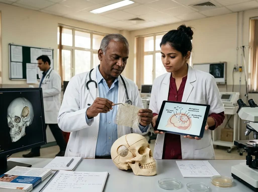
Researchers have engineered a self-powered Zn/C micro-galvanic electrospun membrane that couples sustained zinc release with endogenous microcurrents. This bioelectric scaffold stimulates M2 macrophage polarization and accelerates bone deposition to achieve robust cranial defect regeneration.
Today

A meta-analysis of 18 studies reveals that bariatric surgery does not significantly alter circulating homocysteine levels, regardless of follow-up duration. Clinicians must maintain vigilance over post-surgical micronutrient supplementation to prevent hyperhomocysteinemia.
Today
