

Loading, please wait...
Loading, please wait...

Acute mechanical trauma to the spine initiates a catastrophic sequence of cellular events known as secondary neurological injury. In clinical neurotrauma, managing traumatic spinal cord injury requires prompt recognition of molecular disruptions that exacerbate tissue necrosis. Consequently, clinicians must look beyond static radiographic imaging to evaluate real-time biochemical deterioration. Furthermore, recent clinical investigations highlight how excitatory amino acids drive sustained secondary tissue destruction. Understanding these neurochemical alterations helps surgical and critical care teams anticipate functional deficits and refine individualized protective strategies.
Primary mechanical trauma disrupts microvascular integrity, collapses local perfusion, and destabilizes neuronal membranes within minutes of impact. Consequently, damaged cells spill excessive concentrations of glutamic acid and aspartic acid into the extracellular space. These excitatory neurotransmitters persistently overstimulate post-synaptic N-methyl-D-aspartate and alpha-amino-3-hydroxy-5-methyl-4-isoxazolepropionic acid receptors. Therefore, massive calcium ion influx overwhelms mitochondrial buffering capacity and triggers destructive enzymatic pathways. Intracellular proteases, calpains, and endonucleases rapidly degrade the structural cytoskeleton of surviving spinal neurons. In addition, dysfunctional astrocytes fail to reabsorb ambient glutamate due to severe energy depletion. This failure creates a toxic self-amplifying cascade that expands the original zone of spinal parenchymal infarction. Meanwhile, microglial cells release inflammatory cytokines, reactive oxygen species, and secondary neurotoxins that worsen localized spinal edema. Consequently, secondary excitotoxicity converts partial tissue deficits into permanent neurological injury across neighboring white matter tracts. Experimental and clinical data demonstrate that the intensity of this biochemical insult directly dictates functional survival. Therefore, early quantification of excitatory neurotransmitters offers invaluable insight into secondary lesion development and long-term functional recovery. Thus, interrupting early excitotoxic signaling remains a paramount objective in acute spine management.
A landmark clinical study prospectively enrolled 62 consecutive patients presenting with secondary traumatic spinal damage between January 2021 and December 2023. Investigators gathered paired peripheral blood and cerebrospinal fluid samples at days 1, 3, 7, 14, and 28 post-injury. Furthermore, technicians quantified glutamic acid and aspartic acid concentrations using high-performance liquid chromatography with standardized fluorometric detection. This sophisticated analytical approach ensured precise measurement of amino acid concentrations even at minute nanomolar thresholds. Simultaneously, neurosurgeons categorized neurological deficits using the American Spinal Injury Association impairment scale at initial hospital admission. Researchers subsequently completed rigorous neurological follow-up evaluations at three months after the initial trauma. Consequently, the team stratified patients into two distinct prognostic cohorts based on functional improvement. The good prognosis group achieved an increase of 10 or more points on their neurological recovery score. Conversely, the poor prognosis group demonstrated minimal functional recovery with score improvements falling strictly below 10 points. Thus, the clinical protocol successfully correlated dynamic temporal amino acid trajectories with validated functional recovery parameters. This systematic methodology highlights the utility of serial laboratory tracking in acute neurotrauma wards.
The laboratory analysis revealed striking temporal fluctuations in excitatory amino acid concentrations throughout the twenty-eight-day observation period. Specifically, both glutamic acid and aspartic acid peaked rapidly within the first 72 hours after initial trauma. Cerebrospinal fluid concentrations reached levels several-fold higher than baseline physiologic parameters during this hyperacute phase. In addition, peripheral venous concentrations mirrored central neurochemical surges, confirming significant disruption of the blood-spinal cord barrier. Concentrations gradually declined over days 7 and 14 as endogenous homeostatic mechanisms regained partial clearance capacity. Nevertheless, sustained elevation persisted through day 28 in patients suffering from extensive cord compression. Notably, patients in the poor prognosis cohort maintained significantly higher concentrations of both neurotransmitters across every single sampling milestone. Conversely, patients achieving favorable motor recovery demonstrated a swift return toward baseline transmitter levels by the end of the second week. Therefore, the duration of biomarker elevation reflects persistent secondary pathological processes rather than transient mechanical disturbance alone. Consequently, measuring these continuous curves provides clinicians with superior longitudinal resolution compared to single admission titers. Furthermore, persistently elevated blood levels correlated tightly with ongoing central excitotoxicity.
Statistical analysis confirmed a robust inverse correlation between peak excitatory amino acid levels and neurological improvement at three months. Specifically, patients with marked cerebrospinal fluid glutamate elevation during week one exhibited significantly worse sensory scores. Aspartic acid concentrations displayed an identical predictive capacity regarding independent ambulation and bladder control. Moreover, receiver operating characteristic curve analysis revealed excellent sensitivity and specificity for early transmitter surges in predicting poor outcomes. Elevated blood aspartate at day three accurately identified patients at elevated risk for permanent quadriplegia. Furthermore, combined assessment of both amino acids generated higher prognostic accuracy than either individual metabolic marker alone. Clinicians previously relied exclusively on baseline motor scoring to forecast long-term functional independence. However, severe spinal shock frequently clouds acute clinical examinations during the initial resuscitations. Therefore, objective biochemical measurements provide critical clarity when clinical sensory testing remains unreliable or inconclusive. Additionally, linear regression models confirmed that prolonged transmitter elevation independently predicted diminished functional independence ratings. Ultimately, persistent excitotoxicity correlates directly with irreversible axonal disruption and enduring motor deficit.
These clinical findings present profound therapeutic implications for intensive care specialists managing acute spinal trauma. First, routine quantification of excitatory neurotransmitters can identify candidates for aggressive secondary neuroprotective protocols. For example, neurointensivists can titrate hypothermia, mean arterial pressure augmentation, or experimental glutamate receptor antagonists based on molecular severity. In addition, serial sampling allows clinical teams to monitor therapeutic efficacy in real time. If excitatory biomarkers remain persistently elevated, clinicians must suspect ongoing ischemia, unstable alignment, or expanding subdural hematoma. Furthermore, identifying unfavorable metabolic trajectories early encourages proactive dialogue with families regarding rehabilitation expectations and functional prognosis. Consequently, neurosurgeons can allocate specialized rehabilitation resources and adaptive equipment far more efficiently during early hospitalization. Modern spine centers must therefore integrate biochemical profiling alongside advanced magnetic resonance imaging protocols. Moreover, precise metabolic stratification minimizes futile interventions while maximizing targeted physiological support during critical window periods. Ultimately, bridging molecular diagnostics with acute surgical intervention optimizes patient outcomes following traumatic neural damage.
In Indian tertiary centers, neurotrauma units handle an enormous volume of vehicular accidents and construction falls. However, limited access to advanced intraoperative monitoring often hampers precise prognostication in overcrowded trauma intensive care wards. Therefore, validating accessible blood-based biomarkers of excitotoxicity offers tremendous practical value for Indian healthcare systems. High-performance liquid chromatography infrastructure already exists across many diagnostic teaching hospitals and accredited commercial pathology networks. Consequently, establishing standardized referral testing for blood glutamate and aspartate requires minimal novel capital investment. Furthermore, early prognostic stratification helps Indian critical care physicians optimize bed allocation in resource-constrained environments. Families can also make informed decisions regarding expensive prolonged intensive therapy and long-term home nursing needs. Moreover, monitoring systemic amino acid trajectories empowers medical teams to refine supportive hemodynamics without requiring serial invasive spinal punctures. In addition, early prognostic clarity provides families with objective biological evidence of injury severity. Thus, implementing neurochemical monitoring represents a practical, scalable innovation for acute trauma care across Indian hospitals.
Glutamic acid and aspartic acid act as major excitatory neurotransmitters in the human central nervous system. Following mechanical spinal injury, dying cells release excessive amounts of these transmitters into extracellular fluid. This massive excess overactivates glutamate receptors, triggering toxic intracellular calcium influx, mitochondrial breakdown, free radical synthesis, and irreversible neuronal apoptosis.
Blood testing offers an accessible, non-invasive method that closely reflects central excitotoxicity because spinal trauma severely compromises the blood-spinal cord barrier. Although cerebrospinal fluid testing provides direct central measurements, lumbar punctures carry significant procedural risks in unstable spine injuries. Therefore, serial peripheral blood sampling serves as a reliable surrogate for clinical monitoring.
Serial tracking reveals the exact duration and magnitude of ongoing secondary neural injury. Patients demonstrating swift biomarker normalization within fourteen days consistently achieve superior neurological recovery. Conversely, persistently high glutamate and aspartate levels indicate ongoing secondary tissue destruction, accurately warning clinical teams of poor motor recovery and potential long-term functional impairment.
Disclaimer: This content is for informational and educational purposes only... Refer to the latest local and national guidelines for clinical practice.
References

Read summarized clinical updates, watch expert medical content, and earn CME certifications right from your smartphone.
Open your smartphone camera to scan and install.





Recent clinical research demonstrates that dynamic fluctuations of glutamate and aspartate in blood and cerebrospinal fluid correlate with secondary injury severity and functional recovery after traumatic spinal cord injury, offering vital prognostic insight for acute neurotrauma care.
Today

A retrospective CAHAL study comparing Amplatzer and Occlutech devices in 413 patients found no significant difference in moderate-to-large residual shunt rates at 6 months follow-up after percutaneous PFO closure, supporting clinical equivalence between both platforms.
Today

AIIMS researchers suggest yoga may alleviate gut dysbiosis in Polyendocrine Metabolic Ovarian Syndrome (PMOS) by modulating the gut-brain axis, lowering chronic inflammation, and enhancing parasympathetic tone to support metabolic and hormonal equilibrium.
Today
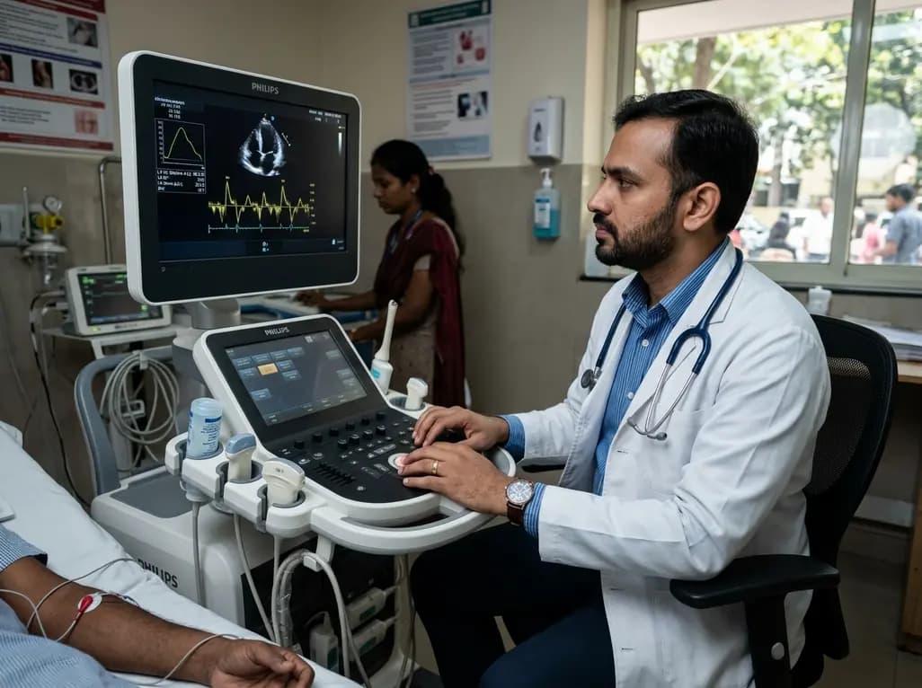
A structured practical guide to left atrial echocardiographic assessment covering biplane volumetry, phasic physiology, speckle-tracking strain, and hemodynamic filling pressure estimation for routine cardiovascular practice.
Today

Age-related spinal kyphosis is driven by vertebral fragility, disc degeneration, and paraspinal muscle dysfunction. Coexisting with locomotive syndrome and frailty, its management requires combined anti-osteoporotic therapy, spinal extensor rehabilitation, and comprehensive nutritional optimization.
Today

A longitudinal feasibility pilot study highlights that combining polygenic risk score disclosure with in-person lifestyle counseling significantly enhances genomic literacy in individuals at risk for type 2 diabetes, overcoming educational disparities and reinforcing patient motivation.
Today
