

Loading, please wait...
Loading, please wait...

Understanding intratumoral heterogeneity remains a central challenge in modern clinical oncology. Malignant tumors do not exist as isolated cellular clones. Instead, they operate as complex ecosystems composed of malignant cells, immune infiltrates, and vascular networks. Within this cellular architecture, discrete microenvironments govern disease behavior, therapy evasion, and metastatic dissemination. Investigating tumor niche dynamics provides essential insights into how regional cellular neighborhoods evolve over time. Recently developed spatial sequencing tools capture these intricate spatial relationships without sacrificing anatomic context. By cataloging these regional patterns, researchers can illuminate critical biological transitions driving therapeutic resistance.
Conventional bulk molecular profiling averages transcriptional signatures across millions of disparate cells. Consequently, bulk techniques completely obscure rare subclonal populations and localized cellular crosstalk. While single-cell RNA sequencing resolved individual cellular identities, it inevitably sacrificed tissue topography during sample dissociation. Therefore, clinicians and translational researchers struggled to pinpoint exactly where critical immune-suppressive signals originated within solid tumors. Spatial transcriptomics effectively bridged this divide by preserving tissue architecture while measuring deep transcriptomic programs. However, analyzing spatial datasets across diverse cohorts presents immense computational hurdles. Investigators often encounter incompatible platforms, variable resolution thresholds, and fragmented data repositories. Moreover, tracking how microenvironments remodel during progression requires integrated longitudinal frameworks rather than static snapshots. Clinicians need standardized platforms that bridge single-cell resolution with histological tissue coordinates. By connecting cellular identities to physical coordinates, pathologists can better understand why specific tumor regions resist chemotherapy or radiotherapy. Thus, harmonized pan-cancer repositories are vital to transform raw spatial data into actionable mechanistic insights.
To address the limitations of fragmented data, investigators established EvoNiche as an open-access knowledge base. The platform systematically integrates ninety-one matched spatial and single-cell transcriptomic datasets spanning thirty-five distinct cancer malignancies. Overall, the repository encompasses 712 tissue sections containing nearly two million spatial spots. In addition, the creators curated 58 single-cell transcriptomic datasets covering nearly three million individual cells. This rigorous integration establishes an unprecedented biological atlas of human malignancies. Through harmonized processing pipelines, EvoNiche delineates 5609 unique spatial niches across varied anatomic sites. Furthermore, the platform reconstructs both tissue-level and single-cell-level evolutionary trajectories. Users can explore high-resolution spatial maps that link histopathological morphology directly to transcriptional phenotypes. Because the resource standardizes disparate profiling methods, investigators can evaluate cross-cohort similarities without cumbersome pre-processing steps. Additionally, intuitive data exploration interfaces allow oncologists to inspect specific gene expression signatures across matched spatial coordinates. As a result, the database converts complex multi-omic data into interpretable visualizations of malignant ecosystems.
Tumors constantly reorganize their internal neighborhoods in response to nutrient deprivation, hypoxia, and immunological attacks. Therefore, understanding tumor niche dynamics requires tracking continuous evolutionary paths rather than static histological zones. EvoNiche achieves this capability by deploying advanced spatial pseudotime modeling and trajectory reconstruction tools. Specifically, the framework models how localized cellular zones transition into aggressive, invasive margins. Along these computational trajectories, the database resolves dynamic shifts in cellular proportions and functional gene networks. For instance, researchers can follow the gradual accumulation of cancer-associated fibroblasts and exhausted lymphocytes along regional progression vectors. Moreover, the platform highlights trajectory-associated biological functions, including epithelial-mesenchymal transition and metabolic reprogramming. Consequently, clinicians gain clear visual representations of regional tumor evolution across different stages of malignancy. In addition, the analytical framework quantifies subtle microenvironmental alterations that precede gross histological invasion. Because these transitions occur before metastatic spread becomes clinically evident, mapping trajectory paths uncovers early intervention opportunities. Thus, trajectory mapping clarifies the fundamental rules governing regional cancer evolution.
Within the malignant architecture, localized cell-to-cell communication dictates therapeutic susceptibility and tumor invasion. EvoNiche directly investigates these interactions by calculating spatially proximal ligand-receptor pairings across defined ecological niches. Consequently, the database uncovers how malignant cells reprogram surrounding stromal cells through paracrine and juxtacrine signals. For example, the interface between the tumor invasive margin and adjacent healthy parenchyma exhibits unique immunosuppressive signaling cascades. EvoNiche characterizes these boundary microenvironments in detail, highlighting specialized molecular programs that protect malignant cells from cytotoxic surveillance. Furthermore, the platform evaluates how dense extracellular matrix deposition creates physical barriers against therapeutic agents and infiltrating lymphocytes. Because spatial proximities determine whether signaling actually occurs in vivo, this spatial mapping eliminates the false-positive interactions common in dissociated single-cell data. Additionally, the database links distinct communication networks to localized functional outputs, such as angiogenesis and stemness maintenance. As a result, researchers can identify actionable ligand-receptor pairs that maintain immunosuppressive niches. Therefore, targeting these local spatial circuits may restore immune infiltration and enhance therapeutic responses.
The clinical value of spatial transcriptomics lies in its ability to discover robust diagnostic and prognostic biomarkers. EvoNiche prioritizes region-specific and transition-associated genes as spatially derived candidate biomarkers across pan-cancer cohorts. Furthermore, the developers systematically validated these candidates against clinical outcomes and immunotherapy response datasets. Because standard prognostic models frequently ignore tissue organization, spatially informed biomarkers offer superior discriminatory accuracy for patient stratification. For instance, specific transition genes identified by the platform correlate closely with immune checkpoint inhibitor resistance. Consequently, oncologists can leverage these spatial signatures to identify patients unlikely to benefit from conventional PD-1 or CTLA-4 blockade. Moreover, identifying specialized niches, such as tertiary lymphoid structures, provides vital insights into anti-tumor immunity and patient survival. Clinicians can also identify localized enzymatic targets that promote immune exclusion within solid tumors. Therefore, these spatial candidate genes represent promising targets for novel combination therapies. In conclusion, EvoNiche bridges high-throughput spatial discovery with practical clinical translation, paving the way for spatially guided precision oncology.
Conventional single-cell RNA sequencing dissociates intact tissue into isolated cell suspensions. Consequently, it discards critical information regarding tissue histology and exact cellular coordinates. In contrast, spatial transcriptomics quantifies gene expression patterns directly on preserved histological sections. Therefore, spatial profiling preserves the native anatomic architecture. This capability allows researchers and oncologists to observe localized cellular interactions, map regional gradient shifts, and identify specialized spatial niches that govern tumor growth and therapeutic resistance.
Clinicians in India can utilize EvoNiche as an open reference atlas to benchmark localized biopsy specimens and validate emerging biomarkers. Furthermore, the database helps oncologists understand treatment failure mechanisms without requiring prohibitive spatial profiling infrastructure. By analyzing publicly available spatiotemporal trajectory data, clinical researchers can identify novel therapeutic targets, design biomarker-driven clinical trials, and explore tailored combination regimens for diverse cancer populations across domestic healthcare centers.
Spatial transition genes mark dynamic cellular states that emerge as tumors invade adjacent healthy tissues or resist therapeutic interventions. Consequently, these genes highlight localized mechanisms of immune evasion, such as T-cell exclusion or fibroblast activation. By evaluating spatial transition signatures, clinicians can identify immunosuppressive barriers that standard bulk profiling overlooks. Therefore, these genes provide highly reliable predictive biomarkers for stratifying immunotherapy candidates and overcoming checkpoint resistance.
Disclaimer: This content is for informational and educational purposes only, and does not substitute professional medical advice, diagnosis, or treatment. Refer to the latest local and national guidelines for clinical practice.
References

Read summarized clinical updates, watch expert medical content, and earn CME certifications right from your smartphone.
Open your smartphone camera to scan and install.





EvoNiche is a comprehensive resource integrating spatial transcriptomics and single-cell profiling across 35 cancer types to decode tumor niche dynamics, spatiotemporal heterogeneity, and therapeutic targets.
Today
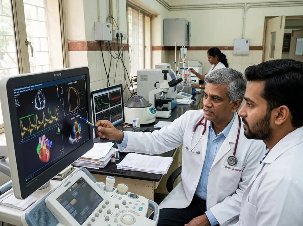
A study evaluates a cardiometabolic HFpEF model combining metabolic stress with chronic mineralocorticoid excess. The findings reveal profound diastolic stiffness, endothelial dysfunction, and pulmonary hypertension, illuminating key pathophysiological mechanisms for targeted cardiopulmonary therapies.
Today

The Human Reproduction and Development Database (HRD) consolidates bulk and single-cell transcriptomics, epigenomics, metabolomics, proteomics, and clinical trial records into a unified platform to advance reproductive health research.
Today
Partial heart transplantation offers living, growth-capable cardiac valve replacements for children. While orthotopic heart transplantation faces severe logistical barriers in low-resource settings, partial transplantation presents lower graft ischemia sensitivity and simplified immunosuppression protocols.
Today

A single-cell transcriptomic atlas reveals how peripheral nerve injuries cause dorsal root ganglion remodeling. By evaluating proximal nerve crush and resection, researchers identify distinct glial and immune programs that resolve neuropathic pain and promote regeneration.
Today
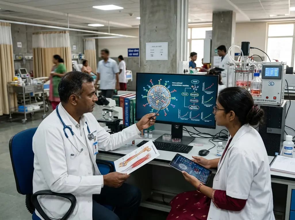
A breakthrough study demonstrates that fullerol-primed extracellular vesicles generated in a 3D SECCS bioreactor attenuate ferroptosis and restore myogenic regeneration in diabetes via the FOXO3 pathway, offering a cell-free therapeutic strategy for diabetic musculoskeletal degeneration.
Today
