.webp&w=384&q=75)

Loading, please wait...
Loading, please wait...

Minimally invasive transforaminal lumbar interbody fusion has transformed surgical management for degenerative lumbar spine conditions over the past two decades. However, the integration of advanced image guidance has redefined the procedural workflow, establishing navigated MIS-TLIF as a modern benchmark for technical precision. While initial adoption often raised concerns regarding operating room setup time and technological complexity, contemporary longitudinal evidence illustrates how progressive technical mastery ultimately streamlines surgical efficiency and enhances procedural safety.
Historically, conventional minimally invasive spine stabilization relied extensively on two-dimensional fluoroscopy. Although percutaneous screw insertion and tubular retractor systems reduced muscular trauma compared to open arthrodesis, reliance on repetitive ionizing imaging introduced significant occupational hazards. Surgeons and theater teams faced cumulative radiation exposure throughout prolonged procedures. Furthermore, relying purely on planar fluoroscopy created visual challenges when identifying pedicle trajectories across dysplastic or severely osteopenic anatomy.
Consequently, image-guided navigation platforms emerged to mitigate these structural limitations. Computer-assisted three-dimensional navigation systems integrate real-time tracking arrays with intraoperative computed tomography. This setup provides continuous anatomical orientation in axial, coronal, and sagittal planes simultaneously. As a result, early adopters reported heightened accuracy during pedicle instrumentation and reduced breach rates. Nevertheless, early clinical reports often identified longer total operative duration as an initial trade-off. This delay stemmed from intraoperative registration steps, device calibration, and learning curves. Therefore, understanding the longitudinal evolution from conventional fluoroscopy to full technological integration remained a critical clinical objective.
To evaluate this technological transition, investigators conducted a comprehensive 15-year retrospective observational study evaluating 823 consecutive patients undergoing MIS-TLIF between 2008 and 2022. The investigators stratified the entire cohort into two primary clinical cohorts: a non-navigated cohort comprising 427 patients treated under traditional fluoroscopic guidance, and an image-guided navigation cohort comprising 396 patients who underwent navigated MIS-TLIF protocols.
Furthermore, researchers categorized patients across four distinct historical epochs to analyze technical maturation. The earliest epochs, 2008 to 2011 and 2012 to 2015, utilized conventional fluoroscopic methods without computer assistance. Subsequently, the transitional era between 2016 and 2019 introduced navigation specifically for pedicle screw insertion, representing partial navigation. Finally, the modern era from 2020 through 2022 established total navigation, expanding real-time tracking beyond screw placement to encompass neural decompression, disc preparation, and endplate interbody grafting. Clinical endpoints encompassed total surgical duration, estimated intraoperative blood loss, targeted disc levels, and revision rates associated with screw malposition, surgical site infection, implant subsidence, or adjacent segment disease.
The longitudinal data demonstrated marked procedural improvements over the study period. In non-navigated single-level procedures, surgical duration declined steadily as surgical teams accumulated general minimally invasive experience. Operative time decreased from 266.0 minutes during the baseline 2008 to 2011 period to 215.5 minutes between 2012 and 2015, and finally settled at 191.7 minutes during the 2016 to 2019 era. This steady reduction highlights the natural technical learning curve inherent to tubular spine approaches.
Crucially, the introduction of intraoperative navigation altered this trajectory positively once institutional familiarity was established. Although critics anticipated that registration steps would lengthen operative times, total operative duration in navigated cases shortened markedly as workflows matured. Navigated instrument tracking streamlined pedicle cannulation, eliminating tedious alternating fluoroscopy views between lateral and anteroposterior trajectories. Additionally, automated trajectory planning removed guesswork during screw tapping and insertion. Consequently, operative workflows achieved high predictability, disproving the historical assumption that navigation inherently prolongs operating room duration in established programs.
Beyond single-segment stabilization, navigation technology fundamentally transformed the feasibility of complex interventions. During the earliest study periods, non-navigated approaches focused predominantly on single-level degenerative instability or isolated spondylolisthesis. Multilevel minimally invasive interventions remained uncommon because cumulative fluoroscopy time and extensive mental reconstruction placed prohibitive cognitive demands on the operating surgeon.
However, the establishment of comprehensive navigation expanded surgical indications to multilevel degenerative disc pathology. Surgeons comfortably addressed two or more adjacent disc spaces through minimally invasive corridors without increasing complication rates. Specifically, navigation accelerated procedural speed across multilevel interventions because a single intraoperative scan provided continuous spatial orientation across several vertebral levels. Surgeons placed multiple bilateral pedicle screws rapidly using calibrated instruments without repeating intraoperative scans. Moreover, revision rates attributable to hardware malposition, neurological injury, or mechanical implant failure remained consistently low across all temporal cohorts, confirming that procedural expansion did not compromise structural safety.
The progression toward full navigation culminated in an innovative fluoro-less surgical paradigm. In traditional workflows, spine surgeons relied heavily on repeated fluoroscopic bursts to verify retractor docking, guide annulotomy knives, assess disc clearing, and inspect interbody cage expansion. This continuous imaging exposed operating personnel to persistent radiation and interrupted surgical cadence.
In contrast, the total navigation protocol from 2020 to 2022 integrated navigated debridement instruments, tracked disc shavers, and calibrated trial cages. Surgeons verified decompression margins and monitored cage trajectory in real time on three-dimensional displays without activating fluoroscopy units. Consequently, intraoperative fluoroscopy was eliminated entirely from the procedure. This shift established substantial radiation safety benefits for surgical staff while maintaining uncompromising accuracy during endplate decortication. Furthermore, eliminating mobile C-arm manipulation improved theater ergonomics and reduced operative field contamination risks.
These long-term findings offer compelling actionable guidance for orthopedic spine surgeons and neurosurgeons evaluating technology adoption. While institutional acquisition costs and initial setup hurdles require planning, modern navigation delivers profound dividends in procedural efficiency, safety, and occupational health. Overcoming the initial learning curve enables surgical teams to complete complex reconstructions with heightened confidence.
Additionally, institutional protocols should emphasize standardized team training to optimize operating room turnover and array calibration. Standardizing patient positioning, pin placement, and imaging capture ensures rapid registration. As surgical centers prioritize tissue-sparing interventions and low revision rates, transitioning toward comprehensive navigation represents a reliable pathway for modern spine programs. Navigated approaches safely extend the benefits of minimally invasive fusion to broader patient cohorts while protecting healthcare workers from ionizing exposure.
Although navigation requires initial setup and instrument calibration, experienced surgical teams achieve shorter operative times. Navigation eliminates repetitive intraoperative fluoroscopic checks and streamlines pedicle cannulation. Consequently, procedural efficiency improves significantly once teams overcome the initial technological learning curve, especially in complex multilevel cases.
Partial navigation utilizes computer guidance solely for percutaneous pedicle screw trajectory and insertion, whereas total navigation expands real-time tracking to include tubular retractor docking, neural decompression, disc preparation, and interbody cage delivery. This comprehensive integration entirely removes the requirement for conventional intraoperative fluoroscopy.
Yes. Longitudinal evidence confirms that navigated MIS-TLIF maintains remarkably low rates of screw malposition, mechanical implant failure, and revision surgery. Real-time spatial tracking permits safe expansion to multilevel pathologies, allowing surgeons to achieve dependable neural decompression and accurate graft placement without raising complication rates.
Disclaimer: This content is for informational and educational purposes only and should not be construed as medical advice. Always consult with a qualified healthcare professional before making any changes to your health or wellness routine. Refer to the latest local and national guidelines for clinical practice.
References

Read summarized clinical updates, watch expert medical content, and earn CME certifications right from your smartphone.
Open your smartphone camera to scan and install.





A 15-year retrospective cohort study of 823 patients reveals how navigation technology transformed minimally invasive transforaminal lumbar interbody fusion (MIS-TLIF), expanding indications to multilevel spine disease, optimizing operative times, and establishing a safe, fluoro-less surgical workflow.
Today
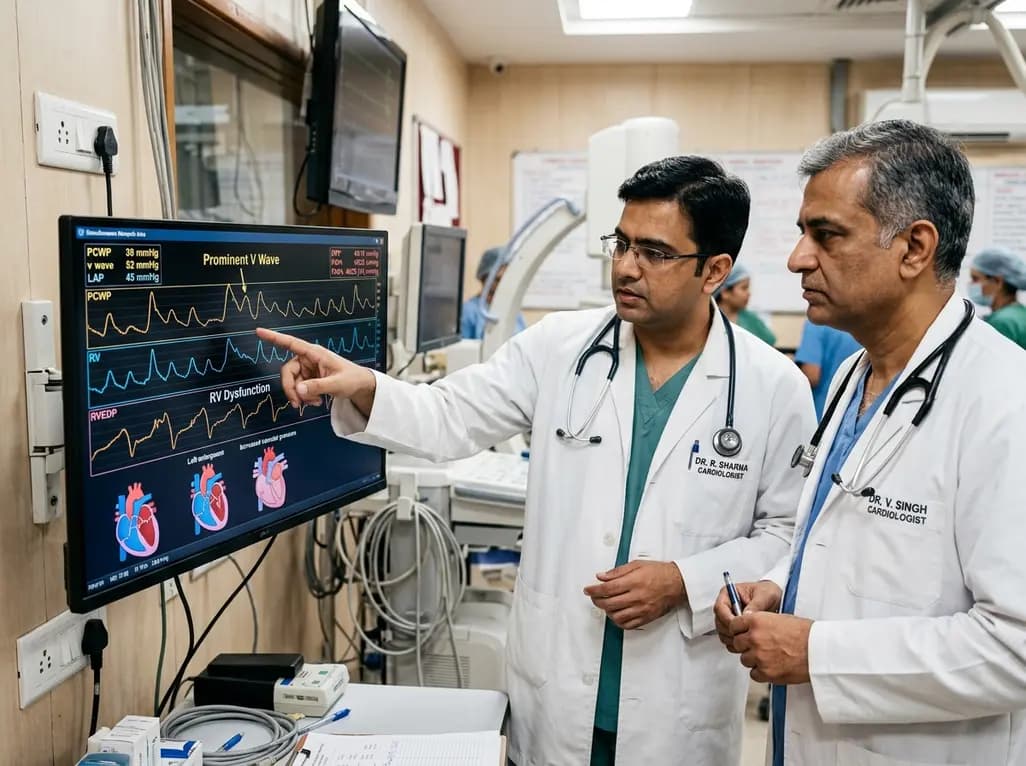
Left atrial stiffening generates prominent V waves that pressurize the pulmonary circulation and augment right ventricular afterload independently of intrinsic pulmonary vascular disease in patients with heart failure with preserved ejection fraction.
Today

A meta-analysis demonstrates that secondhand smoke exposure significantly elevates fetal loss risk by 21% among nonsmoking pregnant women. Clinicians must implement household screening and family-centered cessation interventions to prevent avoidable miscarriages and stillbirths.
Today

Preclinical validation demonstrates that a novel contact force-integrated pulsed field quantization index accurately predicts lesion depth in real time, achieving 100% durable pulmonary vein isolation without collateral injury.
Today
A new cross-sectional evaluation reveals how regional lean mass and bone mineral content interact in resistance-trained men, showing that whole-body size and allometric scaling explain shared musculoskeletal phenotypes and strength relationships.
Today
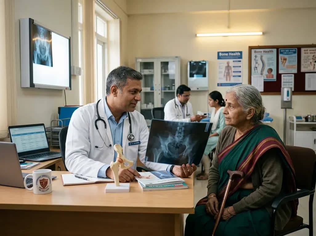
Explore the emerging role of glucose metabolic reprogramming in exercise-attenuated osteoporosis, examining how physical activity influences osteoblast glycolysis, osteoclast bioenergetics, and skeletal remodeling pathways to improve bone mineral density.
Today
