.webp&w=384&q=75)

Loading, please wait...
Loading, please wait...
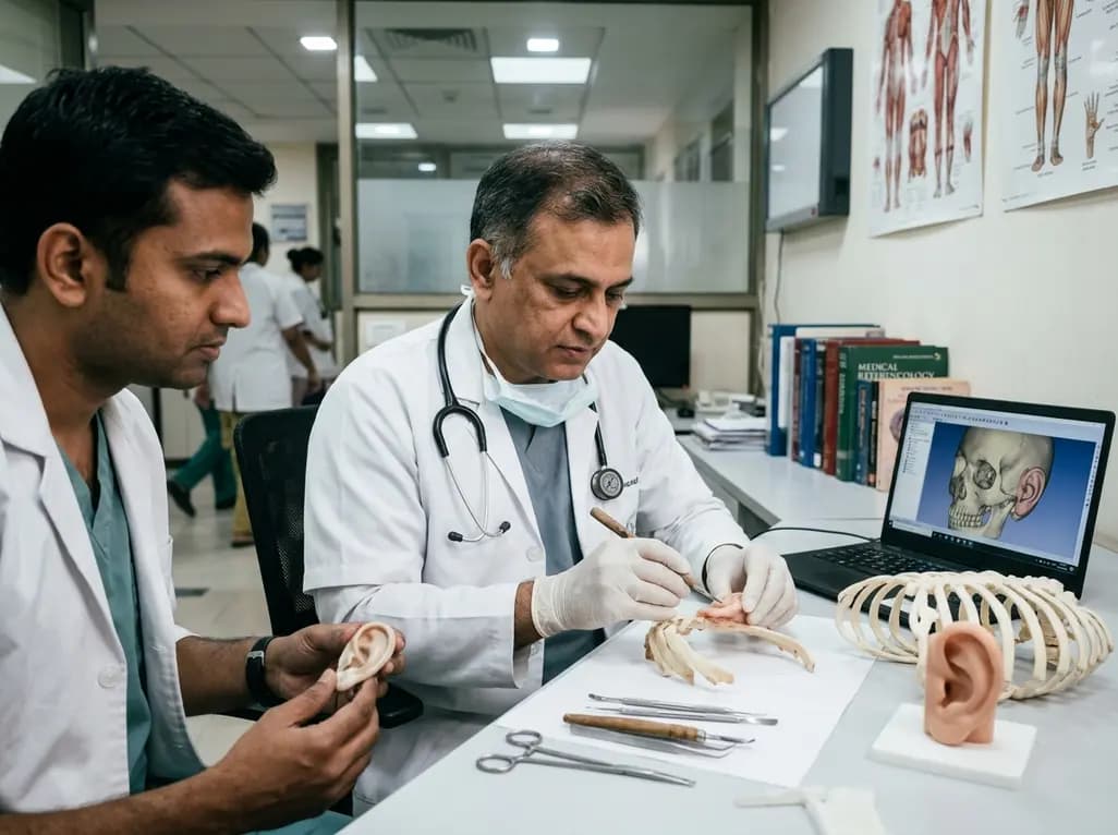
Auricular defects present complex structural, functional, and psychological dilemmas for patients and healthcare providers. Successful ear reconstruction demands a delicate balance of biological science, meticulous manual dexterity, and aesthetic perception. Clinicians address these deformities through two primary disciplines: surgical intervention and prosthetic rehabilitation. While plastic surgeons harvest tissue or carve biocompatible structures, anaplastologists handcraft detailed silicone epitheses. Historically, both pathways evolved along parallel tracks of relentless technical innovation. Today, the clinical community recognizes that technological advancements cannot substitute for refined artisan expertise. Consequently, restoring the delicate curves of the auricle remains one of the ultimate tests of craft skill in modern reconstructive medicine.
Historical records indicate that the pursuit of external ear repair spans millennia. Ancient Indian surgeons in the Sushruta Samhita first described local flap techniques to mend severed earlobes. Centuries later, Renaissance surgeons utilized retroauricular tissue transfers to reconstruct traumatic ear defects. However, complete auricular restoration remained elusive until the twentieth century, when Harold Gillies pioneered cartilage grafting. Initially, Gillies experimented with maternal and banked cartilage, but progressive biological resorption hindered consistent long-term results.
Meanwhile, anaplastologists developed prosthetic alternatives for patients who lacked adequate local tissue or declined operative risks. Early prostheses incorporated rigid substances, including carved ivory, vulcanite, and tinted wax. Although these pioneering appliances masked disfigurement, they degraded rapidly, suffered chromatic instability, and adhered poorly to mobile craniofacial tissue. Therefore, prosthodontists and medical sculptors continuously sought resilient, skin-safe compounds. The mid-twentieth century introduced vinyl plastisols and early acrylics, marking significant progress in durability. Ultimately, the development of modern medical-grade silicones transformed the field entirely. These flexible polymers offered lifelike texture, fine feather-edge margins, and remarkable resistance to environmental weathering. Thus, both surgical and prosthetic traditions systematically advanced their foundational craft by refining raw materials and surgical maneuvers.
Autologous rib cartilage remains the gold standard framework for total ear reconstruction worldwide. Radford Tanzer revolutionized the surgical landscape in 1959 by carving a single block of autogenous costal cartilage. Subsequently, Burt Brent standardized a four-stage protocol that dramatically improved structural safety, skin expansion, and aesthetic consistency. Brent emphasized gentle contouring, utilizing synchondrotic segments from the sixth and seventh ribs alongside a distinct floating eighth rib helix.
Decades later, Satoru Nagata introduced a sophisticated two-stage approach that demanded even greater technical proficiency. Nagata incorporated a complex 3D framework featuring an accentuated antihelix, an integrated tragus, and a distinct intertragic notch. Furthermore, Françoise Firmin refined these sculpting protocols by establishing standardized classification systems and projection projections. Carving autologous cartilage requires extraordinary three-dimensional spatial visualization. The reconstructive surgeon acts as an anatomical sculptor, carving rigid cartilage blocks into delicate helices, scaphae, and conchal walls. Moreover, surgeons must preserve crucial perichondrial vascularization while preventing full-thickness tears during structural carving. Meticulous wire fixation ensures framework integrity without compromising delicate overlying cutaneous flaps. Consequently, autologous ear reconstruction demands hours of deliberate manual practice that few standard surgical training programs can replicate completely.
Although autologous cartilage provides enduring durability, the donor-site morbidity associated with rib harvest poses tangible clinical challenges. Therefore, investigators pioneered synthetic framework substitutes to streamline operative times and eliminate thoracic morbidity. Early trials by Cronin with silicone frameworks suffered catastrophic rates of soft tissue extrusion and recurrent infection. However, John Reinisch achieved a major breakthrough by introducing porous high-density polyethylene implants covered with robust vascular flaps.
By transposing an expansive temporoparietal fascia flap over the porous scaffold, surgeons significantly reduced extrusion rates. Furthermore, this method permits ear reconstruction at a younger age because patients do not need mature rib cartilage. Nevertheless, alloplastic constructs demand rigorous vascular judgment and delicate microvascular handling to avoid catastrophic exposure.
Concurrently, prosthetic rehabilitation remains an invaluable treatment arm, particularly for cancer resections and failed surgical cases. Modern anaplastologists utilize silicone elastomeric systems that replicate dermal translucency and subtle contralateral vascular patterns. Moreover, the advent of titanium craniofacial osseointegrated implants provides dependable mechanical retention, replacing troublesome dermal adhesives. Anaplastologists paint intrinsic pigments directly into layered silicone, reproducing authentic cutaneous melanin and vascular undertones. Thus, modern prosthetic craftsmanship restores anatomical harmony with extraordinary realism while avoiding surgical donor morbidity.
Contemporary digital engineering has introduced powerful tools into both surgical suites and anaplastology laboratories. Specifically, clinicians now routinely employ computer-aided design and computer-aided manufacturing alongside high-resolution surface scanning. Three-dimensional photography accurately captures the contralateral normal ear, generating mirrored virtual models with sub-millimeter precision. Clinicians convert these digital surface templates into physical resin stereolithographic guides for operative carving.
In anaplastology, digital workflows allow practitioners to fabricate rapid three-dimensional prototype molds for vulcanized silicone. Consequently, these advances markedly streamline preliminary sculpting stages and reduce manual fabrication errors. Furthermore, emerging tissue-engineering investigations aim to print living chondrocyte-seeded scaffolds that may eventually eliminate both donor-site harvesting and synthetic foreign materials.
However, modern digital instruments cannot replace core clinical acumen or manual artisan capability. While a three-dimensional guide provides exact geometric proportions, it cannot manipulate cartilage elasticity or gauge local dermal perfusion. The surgeon must still assess tension, trim fragile edges, and adapt to unexpected anatomical variations. Similarly, digital milling cannot duplicate the delicate hand-painted tinting and feathered edge blending that anaplastologists perform. Therefore, emerging technologies enhance efficiency and surgical planning, but fundamental craft skills remain indispensable.
Patient-centered clinical care requires clinicians to tailor reconstructive pathways to the individual's anatomy, age, and systemic health. Pediatric patients with congenital microtia often achieve optimal outcomes with autologous cartilage reconstructions once their thoracic circumference surpasses sixty centimeters. This milestone typically occurs around age ten, ensuring adequate donor cartilage volume for multi-tier framework construction. Conversely, alloplastic frameworks offer an attractive option for younger children or families who prioritize single-stage procedures.
Meanwhile, adult patients who suffer radical auriculectomy following aggressive cutaneous malignancies frequently benefit most from prosthetic rehabilitation. Radiation therapy often compromises the vascular bed of the temporoparietal region, creating high risks for alloplastic or autologous failure. Under these compromised circumstances, an osseointegrated silicone prosthesis offers minimal operative risk and exceptional cosmetic fidelity. In addition, an easily removable prosthesis allows direct oncological surveillance of the mastoid bed.
Ultimately, successful auricular rehabilitation demands close interdisciplinary collaboration between plastic surgeons, otolaryngologists, and anaplastologists. Clinicians must evaluate hearing rehabilitation needs, such as simultaneous bone-anchored hearing device placement, within the master reconstructive timeline. By respecting the unique indications of each craft, multidisciplinary teams consistently deliver individualized, functionally sound, and aesthetically superior patient outcomes.
Prosthetic rehabilitation is generally preferred when severe trauma, extensive oncologic resection, or previous radiation therapy compromises local vascular tissue. Furthermore, elderly patients or individuals with major systemic comorbidities often cannot tolerate the prolonged anesthesia and donor-site morbidity required for autologous rib cartilage harvesting. In these clinical scenarios, an osseointegrated or adhesive-retained silicone prosthesis provides rapid, aesthetically faithful restoration while allowing direct visual surveillance of underlying tissue beds.
The Brent technique typically requires three to four operative stages, beginning with framework implantation followed by lobule transposition, framework elevation, and tragus construction. In contrast, the Nagata method condenses the reconstruction into two stages by harvesting more cartilage to create an integrated tragus and antihelix unit simultaneously. Consequently, the Nagata protocol demands exceptional sculpting precision and larger chest growth, whereas the Brent approach offers conservative staged safety with smaller initial grafts.
Modern CAD-CAM and three-dimensional printing technologies provide exquisite anatomical models and cutting guides, but they cannot replace traditional craft skills. These digital modalities assist pre-surgical planning and streamline mold fabrication, yet surgeons must still evaluate soft tissue compliance, carve delicate cartilaginous frameworks, and ensure flap perfusion. Similarly, anaplastologists must handcraft lifelike silicone feathering and intrinsic coloring. Thus, digital tools merely assist, rather than displace, fundamental manual artistry and clinical judgment.
Disclaimer: This content is for informational and educational purposes only and should not be considered medical advice. Always consult a qualified healthcare professional regarding any medical condition or treatment. Refer to the latest local and national guidelines for clinical practice.
References

Read summarized clinical updates, watch expert medical content, and earn CME certifications right from your smartphone.
Open your smartphone camera to scan and install.





Auricular reconstruction bridges surgical craftsmanship and prosthetic anaplastology. This review analyzes autologous rib carving, alloplastic implants, and digital modeling, highlighting that emerging CAD-CAM and 3D printing technologies complement but cannot replace manual surgical dexterity and clinical judgment.
Today
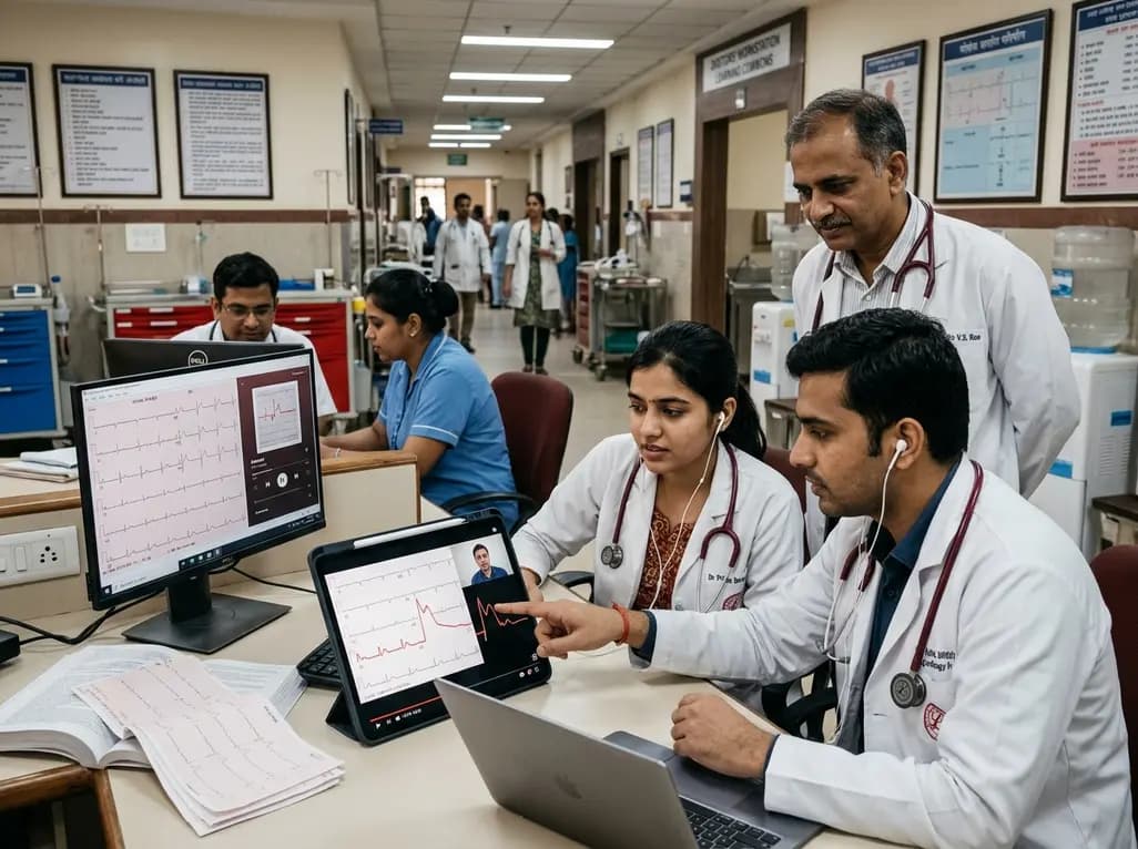
A randomized controlled trial demonstrates that curated FOAMed videos and podcasts produce superior gains in high-risk ECG interpretation and acute coronary syndrome clinical decision-making compared with print-based materials among interprofessional emergency care teams.
Today
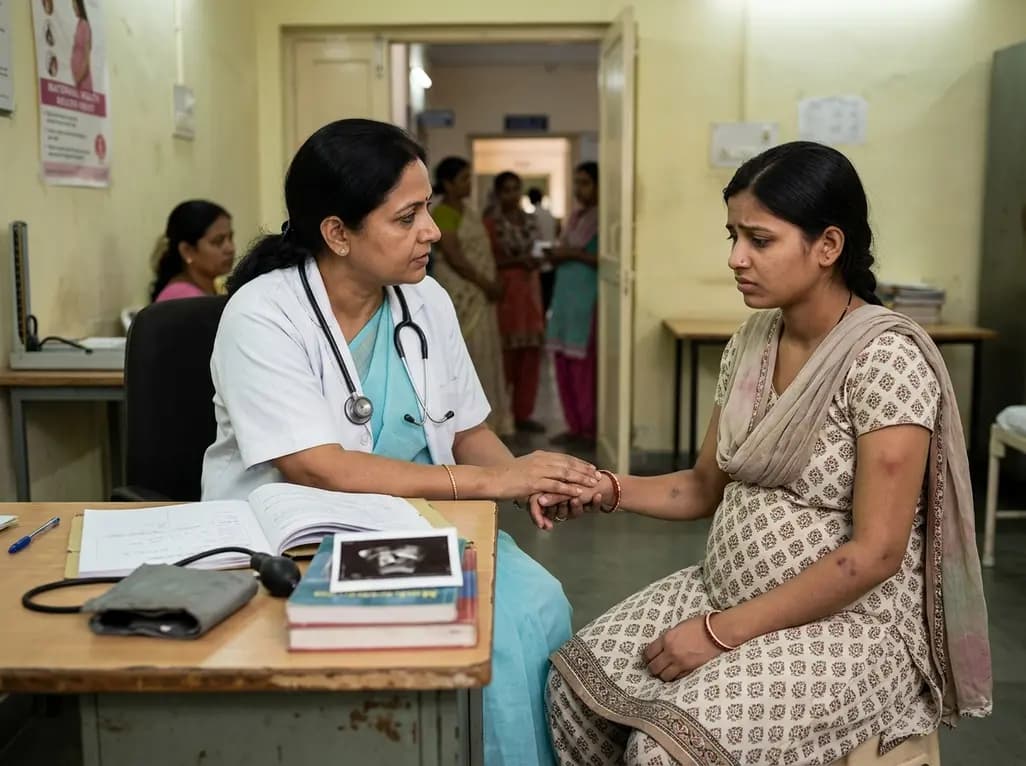
Intimate partner violence during pregnancy significantly increases the risk of miscarriage, premature membrane rupture, and fetal growth restriction. This clinical review explores epidemiological insights, maternal depression, medico-legal documentation, and universal antenatal screening strategies.
Today
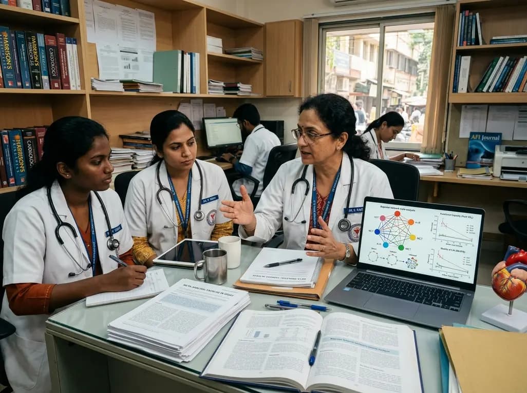
A Bayesian network meta-analysis evaluates exercise training in HFpEF, demonstrating that combined aerobic and resistance training, HIIT, and moderate continuous training significantly enhance peak VO2, while functional electrical stimulation uniquely improves quality of life without altering cardiac remodeling.
Today
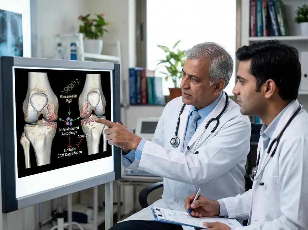
A study reveals that Ginsenoside Rg3 attenuates osteoarthritis progression by modulating the Nrf2-mediated autophagy pathway, reducing chondrocyte apoptosis, and curbing extracellular matrix degradation, offering promising chondroprotective potential.
Today
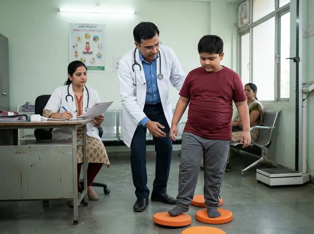
A cross-sectional study demonstrates that lower pediatric motor competence strongly correlates with adverse metabolic syndrome markers, including central adiposity, elevated triglycerides, and low HDL cholesterol, emphasizing early neuromuscular interventions in youth with overweight and obesity.
Today
