

Loading, please wait...
Loading, please wait...
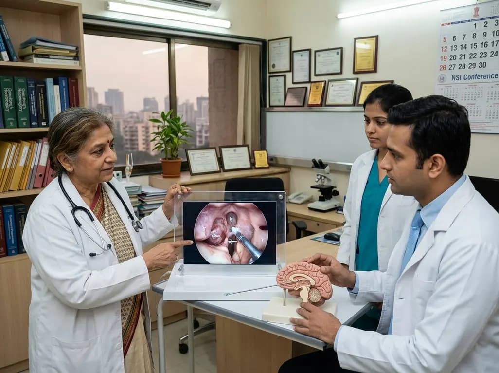
Pathologies situated within the deep ventricular system remain among the most formidable challenges in modern neurosurgery. Specifically, the endoscopic SCIT approach has emerged as an innovative, tissue-preserving operative corridor for addressing deep-seated pathologies within the posterior and ventral compartments of the third ventricle. By exploiting natural anatomical corridors below the tentorium, this method offers neurosurgeons direct visualization while significantly minimizing parenchymal disruption.
Third ventricle colloid cysts and pineal pathologies represent complex technical hurdles due to their central location. Although colloid cysts are histologically benign, they can acutely obstruct cerebrospinal fluid pathways at the foramen of Monro. Consequently, patients frequently present with severe paroxysmal headaches, progressive hydrocephalus, or sudden neurological deterioration. Historically, neurosurgeons relied primarily on microsurgical transcallosal or transcortical transventricular trajectories. While these traditional corridors offer wide exposure, they necessitate traversing healthy cerebral cortex or dividing the anterior corpus callosum. Therefore, patients often face potential postoperative risks, including memory deficits from fornix manipulation, venous infarction, and new-onset seizures. Furthermore, transventricular endoscopic techniques often encounter technical limits when lateral ventricles are small or non-dilated. In this challenging setting, alternative surgical avenues that bypass supratentorial brain retraction are invaluable. The infratentorial corridor circumvents cerebral corticotomy entirely, thereby preserving critical hemispheric white matter tracts and cognitive networks.
The successful execution of this infratentorial corridor demands rigorous patient positioning and precise instrument handling. Surgeons typically place the patient in a prone or Concorde position, taking care to flex the head appropriately. This positioning promotes gravity-dependent cerebellar relaxation, which naturally expands the supracerebellar working space without aggressive retractors. Subsequently, a suboccipital keyhole craniotomy or craniectomy exposes the transverse sinus and the underlying cerebellar vermis. After durotomy, the surgeon introduces rigid neuroendoscopes, often using angled lenses to inspect the quadrigeminal cistern and incisural spaces. Careful arachnoid dissection liberates the precentral cerebellar vein and reveals the posterior incisural window. Crucially, the endoscope brings bright illumination and high-definition magnification directly to the posterior third ventricle wall. Working through a parapineal or suprapineal trajectory, the operator gently accesses the ventricular cavity. Bimanual dissection techniques then permit precise cyst aspiration, capsular mobilization, and hemostasis, thereby protecting the delicate internal cerebral veins and the vein of Galen.
Clinical decision-making requires weighing the distinct biomechanical advantages of each surgical corridor. Transcortical routes provide an expansive corridor into dilated lateral ventricles; however, they inherently require a corticotomy that risks cortical damage and postoperative epilepsy. In contrast, interhemispheric transcallosal approaches prevent cortical transgression, yet they risk venous compromise along the superior sagittal sinus and mutism from anterior callosal disruption. In comparison, the infratentorial approach avoids both cerebral cortex incision and callosal division. Because the corridor traverses the cisterns over the cerebellum, it eliminates retraction on the cingulate gyri and frontal lobes. In addition, the technique offers a direct trajectory toward lesions seated in the ventral and posterior third ventricle. However, the corridor remains narrow, demanding specialized long-shaft micro-instruments and advanced endoscope maneuvering. Thus, surgeons must select candidates judiciously, reserving this route for configurations where lesion geometry aligns naturally with the infratentorial trajectory.
A recent surgical series by Aydemir and colleagues demonstrated encouraging clinical and radiological outcomes using this operative technique. In their cohort of patients treated for colloid cysts, pineal apoplexy, and metastases, the team achieved gross total or near-total resections without intraoperative catastrophes. Long-term follow-up confirmed stable symptom relief and no tumor recurrence. Notably, one patient successfully underwent removal of a preexisting ventriculoperitoneal shunt early in the postoperative period. Postoperative neuroimaging confirmed that minimal cerebellar manipulation caused no secondary ischemic injury or vermian ataxia. Furthermore, the absence of direct traction on the columns of the fornix successfully prevented immediate and delayed neurocognitive deficits. Although air embolism remains a recognized theoretical risk in elevated positioning, proper intraoperative hydration, meticulous surgical technique, and prone positioning mitigate this hazard. Cerebellar swelling and bridging vein disruption also require vigilant avoidance through delicate arachnoid opening.
Integrating advanced neuroendoscopic procedures into tertiary hospitals across India requires thoughtful clinical protocols. In Indian neurosurgical centers, patients with colloid cysts often present with significant hydrocephalus, making standard transventricular endoscopic fenestration attractive. However, in patients presenting with small or normal-sized lateral ventricles, anterior transventricular entry becomes hazardous and technically demanding. For such patients, particularly those harboring ventral or postero-inferior third ventricle lesions, this infratentorial corridor serves as an ideal solution. Nevertheless, hospital departments must ensure appropriate instrumentation before adopting this technique. High-definition endoscopy systems, specialized micro-instruments, and modern neuro-navigation platforms are indispensable for navigating deep corridors safely. Additionally, neurosurgical teams must complete dedicated training in neuroendoscopy and posterior fossa microanatomy. By establishing rigorous case selection criteria, tertiary centers throughout India can expand their skull base and ventricular armamentarium while minimizing patient morbidity.
This approach specifically benefits patients harboring third ventricle lesions without marked lateral ventricular dilatation. When the lateral ventricles remain small, conventional transventricular endoscopic entry carries substantial parenchymal risk. Additionally, lesions located in the posterior, ventral, or pineal-adjacent third ventricle align exceptionally well with the infratentorial trajectory. Consequently, this surgical trajectory avoids unnecessary cortical disruption while ensuring direct visual access to deep lesions.
Traditional transcallosal and transventricular approaches require working through or adjacent to the columns of the fornix, risking short-term memory deficits. In contrast, the infratentorial corridor enters the posterior third ventricle below the velum interpositum and deep veins. Because the surgeon avoids manipulating the anterior fornices and septal nuclei, the risk of transient or permanent neurocognitive and memory impairment remains exceptionally low.
The primary technical challenge involves navigating a long, narrow anatomical corridor bounded by vital neurovascular structures. Surgeons must manipulate fine instruments alongside high-definition endoscopes without injuring the internal cerebral veins, basal vein of Rosenthal, or trochlear nerve. Furthermore, precise patient positioning is essential to allow gravity-assisted cerebellar relaxation while avoiding excessive venous tension or air embolism during suboccipital dissection.
Disclaimer: This content is for informational and educational purposes only... Refer to the latest local and national guidelines for clinical practice.
References

Read summarized clinical updates, watch expert medical content, and earn CME certifications right from your smartphone.
Open your smartphone camera to scan and install.





A recent clinical series highlights the endoscopic supracerebellar-infratentorial (SCIT) approach as a safe, effective corridor for third ventricle lesions. The technique ensures excellent visualization and minimal neural retraction, offering an alternative to conventional transcallosal and transcortical routes.
Today
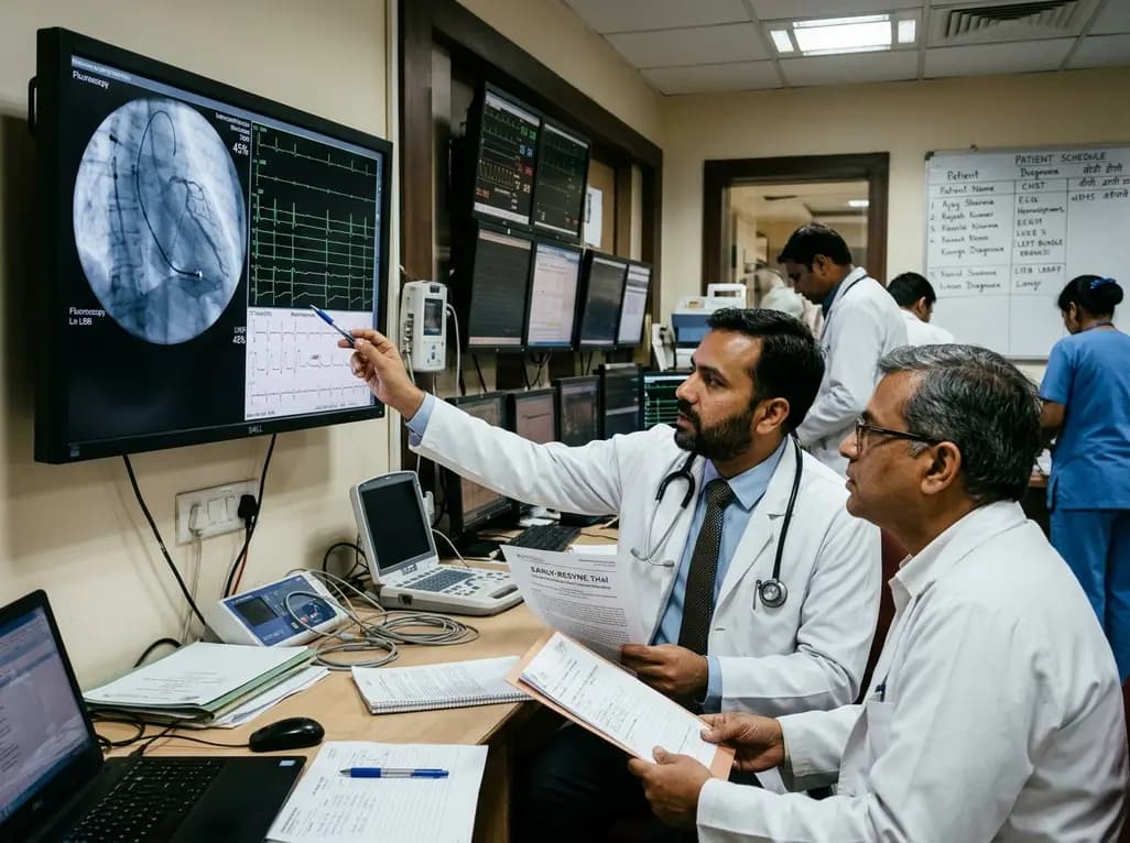
The randomized EARLY-RESYNC trial reveals that early left bundle branch pacing combined with guideline-directed medical therapy significantly improves LVEF and reduces LV end-systolic volume compared to medical therapy alone in patients with complete LBBB and mildly reduced ejection fraction (36%-50%).
Today

A comparative evaluation of INNO-LiPA, Cobas 4800, and Amplicor highlights significant assay-dependent variation in high-risk HPV detection, emphasizing the clinical distinction between analytical sensitivity and screening specificity.
Today

Recent data from PGIMER reveals that young adults diagnosed with type 2 diabetes face an aggressive disease trajectory. They encounter severe cardiovascular and microvascular damage within five years, occurring at higher rates than in type 1 diabetes despite lower mean blood glucose levels.
Today

Discover the six core principles of responsible AI in orthopaedics, emphasizing algorithmic fairness, transparency, clinical reliability, and specialist oversight to safeguard patient trust and outcomes.
Today

A novel smartphone-assisted biosensor utilizing molecularly imprinted polymers and catalytic nanozymes offers antibody-free, enzyme-free urinary microalbumin testing, enabling rapid, low-cost point-of-care screening for early chronic kidney disease.
Today
