

Loading, please wait...
Loading, please wait...

Lumbar spinal stenosis remains one of the most common degenerative spinal conditions encountered by spine specialists globally and in India. When conservative medical management fails, surgical intervention becomes necessary to relieve neural compression and mechanical instability. Over the last two decades, minimally invasive techniques have rapidly evolved to minimize surgical morbidity. Recently, endoscopic lumbar interbody fusion has emerged as a groundbreaking alternative to traditional tubular retractor systems. While tubular retractors successfully reduced tissue trauma, they still inflict significant paraspinal muscle crush injury. A landmark comparative study evaluated a hybrid endoscopic technique—percutaneous uniportal decompression combined with biportal endoscopic fusion—against conventional minimally invasive Quadrant transforaminal lumbar interbody fusion. This comprehensive analysis provides crucial insights into patient recovery and clinical efficacy.
Historically, open lumbar arthrodesis required extensive bilateral subperiosteal detachment of the erector spinae musculature. Although effective for neural decompression, this invasive approach often resulted in severe paraspinal denervation and persistent postoperative back pain. Consequently, spine surgeons developed the minimally invasive Quadrant transforaminal lumbar interbody fusion to reduce muscular disruption. Tubular retractor systems significantly curtailed blood loss and accelerated discharge times. However, continuous retractor blade pressure over several hours can still induce ischemic necrosis of the paraspinal musculature. To overcome these limitations, spine surgery has progressively adopted full-endoscopic and biportal endoscopic platforms. The hybrid endoscopic technique combines the unmatched focal visualization of uniportal endoscopy with the surgical freedom of biportal systems. Specifically, uniportal platforms facilitate delicate nerve root decompression, whereas biportal setups allow independent instrument triangulation for disc preparation. Moreover, continuous fluid irrigation maintains a clear field while clearing surgical debris and mitigating thermal tissue damage. Therefore, endoscopic lumbar interbody fusion represents a sophisticated progression toward minimizing collateral tissue damage during spinal reconstruction.
To establish the clinical efficacy of hybrid endoscopy, researchers conducted a rigorous comparative study involving 68 consecutive patients. All enrolled individuals presented with symptomatic single-level degenerative lumbar spinal stenosis requiring surgical stabilization between June 2019 and June 2021. The investigators divided the cohort into two distinct operative arms. Group A comprised 32 patients who underwent the hybrid endoscopic procedure, while Group B included 36 patients treated via Quadrant-assisted transforaminal interbody fusion. Furthermore, the clinical team prospectively recorded comprehensive perioperative data to evaluate surgical recovery. The primary clinical endpoints included the Visual Analog Scale for back and leg pain, the Oswestry Disability Index, and the Short Form 36 Health Survey. In addition, researchers monitored serum creatine phosphokinase levels to quantify paraspinal muscular trauma objectively. Independent radiologists also evaluated segmental lumbar lordosis, intervertebral disc height restoration, and sagittal Cobb angles across both cohorts. Ultimately, this structured protocol permitted an objective, multiparametric comparison between modern hybrid endoscopic techniques and established tubular retractors.
The clinical findings highlighted substantial benefits in early recovery for patients undergoing the hybrid endoscopic technique. Most notably, at one month postoperatively, Group A demonstrated statistically significant improvements in Visual Analog Scale scores for back and leg pain compared to Group B. Patients in the hybrid endoscopic group also reported superior Short Form 36 scores, reflecting enhanced functional vitality and mental well-being. Conversely, patients treated with the Quadrant retractor experienced greater early wound site soreness and muscular stiffness. Objective laboratory biomarkers corroborated these subjective patient-reported outcomes with convincing clarity. Serum creatine phosphokinase levels were significantly lower in Group A, indicating markedly less paraspinal muscle necrosis. Because the hybrid technique relies on small stab incisions and natural anatomical planes, it preserves multifidus tendon insertions and dorsal ramus innervation. Furthermore, reduced muscular trauma directly translated into decreased reliance on systemic analgesics and expedited inpatient ambulation. Consequently, the hybrid approach substantially improves the immediate postoperative recovery experience without compromising structural stabilization.
Beyond early symptomatic relief, spine specialists must evaluate whether endoscopic techniques achieve durable biomechanical stability. Fortunately, the study demonstrated that both surgical modalities achieved excellent radiological outcomes at long-term follow-up. Patients in both cohorts exhibited significant restoration of intervertebral disc height, segmental lumbar lordosis, and sagittal Cobb angle alignment. Some surgeons initially raised concerns that narrow endoscopic fluid corridors might hinder adequate endplate preparation or restrict cage dimensions. However, biportal endoscopy provides wide triangulation angles, allowing surgeons to use conventional disc raspators and insert large lordotic cages safely. As a result, the radiological fusion rates between the hybrid endoscopic cohort and the Quadrant group were completely comparable. Serial computed tomography and dynamic radiographs confirmed robust bony bridging without signs of early cage subsidence or pedicle screw loosening. Additionally, perioperative complication rates remained equally low across both groups, with zero permanent neurological deficits or deep surgical site infections. Therefore, hybrid endoscopic surgery successfully combines ultra-minimally invasive tissue preservation with reliable long-term fusion success.
Adopting the hybrid endoscopic workflow requires spine surgeons to master unique technical skills and operational nuances. First, surgeons must navigate a steep learning curve involving fluid pressure dynamics and depth perception. Maintaining appropriate continuous saline outflow is vital to prevent elevated epidural pressures and subsequent postoperative headache. Second, the surgeon should leverage the uniportal system during the initial decompressive phase to preserve contralateral facet joints. Once decompressed, switching to the biportal platform facilitates efficient cartilaginous endplate clearance under high-definition visualization. Third, patient selection is crucial for achieving predictable outcomes. While patients with degenerative stenosis and low-grade spondylolisthesis are ideal candidates, cases with severe coronal deformity or high-grade spondylolisthesis still warrant open stabilization. Finally, surgical teams must evaluate institutional costs and operating room equipment setup. Although endoscopic instrumentation requires initial investment, decreased hospital lengths of stay and lower complication rates offset these expenditures. Thus, the hybrid endoscopic technique serves as an invaluable addition to the modern spine surgical armamentarium.
The hybrid endoscopic technique significantly reduces paraspinal muscle injury and early postoperative back pain. By combining uniportal decompression with biportal fusion, surgeons eliminate heavy retractor pressure. Consequently, patients exhibit lower serum creatine phosphokinase levels, require fewer opioid analgesics, and achieve faster functional ambulation during early recovery.
Yes, endoscopic lumbar interbody fusion achieves radiological fusion rates comparable to traditional tubular methods. The biportal visualization portal allows extensive endplate clearing under clear, magnified fluid irrigation. Consequently, surgeons can place large, bone-graft-packed interbody spacers, promoting robust osteointegration, durable sagittal alignment, and solid long-term biomechanical spinal stability.
Serum creatine phosphokinase serves as an objective clinical biomarker for skeletal muscle disruption and ischemic damage. Elevated levels indicate significant mechanical retraction or paraspinal muscle stripping during surgery. Significantly lower postoperative levels after endoscopic procedures objectively validate superior tissue preservation and minimal collateral myofascial trauma.
Disclaimer: This content is for informational and educational purposes only and should not be considered medical advice. Always consult a qualified healthcare professional regarding any medical condition or treatment. Refer to the latest local and national guidelines for clinical practice.
References

Read summarized clinical updates, watch expert medical content, and earn CME certifications right from your smartphone.
Open your smartphone camera to scan and install.





A comparative study evaluated a hybrid endoscopic technique—uniportal decompression with biportal endoscopic lumbar interbody fusion—against minimally invasive Quadrant TLIF for lumbar stenosis, revealing significantly lower early postoperative pain and reduced muscle injury with equivalent fusion rates.
Today

A randomized crossover trial evaluated fifth-year medical students interacting with an AI standardized patient versus a human standardized patient. The study revealed key differences in Mini-CEX scores, active engagement, and interview dynamics.
Today

A comparative evaluation of INNO-LiPA, Cobas 4800, and Amplicor highlights significant assay-dependent variation in high-risk HPV detection, emphasizing the clinical distinction between analytical sensitivity and screening specificity.
Today

A Canadian nutrition study reveals that higher diet costs predict lower dietary energy density and higher fruit intake, but also increase sodium and processed meat consumption.
Today
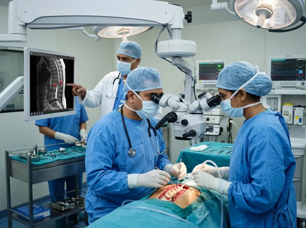
Idiopathic spinal cord herniation is a rare pathology presenting with progressive thoracic myelopathy. Early MRI identification and meticulous microsurgical reduction with dual bolstered dural defect repair prevent irreversible neurological deterioration.
Today

A novel smartphone-assisted biosensor utilizing molecularly imprinted polymers and catalytic nanozymes offers antibody-free, enzyme-free urinary microalbumin testing, enabling rapid, low-cost point-of-care screening for early chronic kidney disease.
Today
