
Loading, please wait...
Loading, please wait...

Managing lesions at the craniovertebral junction presents significant anatomical challenges for surgeons. This region houses the medulla oblongata and vertebral arteries. Recently, clinicians utilized endoscopic endonasal decompression to treat medullary compression caused by an inferior clival osteophyte. This case is particularly notable because the osteophyte enlarged progressively following a previous C1-2 fixation procedure.
The endoscopic endonasal decompression approach provides the shortest and most direct corridor to ventral lesions. Unlike traditional transoral approaches, this technique minimizes trauma to the oral mucosa and reduces recovery time. Furthermore, it allows for excellent visualization of the clivus and upper cervical spine without significant retraction of neurovascular structures.
While standard neuronavigation is common, it relies on preoperative imaging which cannot reflect real-time bone removal. To overcome this limitation, surgeons employed the O-arm system during the procedure. This technology provides real-time intraoperative 3D imaging, allowing for a precise assessment of the decompression progress. Consequently, the surgical team achieved complete bone removal while ensuring the safety of the medulla oblongata.
Mechanical loading after C1-2 fixation may contribute to the enlargement of clival osteophytes. Therefore, surgeons must remain vigilant regarding postoperative changes in patients with prior spinal stabilization. Notably, this case highlights that the integration of advanced imaging with minimally invasive techniques significantly improves outcomes for complex skull base pathologies.
The endoscopic endonasal approach offers a more direct route to the upper craniovertebral junction. It avoids the contamination risks associated with the oral cavity. Additionally, it typically leads to faster patient recovery and fewer swallowing complications compared to traditional methods.
The O-arm provides real-time 3D scans during the operation. This allows surgeons to see exactly how much bone they have removed. This capability is critical when working near sensitive structures like the medulla oblongata, where precision is paramount.
Disclaimer: This content is for informational and educational purposes only and does not constitute medical advice or a professional relationship between the reader and the author. Always consult a qualified healthcare provider for any medical concerns. Refer to the latest local and national guidelines for clinical practice.
References
Muramatsu Y et al. O-arm-assisted endoscopic endonasal decompression for medullary compression caused by a clival osteophyte: illustrative case. J Neurosurg Case Lessons. 2026 May 04. doi: undefined. PMID: 42081833.
Psotka J, et al. The use of an O-arm in endonasal endoscopic operations of the skull base. Rozhl Chir. 2021;100(1):16-22. doi: 10.33699/PIS.2021.100.1.16-22.
Cappello ZJ, et al. Endoscopic Endonasal Surgery for Craniovertebral Junction Pathology. Consult QD Cleveland Clinic. 2018.

Read summarized clinical updates, watch expert medical content, and earn CME certifications right from your smartphone.
Open your smartphone camera to scan and install.


A case report on using O-arm-assisted endoscopic endonasal decompression for medullary compression caused by a clival osteophyte after spinal fixation....
2 months ago
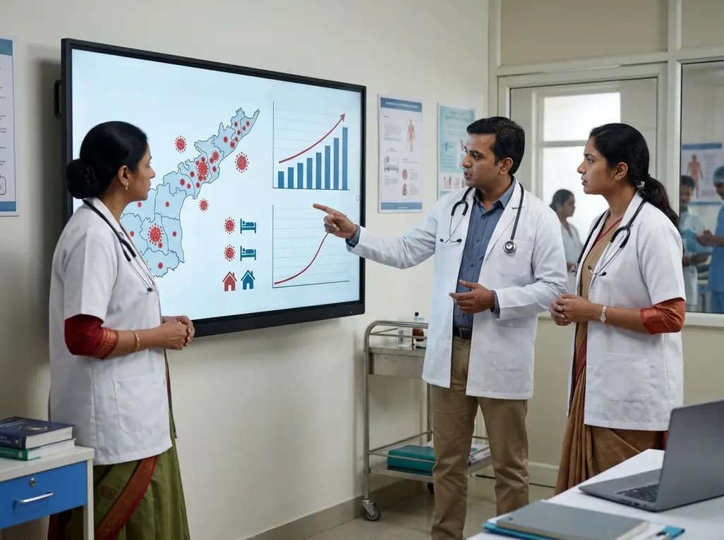
Andhra Pradesh reported 10 new Covid-19 cases, taking the state tally to 49 while deaths remain at four. With 24 patients hospitalized and 16 under home isolation, the Health Department has intensified monitoring. Medical professionals should review regional distribution, diagnostic protocols, and management plans.
Today

An 11-year Swedish registry study of 618 uterine sarcoma patients found that minimally invasive surgery yielded survival comparable to open surgery in early stages. However, adjuvant chemotherapy conferred no survival benefit in localized or advanced disease, highlighting stage and histology as key outcomes.
3 days back
A cross-sectional study evaluates post-intensive care syndrome in cardiac patients 2-4 weeks post-ICU discharge, highlighting cognitive, psychological, and functional impairments and the need for structured multidisciplinary rehabilitation.
3 days back
Anterior cruciate ligament reconstruction failure lacks uniform definition. A narrative review proposes an integrative framework incorporating objective and subjective instability, persistent pain, restricted motion, graft rupture, and secondary meniscal injury to standardize clinical reporting.
3 days back

With World Obesity Atlas data warning that over 41 million Indian children are overweight or obese, ICMR and NIN have unveiled a 10-point policy roadmap. The initiative calls for mandatory front-of-pack labeling, HFSS taxes, strict marketing bans, and healthier school environments to curb non-communicable diseases.
Today
