.webp&w=384&q=75)

Loading, please wait...
Loading, please wait...
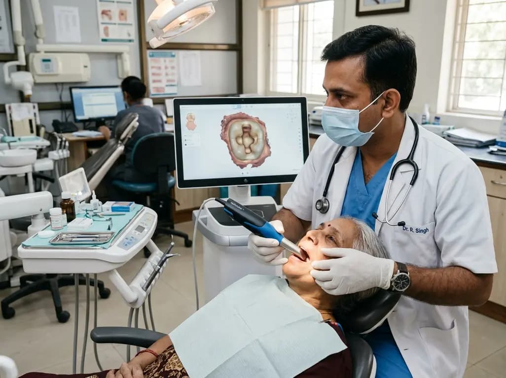
Digital dentistry continues to revolutionize indirect restorative workflows, yet capturing intricate cavity geometry remains challenging. Specifically, achieving high endocrown scanning accuracy is essential when restoring structurally compromised endodontically treated posterior teeth. Clinicians frequently encounter anatomical variations across molars and premolars that complicate optical impression capture. In addition, deeper cavity extensions often limit optical line-of-sight and reduce light reflection. Understanding how prep geometry and intraoral hardware interact allows practitioners to optimize clinical precision. Therefore, evaluating cavity dimensions alongside scanner capabilities provides crucial guidance for contemporary restorative protocols.
Endocrown restorations provide a conservative and reliable alternative to traditional post-and-core supported crowns. Clinicians preserve healthy residual tooth structure by utilizing the natural contours of the pulp chamber for macro-mechanical retention. Consequently, this restorative technique significantly reduces the risk of root perforation and catastrophic biomechanical failure. In addition, adhesive cementation creates a unified monoblock complex that effectively dissipates occlusal loads across the remaining dentin.
However, achieving long-term clinical longevity depends directly on exceptional internal adaptation and marginal fit. If the digital impression fails to capture subtle cavity transitions, the milled restoration will seat poorly. As a result, thick cement layers may develop, which accelerates luting agent degradation and promotes secondary marginal leakage. Furthermore, improper seating can induce localized stress concentrations that compromise restorative longevity. Therefore, dentists must secure precise digital representations of the prepared pulp cavity during computer-aided impression procedures. Modern optical scanning technologies facilitate this process, yet physical constraints within deep, narrow cavities still challenge sensor resolution and optical algorithms.
Tooth morphology plays a fundamental role in determining how intraoral scanners record prepared surfaces. Molars naturally present with broader pulp chambers, wider interaxial dimensions, and accessible surface orientations. Consequently, intraoral scanning wands can easily maintain appropriate focal distances and unobstructed optical angles when capturing molar geometries. Dental practitioners can maneuver the scanning tip smoothly to ensure complete point-cloud acquisition across the entire pulpal floor.
Conversely, premolar teeth present considerably narrower preparation orifices, steeper lateral walls, and restricted optical access. These constricted dimensions frequently create optical shadowing, which impairs the scanner's ability to triangulate depth coordinates accurately. In addition, light scattering increases within tight spaces, leading to software interpolation errors and point-cloud distortion. As a result, premolar endocrown preparations often exhibit lower scanning trueness than molar counterparts. Clinicians must therefore recognize that anatomical constraints inherently limit digital capture efficiency in narrower teeth. When preparing premolars, practitioners should slightly flare axial walls and eliminate sharp line angles. This geometric refinement significantly enhances light penetration, reduces optical artifacts, and improves the overall quality of virtual impressions.
The vertical depth of the pulp chamber significantly influences endocrown scanning accuracy across various restorative scenarios. Shallow preparations with a two-millimeter depth permit unimpeded light entry and optimal optical focal alignment. Therefore, intraoral sensors can record both axial walls and cavity floors with remarkable precision and minimal mesh distortion.
In contrast, deep preparations measuring five millimeters or more introduce substantial challenges for optical triangulation and confocal microscopy. As cavity depth increases, the intensity of reflected light diminishes rapidly, which impairs the sensor's photodetector response. Furthermore, steep cavity walls obstruct peripheral light paths, generating dark visual shadows in sharp line angles. Consequently, intraoral scanning software must mathematically extrapolate missing topographical data, which compromises scan trueness. Studies consistently show that deeper pulpal floors suffer higher deviation values compared to shallow cavity preparations. Clinicians must actively compensate for this optical depth limitation during daily practice. Specifically, applying structured scanning paths and maintaining precise sensor focal distances helps overcome optical attenuation in deep preparations. Practitioners should also consider placing an adhesive resin base to raise deep floors whenever retention remains sufficient.
Intraoral scanners utilize distinct optical principles, camera sensors, and software stitching algorithms to generate digital 3D models. Devices incorporating advanced structured-light projection or high-frequency confocal microscopy often manage deep, narrow cavities more effectively than older systems. Moreover, variations in scanner wand dimensions, focal depths, and processing algorithms directly impact the overall accuracy of complex impressions.
For instance, scanning systems equipped with wider dynamic focal ranges capture deeper cavity floors without losing surface sharpness. In addition, sophisticated proprietary algorithms eliminate noise and filter out optical reflections with greater precision. Conversely, scanners with fixed or narrow focal distances struggle when measuring deep pulp floors beyond their calibrated range. As a result, different intraoral devices produce variable discrepancies when scanning identical endocrown preparations. Clinicians should evaluate the specific hardware capabilities and optical depth-of-field of their chosen scanning systems. Understanding these technical specifications allows practitioners to anticipate scanning difficulties in deep cavities. Furthermore, maintaining up-to-date scanning software ensures that the latest algorithmic improvements optimize mesh reconstruction in difficult clinical environments.
Regional analysis of endocrown digital impressions reveals distinct differences between axial wall surfaces and pulpal cavity bases. In general, axial walls align closely with the scanner lens axis, receiving direct illumination from the light projector. Therefore, intraoral scanners capture vertical axial walls with high trueness, reliable precision, and minimal data loss.
However, the floor of the pulp chamber presents a far more demanding optical target for digital impression devices. Flat pulpal bases frequently reflect light unevenly, while sharp internal line angles scatter light rays erratically. Consequently, digital scanning meshes often exhibit localized artifacts, rounding of sharp margins, or virtual gaps at the cavity floor. These discrepancies alter the calculated insertion axis and impair the passive seating of milled CAD/CAM endocrowns. As a result, premature contact at the cavity base can prevent complete marginal closure along the external butt-joint finish line. To prevent this clinical complication, practitioners must inspect virtual models meticulously before sending files for fabrication. Applying color-coded deviation maps within acquisition software enables dentists to identify and rescan poorly digitized cavity floors before manufacturing restorations.
Dental clinicians can implement several practical techniques to ensure outstanding impression fidelity during endocrown fabrication. First, operators should refine preparations with smooth transitions, rounded internal line angles, and a divergence of six to eight degrees. This geometric configuration eliminates sharp shadows, facilitates optical beam entry, and maximizes data capture. In addition, practitioners must maintain absolute moisture control using rubber dam isolation to eliminate saliva reflections.
Furthermore, following a disciplined, manufacturer-recommended scanning trajectory significantly enhances point-cloud registration. Dentists should begin by capturing occlusal anatomical landmarks before tilting the wand smoothly toward the internal cavity surfaces. Maintaining a steady, consistent scanning distance ensures the optical sensor remains within its optimal focal range throughout data acquisition. If deep pulpal floors fail to scan accurately, dentists can use thin layers of scan spray to reduce glare. Finally, verifying the digital impression using high-magnification cross-sectional views ensures that no critical geometric data is missing. By combining meticulous cavity preparation with disciplined scanning protocols, dental professionals achieve consistent accuracy, excellent fit, and superior restorative longevity.
Pulp chamber depth directly influences how well intraoral scanners capture deep cavity contours. As the preparation depth increases beyond three millimeters, optical light weakens and internal shadowing increases. Consequently, intraoral scanners may produce inaccurate virtual models with interpolated pulpal floors. These digital discrepancies cause internal premature contacts in milled restorations, preventing full seating and creating wider marginal gaps that increase the risk of microleakage and cement failure.
Premolars possess significantly narrower pulp chambers and steeper internal walls compared to molars. This restricted anatomical space limits optical wand access and blocks peripheral scanning illumination. As a result, deep shadows and light scattering frequently occur in premolar preparations, causing point-cloud triangulation errors. Clinicians must slightly widen axial wall divergence and smooth internal line angles to allow intraoral scanners to capture these constricted geometries accurately.
Dentists can improve digital impression accuracy by creating rounded internal angles and maintaining a six-degree axial wall taper. In addition, strict moisture control using rubber dam isolation prevents optical reflections from saliva and blood. Clinicians should follow a steady, calibrated scanning path, holding the scanner wand at the optimal focal distance. Finally, evaluating virtual cross-sections before milling ensures the pulpal floor is completely digitized without artifacts.
Disclaimer: This content is for informational and educational purposes only. It is not intended to provide medical advice or replace professional healthcare judgment. Clinical decisions must always be made by qualified healthcare professionals based on individual patient circumstances and valid diagnostic evaluations. Refer to the latest local and national guidelines for clinical practice.
References

Read summarized clinical updates, watch expert medical content, and earn CME certifications right from your smartphone.
Open your smartphone camera to scan and install.





This comparative review examines how pulp chamber depth, tooth anatomy, and intraoral scanner selection affect endocrown scanning accuracy, providing practical strategies for clinicians to optimize digital impressions.
last month

A large UK cohort study of 1.4 million women shows that pregnancy complications, including gestational diabetes, hypertension, and preterm birth, substantially raise long-term risks for hypertension, chronic kidney disease, type 2 diabetes, and cardiovascular events, highlighting early postpartum intervention needs.
Today
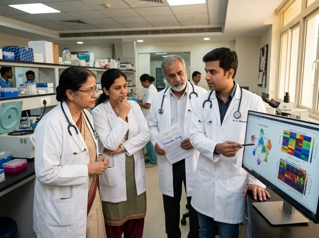
A landmark single-cell study reveals that MEHP, a major phthalate metabolite, damages human ovarian tissue beyond follicles. By disrupting glial-stromal communication, cellular adhesion, and mitochondrial respiration, environmental plasticizers impair ovarian integrity at realistic exposure levels.
Today

The Delhi High Court granted interim relief to beverage manufacturers against the FSSAI ban on the energy drink descriptor. Discover the regulatory dispute, caffeine safety thresholds, metabolic concerns, and the vital clinical recommendations for healthcare practitioners advising young consumers in India.
Today

A scoping review reveals severe gaps in long-term assistive technology provision following humanitarian emergencies. Addressing post-disaster rehabilitation requires systematic follow-up, workforce training, and integration into national health systems to sustain mobility and recovery.
Today

The World Health Organization has issued its first global guidelines for managing child and adolescent obesity. The recommendations strongly endorse family-based dietary interventions, regular physical activity, and behavioral support as frontline therapy, reserving GLP-1 therapies and surgery for select cases.
Today
