.webp&w=384&q=75)

Loading, please wait...
Loading, please wait...

Pediatric soft tissue sarcomas exhibit remarkable morphological and phenotypic diversity. Among these childhood malignancies, embryonal rhabdomyosarcoma represents the most common soft tissue cancer diagnosed in early life. Occasionally, surgical pathologists encounter rare biphasic tumors that combine classical myogenic elements with divergent neuroectodermal differentiation. This entity, often characterized as embryonal rhabdomyosarcoma with heterologous neuroectodermal differentiation or malignant ectomesenchymoma, creates significant diagnostic confusion. Consequently, clinicians require precise clinicopathological criteria to ensure accurate diagnosis and management. A recent investigation evaluated seventeen pediatric patients diagnosed with this rare condition at Foshan Hospital of Traditional Chinese Medicine. The investigators examined patient demographics, detailed histopathology, immunohistochemical profiles, and targeted molecular alterations. Their findings provide critical diagnostic guidance for surgical pathologists and pediatric oncologists handling complex soft tissue lesions. Because the neuroectodermal component closely mimics independent neural neoplasms, standard biopsies risk serious misdiagnosis. Therefore, recognizing biphasic differentiation prevents misclassification and guides appropriate clinical management. Furthermore, understanding the complete spectrum of neural crest lineages within these sarcomas clarifies their biological behavior and therapeutic sensitivity. Multidisciplinary teams benefit immensely from recognizing these dual characteristics during diagnostic evaluations.
Epidemiological findings from the seventeen-patient cohort reveal a striking male preponderance in clinical practice. Specifically, the study identified fifteen boys and only two girls among the seventeen evaluated cases. The median patient age stood at twenty-four months, with ages spanning from two weeks to twenty-two years. Moreover, fourteen of the seventeen children were five years old or younger at initial diagnosis. This pediatric age distribution aligns closely with conventional embryonal myogenic neoplasms. Anatomically, the pelvic and genitourinary tract represented the most common site, accounting for nearly half of all cases. Additionally, tumors arose within retroperitoneal spaces and deep somatic soft tissues. Patients typically present with rapidly enlarging, painless pelvic masses. As a result, presenting symptoms include palpable abdominal distension, acute urinary retention, or gastrointestinal obstruction. Because pelvic tumors expand quickly, cross-sectional imaging with computed tomography or magnetic resonance remains essential. Imaging generally reveals large, heterogeneous, locally invasive soft tissue lesions. Therefore, clinicians must obtain generous core needle or incisional biopsies to capture both cellular elements. Accurate sampling prevents diagnostic errors and allows prompt initiation of multidisciplinary pediatric oncology care.
Histological analysis confirmed biphasic differentiation across all seventeen patient specimens. In sixteen cases, the mesenchymal component displayed classic embryonal rhabdomyosarcoma morphology. Microscopically, these areas featured alternating cellular and hypocellular zones set within a loose myxoid stroma. Rhabdomyoblasts presented primitive spindle and rounded shapes with dense eosinophilic cytoplasm. In contrast, the neuroectodermal component exhibited a broad spectrum of neural crest differentiation. Specifically, twelve tumors contained distinct neuroblastoma elements with primitive round cells and fibrillary neuropil. Two cases demonstrated mature ganglion cells, one showed composite ganglioneuroblastoma, and two featured differentiated Schwannian stroma. Beyond standard patterns, pathologists identified novel micronest and inverted architectural arrangements. These unique histological architectures may confuse surgical pathologists during intraoperative frozen evaluations. However, careful microscopic examination reveals intimate intermingling between myogenic and neural elements rather than distinct collision neoplasms. Furthermore, adequate sampling across multiple tissue blocks exposes both diagnostic lineages. Thus, thorough histopathological analysis remains the foundation for confirming heterologous neural crest differentiation in challenging pediatric sarcomas. Pathologists must recognize this morphological diversity to avoid misinterpreting biphasic tumors as pure neural or muscular lesions.
Immunohistochemical profiling provides crucial confirmation when evaluating biphasic pediatric sarcomas. The mesenchymal component uniformly expressed skeletal muscle markers, including desmin, MyoD1, and myogenin. Conversely, the neuroectodermal component showed distinct marker profiles depending on its specific lineage maturation. Neuroblastoma areas stained positively for synaptophysin, neuron-specific enolase, chromogranin A, and CD56. Meanwhile, Schwannian elements demonstrated strong positivity only for S-100 protein and SOX10. Interestingly, negative neuroendocrine markers do not exclude neuroectodermal differentiation when Schwann cell markers remain strongly positive. Notably, only two cases exhibited focal PHOX2B positivity in neuroblastic areas. Because genuine primary neuroblastomas show diffuse and strong PHOX2B expression, low expression aids differential diagnosis. Consequently, minimal PHOX2B staining serves as a valuable diagnostic tool to distinguish divergent elements from metastatic neuroblastoma. Furthermore, pathologists must utilize comprehensive antibody panels rather than single markers to prevent diagnostic pitfalls. Therefore, combining muscle and neural markers ensures accurate classification of complex soft tissue specimens. This systematic approach allows surgical pathologists to establish a secure and reproducible diagnosis. Overall, incorporating these complementary biomarkers provides objective confirmation of divergent neuroectodermal differentiation.
Molecular testing offers vital genetic insights to confirm the tumor subtype. In this series, genetic assays identified activating RAS mutations in three cases, including one HRAS and two NRAS alterations. Importantly, all seventeen tumors tested negative for FOXO1, PAX3, and PAX7 rearrangements, excluding alveolar rhabdomyosarcoma. A key differential diagnosis is malignant triton tumor, which exhibits malignant Schwannian stroma with rhabdomyoblastic differentiation. However, comprehensive genomic testing in Case 17 showed no PRC2 mutations, NF1 alterations, or CDKN2A deletions. Most importantly, the Schwannian elements retained trimethylated histone H3 lysine 27 (H3K27me3) expression. Malignant triton tumors lose H3K27me3 due to PRC2 disruption, whereas this entity maintains epigenetic expression. Additionally, RAS mutations suggest a single clonal origin driving divergent myogenic and neuroectodermal development. Thus, molecular diagnostics confirm that these tumors represent an embryonal rhabdomyosarcoma variant rather than triton tumors. Consequently, integrating histology, immunohistochemistry, and molecular profiling ensures accurate diagnosis and optimized systemic therapy. Pathologists and pediatric oncologists rely on this molecular data to guide personalized therapeutic strategies. Ultimately, comprehensive genetic analysis refines risk stratification.
This variant exhibits a biphasic microscopic pattern combining classic embryonal skeletal muscle differentiation with heterologous neural crest components. While typical embryonal rhabdomyosarcoma contains only mesenchymal rhabdomyoblasts, this tumor also displays neuroblastoma, ganglioneuroma, or Schwann cell elements. Pathologists confirm this dual nature through positive staining for both myogenic markers like desmin and neural markers like S-100 or synaptophysin. Identifying both components prevents misclassifying the tumor as a collision neoplasm or separate neural malignancy.
Malignant triton tumors represent aggressive malignant peripheral nerve sheath tumors that develop heterologous rhabdomyoblastic differentiation. They frequently harbor alterations in PRC2 core components, leading to a diagnostic loss of H3K27me3 nuclear expression. In contrast, embryonal rhabdomyosarcoma with Schwannian differentiation retains normal H3K27me3 staining and lacks PRC2 or NF1 alterations. Evaluating H3K27me3 expression provides pathologists with an objective biomarker to separate this primary myogenic tumor from aggressively lethal malignant nerve sheath malignancies.
Genuine primary neuroblastomas display diffuse, high-intensity nuclear PHOX2B expression as a hallmark of autonomic nervous system origin. Conversely, the neuroectodermal component in embryonal rhabdomyosarcoma shows absent or merely focal PHOX2B positivity. Pathologists utilize this markedly restricted or absent staining pattern to distinguish secondary neural crest differentiation from true neuroblastoma metastatic invasion. This subtle immunohistochemical difference prevents misdiagnosis and helps clinicians select chemotherapy regimens optimized for fusion-negative soft tissue rhabdomyosarcoma protocols.
Disclaimer: This content is for informational and educational purposes only... Refer to the latest local and national guidelines for clinical practice.
References

Read summarized clinical updates, watch expert medical content, and earn CME certifications right from your smartphone.
Open your smartphone camera to scan and install.





A retrospective analysis of 17 pediatric cases reveals the clinicopathological, immunohistochemical, and molecular features of embryonal rhabdomyosarcoma with divergent neuroectodermal differentiation, emphasizing diagnostic biomarkers like S-100, SOX10, and retained H3K27me3.
Today
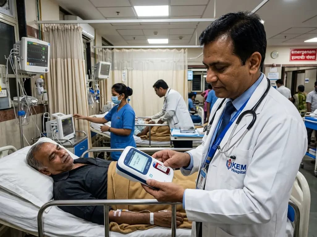
A real-world emergency department evaluation demonstrates that the Abbott i-STAT Alinity point-of-care high-sensitivity troponin I assay achieves excellent analytical precision and safely rules out myocardial infarction without false negatives, offering rapid bedside results to decongest acute care pathways.
Today

Integrating adult genetic services into reproductive and pregnancy care substantially alters clinical management. A study reveals that diagnostic genetic evaluations directly inform maternal, fetal, and neonatal care in nearly 57% of tested patients, highlighting the value of preconception and antenatal genomics.
Today

A cross-sectional study in Nepal evaluates cardiovascular risk factors among adults working above 4,000 meters. The findings reveal unexpected rates of diabetes, hypertension, and mountain sickness, underscoring the urgent need for targeted cardiometabolic screening in high-altitude occupational cohorts.
Today

A recent scoping review highlights the efficacy, usability, and patient satisfaction of virtual reality pain management for chronic musculoskeletal pain, emphasizing psychotherapy-based VR protocols.
Today

Preclinical findings demonstrate that dietary flavonoid hyperoside ameliorates high-fat diet-induced type 2 diabetes by stimulating brown adipose tissue thermogenesis, promoting white fat browning, dampening adipose macrophage inflammation, and restoring gut microbiota homeostasis.
Today
