

Loading, please wait...
Loading, please wait...

Surgical management of advanced oral cancer frequently requires extensive resection, leaving patients with catastrophic speech and swallowing deficits. In recent years, dynamic tongue reconstruction has emerged as a groundbreaking reconstructive technique to restore functional oral mobility after subtotal or total glossectomy. Traditionally, reconstructive surgeons relied on passive soft-tissue flaps, such as the anterolateral thigh or radial forearm flaps. Although these conventional options provide volume, they fail to reproduce muscular motility. Consequently, patients often experience severe dysphagia, persistent aspiration risk, and unintelligible speech. Reinnervated muscular transfers directly address these limitations by re-establishing active contractile movement in the oral cavity. Surgical teams can now achieve remarkable functional restoration using functioning free muscle transplantation. This milestone report highlights the innovative application of the reinnervated free gracilis flap, offering new hope for post-glossectomy rehabilitation.
Carcinoma of the oral tongue represents a major surgical oncological challenge worldwide, especially across India and South Asia. Extensive surgical resection remains the cornerstone of curative therapy for locally advanced lesions. However, subtotal and total glossectomies remove critical intrinsic and extrinsic lingual musculature. As a result, patients lose the dynamic coordinated movement necessary for forming and propelling a food bolus. Furthermore, glossectomy impairs airway protection, causing significant post-deglutition aspiration and chronic nutritional compromise. The tongue also plays an indispensable role in articulating consonants and modulating phonetic sounds. Without structural elevation against the hard palate, speech intelligibility drops precipitously. Traditional static reconstructions restore oral contour but remain essentially motionless inert tissue pillows. Over time, these static reconstructions suffer from gravitational sagging and progressive muscular atrophy. Consequently, saliva pools in the dependent floor of the mouth, worsening aspiration risks and social isolation. Head and neck surgeons have therefore sought active reconstructive alternatives that preserve tissue bulk while restoring kinetic motor control.
To overcome static limitations, microsurgeons introduced dynamic tongue reconstruction using innervated functional free muscle transfers. The gracilis muscle serves as an ideal donor for oral cavity reanimation. First, the gracilis offers reliable vascular anatomy and a consistent motor nerve branch from the obturator nerve. Second, surgeons can harvest the muscle simultaneously with tumor ablation, reducing operative time. In addition, the muscle can carry a tailored skin island to restore mucosal lining and seal mucosal boundaries. By transferring vascularized muscle alongside its neural supply, the surgeon prevents denervation atrophy. Furthermore, surgeons fold and orient the muscle fibers along specific vectors to mimic native tongue motions. Specifically, orienting the gracilis longitudinally simulates the genioglossus, which drives forward propulsion and base elevation. Therefore, when the reinnervated muscle contracts, it generates genuine cranio-caudal motility. This coordinated kinetic action pushes food boluses toward the oropharynx during the pharyngeal phase of swallowing. Consequently, this innovation transforms a passive reconstructed oral defect into an active motor organ.
Successful functional muscle transplantation demands meticulous microvascular and microneural techniques under high surgical magnification. Initially, the surgical team isolates recipient vessels in the neck, commonly selecting branches of the external carotid artery. For venous drainage, surgeons anastomose the flap veins to the internal jugular system or facial vein tributaries. Reliable microvascular perfusion ensures early flap viability and supports subsequent neural axonal regeneration. Next, the reconstructive surgeon performs precise perineural coaptation between the flap nerve and a recipient motor nerve. The donor obturator nerve branch aligns end-to-end with the transected hypoglossal nerve stump. Because the hypoglossal nerve provides native central motor control for swallowing, it provides ideal physiologic innervation. However, if radical tumor clearance sacrificed the ipsilateral hypoglossal nerve, surgeons can use the masseteric nerve. Furthermore, contralateral nerve transfers offer another reliable motor source. Reinnervation begins within weeks as regenerating axons traverse the suture line. Ultimately, successful neurotization maintains muscular trophism, preventing long-term volume loss and contracture.
Postoperative management protocols play a crucial role in securing optimal functional outcomes after microvascular reconstruction. In the immediate postoperative setting, clinicians closely monitor flap perfusion using clinical inspection and handheld Doppler ultrasonography. Meanwhile, enteral nutrition through a nasogastric or gastrostomy tube maintains caloric intake while intraoral incisions heal. Speech and language pathologists initiate early swallowing rehabilitation as tissue edema resolves. Interestingly, the clinical report demonstrated uneventful mucosal healing and full resumption of independent oral feeding by postoperative day twenty-eight. Furthermore, objective swallowing examinations and dynamic radiologic imaging at eight months confirmed impressive functional restoration. Videofluoroscopy demonstrated that the neotongue moved actively in a distinct cranio-caudal direction during deglutition. This dynamic elevation propelled liquids and purees effectively into the pharynx without tracheal penetration. In addition, patients showed significant improvements in speech articulation, producing intelligible phonetic sounds. Thus, structured postoperative rehabilitation capitalizes on dynamic flap reinnervation to rebuild patient confidence and independent oral intake.
The successful implementation of reinnervated free gracilis transfers marks a significant milestone in head and neck surgical oncology. For oncologists and reconstructive surgeons, functional preservation must accompany complete oncological ablation. In regions with high oral cancer burdens, such as India, advanced tongue tumors frequently present in younger working individuals. Consequently, restoring speech and swallowing capabilities directly impacts social reintegration, mental health, and return to employment. Although microvascular gracilis transfer requires advanced surgical expertise and longer operative planning, the functional payoffs are substantial. The flap offers minimal donor site morbidity, because the remaining thigh adductor muscles fully compensate for gracilis loss. Moreover, maintaining long-term muscle volume prevents secondary pharyngeal dead spaces where saliva and debris stagnate. Multidisciplinary head and neck teams should therefore evaluate dynamic reconstruction protocols for eligible glossectomy candidates. By integrating dynamic free tissue transfers into standard oncological care, surgeons can significantly elevate post-treatment quality of life.
Static reconstruction relies on passive soft-tissue volume from fasciocutaneous flaps to fill the oral cavity defect. Consequently, these static flaps lack active muscular contraction and cannot assist in bolus propulsion. In contrast, dynamic reconstruction transfers functional skeletal muscle innervated by a motor donor nerve. Therefore, the reinnervated muscle contracts actively during deglutition. This active movement improves speech articulation, elevates the neotongue, and reduces oral stasis significantly.
Surgeons most frequently coapt the obturator nerve branch of the gracilis muscle to the recipient hypoglossal nerve. However, when surgeons sacrifice the ipsilateral hypoglossal nerve during radical neck dissection, they can utilize the contralateral hypoglossal nerve or the masseteric nerve. Consequently, direct neural coaptation allows coordinated motor signals to reach the transplanted muscle fibers. This neurotization prevents denervation muscular atrophy and preserves long-term flap volume and functional dynamic mobility.
Clinicians typically initiate cautious oral feeding trials around the fourth postoperative week, often near post-op day twenty-eight. First, clinicians perform objective videofluoroscopic swallowing studies to confirm competent airway protection and rule out silent aspiration. Subsequently, dedicated speech and language therapists guide the patient through progressive dietary textures. Although neural regeneration requires several months to achieve full active contractility, early rehabilitative exercises optimize swallowing coordination and accelerate oral nutritional independence.
Disclaimer: This content is for informational and educational purposes only... Refer to the latest local and national guidelines for clinical practice.
References
Füzes A et al. [Dynamic tongue reconstruction with reinnervated free gracilis muscle flap]. Orv Hetil. 2026 Nov 01. doi: 10.1556/650.2026.33652. PMID: 42967261.
Calabrese L, Tagliabue M, Maffini F, et al. Tongue reconstruction with the gracilis myocutaneous free flap. Microsurgery. 2011;31(5):355-359.
Yoleri L, Mavioğlu H. Total tongue reconstruction with free functional gracilis muscle transplantation: a technical note and review of the literature. Ann Plast Surg. 2000;45(2):181-186.

Read summarized clinical updates, watch expert medical content, and earn CME certifications right from your smartphone.
Open your smartphone camera to scan and install.





Dynamic tongue reconstruction using a reinnervated free gracilis muscle flap offers active oral mobility, preserving muscle volume and restoring vital speech and swallowing functions after subtotal or total glossectomy for oral cancer.
Today
Subjective components in DAS28 often inflate disease activity scores due to central sensitization and emotional distress. Integrating the DAS28-P index, musculoskeletal ultrasound, and emerging digital biomarkers allows clinicians to differentiate true synovitis from non-inflammatory pain and prevent futile escalation.
Today
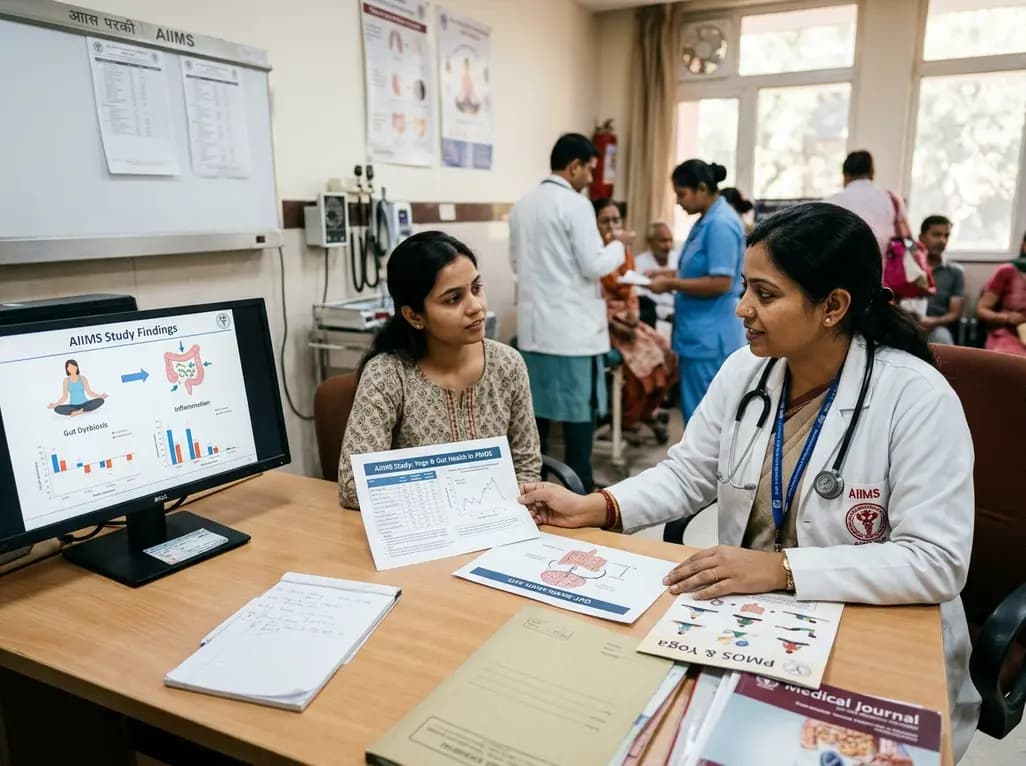
AIIMS researchers suggest yoga may alleviate gut dysbiosis in Polyendocrine Metabolic Ovarian Syndrome (PMOS) by modulating the gut-brain axis, lowering chronic inflammation, and enhancing parasympathetic tone to support metabolic and hormonal equilibrium.
Today
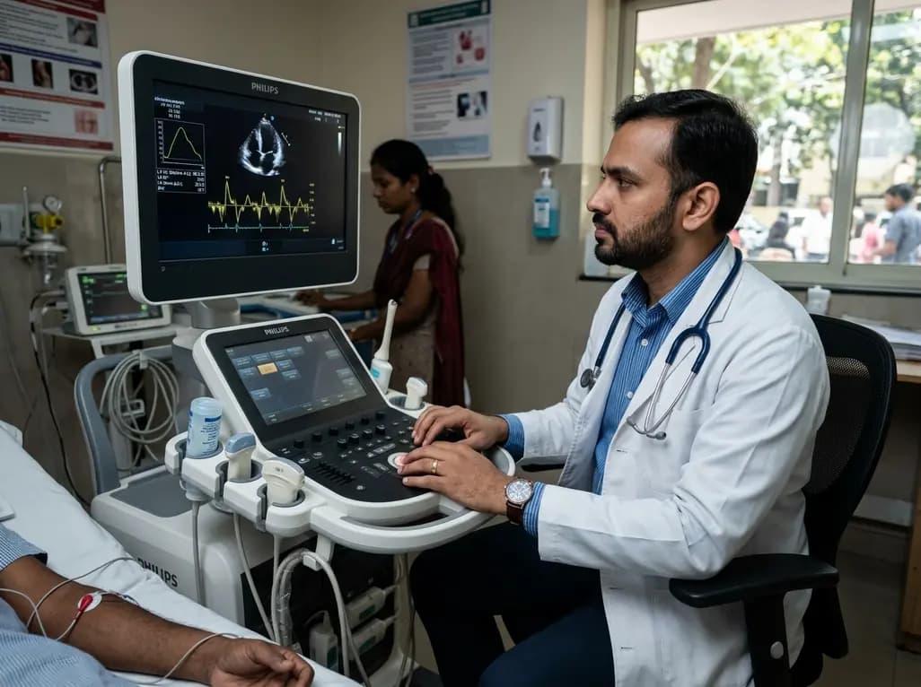
A structured practical guide to left atrial echocardiographic assessment covering biplane volumetry, phasic physiology, speckle-tracking strain, and hemodynamic filling pressure estimation for routine cardiovascular practice.
Today
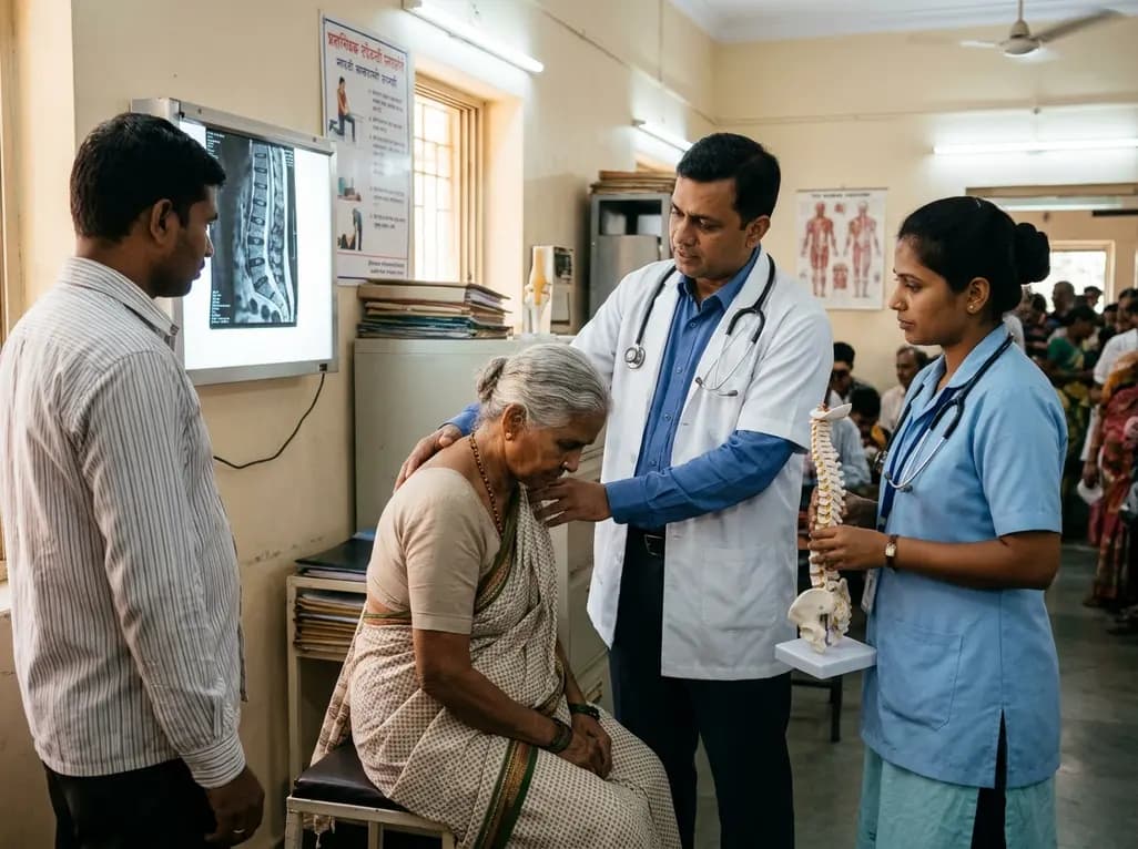
Age-related spinal kyphosis is driven by vertebral fragility, disc degeneration, and paraspinal muscle dysfunction. Coexisting with locomotive syndrome and frailty, its management requires combined anti-osteoporotic therapy, spinal extensor rehabilitation, and comprehensive nutritional optimization.
Today
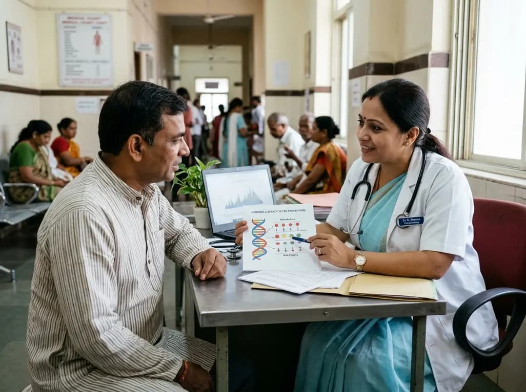
A longitudinal feasibility pilot study highlights that combining polygenic risk score disclosure with in-person lifestyle counseling significantly enhances genomic literacy in individuals at risk for type 2 diabetes, overcoming educational disparities and reinforcing patient motivation.
Today
