

Loading, please wait...
Loading, please wait...

Postoperative cerebral edema and secondary intracranial hypertension remain formidable clinical challenges in cranial neurosurgery. When elevated intracranial pressure threatens cerebral perfusion, neurosurgeons historically face an agonizing binary dilemma between rigid bone flap fixation and decompressive craniectomy. While rigid cranial fixation preserves cosmetic integrity and anatomical skull architecture, it restricts intracranial volume and risks catastrophic brain herniation if refractory swelling develops. Conversely, decompressive craniectomy effectively mitigates intracranial pressure by discarding cranial containment, but it subjects vulnerable brain tissue to the syndrome of the trephined and mandates a mandatory second-stage cranioplasty procedure. To address this persistent gap, translational researchers developed dynamic craniotomy as an adaptive, pressure-responsive cranial reconstruction platform. This approach integrates reversibly expandable hardware that safely accommodates acute cerebral swelling while preserving native bone flap continuity.
The biomechanical foundation of dynamic craniotomy relies on low-profile, reversibly expandable plates and telescopic burr hole covers engineered to respond directly to intracranial volume fluctuations. Unlike conventional rigid miniplates that lock the calvarium into an unyielding shell, these specialized dynamic implants permit controlled outward displacement when intracranial pressure rises above normal thresholds. Furthermore, rigorous static compression testing demonstrates a remarkable inward displacement load resistance of 643.3 ± 26.1 Newtons. Consequently, the dynamic construct establishes absolute structural prevention against inward bone flap collapse or pathological intracranial sinking. In addition, quasi-static shear tension evaluations reveal an expansive tolerance reaching up to 71.6 millimeters of total excursion. Dynamic fatigue testing further confirms exceptional durability, showing zero mechanical structural failures across 500 complete loading cycles at 15 millimeters of displacement. Therefore, this specialized hardware safely balances expansile compliance during severe cerebral edema with robust physiological protection against accidental external cranial trauma.
Preclinical cadaveric models have yielded crucial physiological insights regarding intracranial dynamics during sudden parenchymal volume challenges. During rigorous translational testing, investigators introduced an acute 120-milliliter volumetric infusion challenge to evaluate intracranial compliance across different cranial reconstruction methods. In standard rigid fixation constructs, this acute volume increase caused intracranial pressure to spike precipitously from 11.4 to 100.1 millimeters of mercury. In sharp contrast, cadaveric preparations utilizing the dynamic platform exhibited profound physiological buffering capacity. Specifically, the intracranial pressure rose minimally from 2.8 to merely 13.4 millimeters of mercury under identical volume expansion conditions. Furthermore, this dynamic response yielded a statistically significant buffering superiority over rigid fixation protocols. By providing spontaneous, pressure-triggered volume relief without requiring emergency surgical bone removal, the dynamic platform maintains cerebral perfusion pressure and mitigates fatal trans-tentorial or uncal herniation risks during critical postoperative resuscitation windows.
Clinical translation has advanced through multi-center cohorts encompassing over sixty patients treated for diverse acute neurosurgical emergencies. In a consecutive clinical series of 25 adult patients presenting with severe traumatic brain injury, postoperative swelling prompted outward bone flap migration in 84 percent of individuals. Remarkably, severe parenchymal expansion exceeding 8 millimeters occurred in 40 to 56 percent of cases, producing immediate decreases in midline shift. In addition, comparative evaluations in spontaneous intracerebral hemorrhage and posterior fossa craniotomies revealed similar therapeutic responsiveness. Specifically, the dynamic platform accommodated infratentorial cerebellar swelling, effectively preventing secondary brainstem compression while avoiding extensive suboccipital craniectomy defects. Importantly, none of the evaluated patients required rescue decompressive craniectomies, emergency hematoma evacuations, or second-stage cranioplasty procedures. Once the parenchymal swelling naturally resolved, the bone flap spontaneously migrated inward toward its anatomical baseline without cosmetic contour deformity.
Propensity score-matched comparisons between dynamic craniotomy and conventional fixed craniotomy cohorts provide intriguing preliminary clinical insights. Patients undergoing dynamic reconstruction demonstrated significantly reduced postoperative midline shift on sequential neuroimaging evaluations compared with rigidly fixed controls. Furthermore, investigators observed lower overall mortality and a higher proportion of favorable neurological recoveries on the Glasgow Outcome Scale at three-month postoperative assessments. Notably, surgical teams observed zero fixation hardware failures, no instances of paradoxical bone sinking, and remarkably low rates of wound dehiscence or cerebrospinal fluid leakage. However, clinicians must interpret these initial comparative data with measured scientific caution. Because these preliminary findings derive primarily from retrospective, single-center datasets authored by device developers, selection bias remains a distinct methodological limitation. Therefore, neurosurgeons must view current outcome advantages as hypothesis-generating evidence that warrants validation through independent, prospective clinical trials.
Successfully performing this adaptive surgical procedure requires meticulous operative technique and strict adherence to specific cranial reconstruction principles. First, the neurosurgeon must perform an expansive, tension-free duroplasty using pericranial or synthetic dural substitutes, ensuring adequate room for cerebral outward expansion. Next, the surgical team positions the dynamic plates over strategically drilled burr holes, avoiding mechanical impingement against surrounding osteotomy margins. Moreover, surgeons must execute careful, multi-layered scalp closure without excessive galeal tension to permit external bone flap excursion without compromising microvascular cutaneous perfusion. Looking forward, the dynamic fixation paradigm represents an elegant, low-disruption technological evolution in modern cranial trauma surgery. As ongoing randomized trials emerge, this innovative cranial fixation platform may fundamentally redefine neurotrauma algorithms worldwide by mitigating the secondary morbidity of craniectomy and eliminating avoidable cranioplasty surgeries.
Dynamic craniotomy is an adaptive surgical platform that secures a cranial bone flap using specialized expandable plates and telescopic covers. Rather than locking the skull into a rigid position, these implants expand outward when intracranial pressure rises from postoperative edema. Consequently, this controlled outward movement provides immediate intracranial volume expansion and automatically retracts to anatomical position once swelling subsides.
Standard decompressive craniectomy completely removes the cranial bone flap, leaving the brain unprotected and requiring an expensive second-stage cranioplasty. Conversely, dynamic craniotomy preserves the native bone flap in situ while permitting controlled outward expansion during cerebral edema. As a result, patients avoid secondary surgical skull reconstruction, paradoxical brain herniation, and the long-term cognitive complications of trephined syndrome.
Current published evidence stems primarily from retrospective, single-center cohorts and non-randomized comparisons authored largely by device innovators. While preclinical biomechanical testing confirms exceptional fatigue tolerance and structural safety, broader clinical efficacy remains unproven across diverse surgical centers. Therefore, neurosurgeons require independent, prospective randomized clinical trials to establish definitive guideline-directed indications for traumatic brain injury and hemorrhage.
Disclaimer: This content is for informational and educational purposes only and should not replace professional medical judgment. Refer to the latest local and national guidelines for clinical practice.
References

Read summarized clinical updates, watch expert medical content, and earn CME certifications right from your smartphone.
Open your smartphone camera to scan and install.





Dynamic craniotomy offers an adaptive cranial bone flap fixation platform that bridges standard rigid fixation and decompressive craniectomy. By allowing reversible bone flap excursion, it manages intracranial swelling while avoiding secondary cranioplasty, presenting a promising paradigm for neurosurgical trauma.
Today
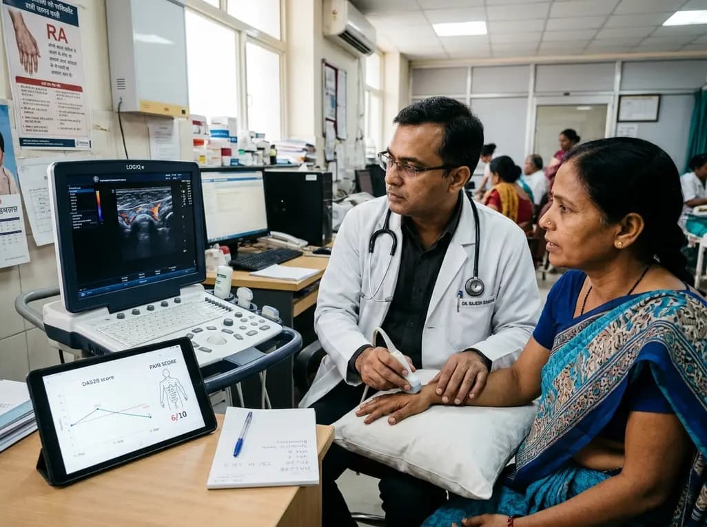
Subjective components in DAS28 often inflate disease activity scores due to central sensitization and emotional distress. Integrating the DAS28-P index, musculoskeletal ultrasound, and emerging digital biomarkers allows clinicians to differentiate true synovitis from non-inflammatory pain and prevent futile escalation.
Today

AIIMS researchers suggest yoga may alleviate gut dysbiosis in Polyendocrine Metabolic Ovarian Syndrome (PMOS) by modulating the gut-brain axis, lowering chronic inflammation, and enhancing parasympathetic tone to support metabolic and hormonal equilibrium.
Today

A structured practical guide to left atrial echocardiographic assessment covering biplane volumetry, phasic physiology, speckle-tracking strain, and hemodynamic filling pressure estimation for routine cardiovascular practice.
Today

Age-related spinal kyphosis is driven by vertebral fragility, disc degeneration, and paraspinal muscle dysfunction. Coexisting with locomotive syndrome and frailty, its management requires combined anti-osteoporotic therapy, spinal extensor rehabilitation, and comprehensive nutritional optimization.
Today

A longitudinal feasibility pilot study highlights that combining polygenic risk score disclosure with in-person lifestyle counseling significantly enhances genomic literacy in individuals at risk for type 2 diabetes, overcoming educational disparities and reinforcing patient motivation.
Today
