.webp&w=384&q=75)

Loading, please wait...
Loading, please wait...

Glioblastoma remains one of the most aggressive and challenging central nervous system malignancies encountered in clinical practice today. Researchers continuously seek novel non-invasive imaging biomarkers to better predict patient clinical outcomes, optimize therapeutic stratifications, and refine survival prognostications. Recently, diffusion tensor imaging analysis along the perivascular space has emerged as a promising non-invasive tool to evaluate glymphatic system integrity. Consequently, evaluating the prognostic utility of DTI-ALPS in glioblastoma has attracted significant attention within neuro-oncology and neuroradiology communities worldwide. Understanding whether impaired glymphatic clearance independently influences overall patient survival is essential before integrating such advanced quantitative magnetic resonance imaging parameters into routine clinical decision-making protocols. Furthermore, clinicians must evaluate these novel imaging parameters alongside well-established prognostic clinical factors. These established variables include the extent of surgical resection, patient age, baseline functional clinical status, O6-methylguanine-DNA methyltransferase promoter methylation status, and completion of standard adjuvant chemoradiotherapy. A thorough appraisal of glymphatic function allows neuro-oncologists to determine whether automated or manual tensor-derived indices truly provide additive prognostic value beyond conventional clinical metrics. As neuro-imaging techniques continue to evolve rapidly, distinguishing true independent prognostic markers from secondary physiological epiphenomena is vital. This comprehensive review examines current evidence surrounding glymphatic function assessment and clarifies the independent prognostic significance of tensor metrics in adult patients with high-grade diffuse gliomas.
The glymphatic system plays a crucial role in maintaining central nervous system homeostasis by facilitating fluid clearance and waste removal through perivascular pathways. Advanced diffusion magnetic resonance imaging techniques, specifically diffusion tensor imaging analysis along the perivascular space, measure water diffusivity parallel to medullary veins relative to perpendicular fiber tracts. This mathematical ratio yields an index that reflects perivascular fluid dynamics and regional glymphatic function. In neuro-oncological conditions, tumor mass effect, vasogenic edema, microvascular proliferation, and disruption of the blood-brain barrier significantly alter localized interstitial fluid flow. Consequently, patients suffering from high-grade gliomas frequently demonstrate significantly lower index values compared to healthy age-matched control subjects. This observation led investigators to hypothesize that severe glymphatic dysfunction might directly correlate with aggressive tumor biology, rapid disease progression, and shortened patient survival. However, measuring glymphatic impairment within tumor-bearing brain parenchyma presents inherent technical challenges. Intratumoral necrosis, peri-tumoral vasogenic edema, and surgical cavity alterations can confound tensor calculations. Therefore, clinicians and radiomic researchers must rigorously analyze whether observed reductions in perivascular diffusion indices reflect intrinsic oncological aggressiveness or merely represent structural tissue distortion caused by mass lesion expansion and localized perilesional edema.
To determine the true clinical utility of perivascular diffusion metrics, recent clinical investigations evaluated cohort populations comparing adult patients with primary glioblastoma against healthy control cohorts. Quantitative analysis consistently demonstrates that hemispherically averaged index scores are lower in glioblastoma patients compared to healthy individuals. Furthermore, diffusivity calculations strictly isolated to the tumor-bearing ipsilateral hemisphere display marked impairment, whereas the uninvolved contralateral hemisphere often maintains near-normal diffusion values. Initial univariable survival analyses suggested that lower perivascular diffusion values correlated with abbreviated overall survival. However, univariable associations can be highly misleading in complex oncological diseases because confounding clinical variables frequently cluster together. For instance, larger tumor volumes, extensive mass effect, and severe perilesional edema cause profound localized glymphatic impairment while simultaneously limiting surgical resectability and performance scores. When rigorous statistical models account for these confounding physiological mechanisms, the univariable relationship between glymphatic impairment and shortened survival diminishes substantially. Consequently, evaluating raw tensor metrics without adjusting for baseline performance status or surgical yield creates an incomplete and potentially inaccurate prognostic assessment for neuro-oncology clinicians.
Establishing true prognostic independence requires multivariate regression modeling that incorporates standard established predictors of survival in neuro-oncology practice. Established determinants of overall survival in glioblastoma include surgical extent of resection, patient baseline functional performance, molecular markers such as promoter methylation status, and administration of standard adjuvant chemoradiation therapy. Recent cohort evaluations using multivariate Cox proportional hazards models revealed that perivascular diffusion metrics fail to maintain independent prognostic significance once these established clinical parameters are included in the model. Extent of surgical cytoreduction and favorable molecular profiles remain the dominant independent predictors of favorable clinical outcomes. While perivascular diffusion metrics reflect local microenvironmental alterations and tissue swelling, they do not independently dictate long-term oncological trajectories. Therefore, incorporating routine tensor-derived glymphatic indices into clinical risk stratification models does not offer additional explanatory power beyond standard clinical and molecular markers. Clinicians should recognize that while advanced magnetic resonance imaging metrics offer fascinating insights into tumor microenvironmental pathophysiology, traditional prognostic indicators remain the primary foundation for patient counseling and therapeutic decision-making in clinical oncology.
For multidisciplinary neuro-oncology teams, integrating advanced neuro-imaging markers into daily clinical workflows requires critical evaluation of additive diagnostic and prognostic value. Current evidence indicates that while perivascular diffusion imaging successfully detects regional fluid dynamics and tissue disorganization, it should not replace validated clinical scoring systems or established molecular diagnostic criteria. Radiologists and neuro-oncologists must exercise caution when interpreting quantitative tensor parameters in individual patient reports. A low index score primarily indicates local structural distortion and impaired fluid transport rather than an intrinsically treatment-resistant tumor phenotype. Consequently, treatment decisions regarding surgical radicality, radiation planning, and systemic alkylating chemotherapy should continue to follow evidence-based national and international guidelines focused on functional status and molecular subtyping. Nevertheless, perivascular diffusion analysis remains a valuable quantitative tool for research settings, longitudinal monitoring of cerebral edema, and exploratory clinical trials. Multidisciplinary teams can utilize these quantitative imaging techniques to better understand treatment-induced physiological changes, radiation-induced brain injury, and peri-tumoral microenvironmental evolution over time without over-interpreting their direct impact on overall survival predictions.
Although current data demonstrate that perivascular diffusion metrics do not independently predict overall survival beyond standard prognostic factors, research into brain fluid dynamics remains an active and important field. Future clinical investigations should focus on refining acquisition protocols, standardizing region-of-interest placements, and employing automated multi-shell diffusion sequences to overcome technical limitations inherent to single-shell tensor models. Additionally, longitudinal studies evaluating dynamic changes in glymphatic function before and after surgical resection, anti-angiogenic therapy, or targeted radiotherapy may uncover valuable predictive applications. For instance, tracking fluid dynamics during anti-edema therapy could assist clinicians in quantitatively monitoring treatment response and perilesional tissue recovery. Furthermore, combining glymphatic imaging metrics with advanced advanced amino acid positron emission tomography or artificial intelligence-based radiomic profiling might enhance predictive models for localized tumor recurrence. As neuro-imaging methodologies achieve higher precision and standardized reproducibility, clinical researchers will gain clearer insights into how glial waste clearance systems interact with high-grade glioma biology, ultimately improving patient management and physiological monitoring strategies in neuro-oncological care.
Recent studies demonstrate that while perivascular diffusion values are significantly reduced in glioblastoma patients compared to healthy controls, the index is not independently associated with overall survival when adjusted for established prognostic factors like resection extent, age, baseline performance score, and promoter methylation status.
Glymphatic function appears impaired due to significant structural distortion, peri-tumoral vasogenic edema, blood-brain barrier disruption, and mass effect caused by high-grade glioma expansion. These physiological alterations disrupt normal perivascular fluid flow, leading to lower diffusion tensor metrics, particularly in the tumor-bearing brain hemisphere.
No, neuro-oncologists should not rely on perivascular diffusion indices for routine clinical survival prediction. Traditional clinical predictors, including extent of surgical resection, performance status, age, and molecular markers like promoter methylation, remain the reliable independent factors for guiding patient prognosis and management decisions.
Disclaimer: This content is for informational and educational purposes only and does not constitute professional medical advice, diagnosis, or treatment. Always seek the advice of your physician or other qualified health provider with any questions you may have regarding a medical condition. Refer to the latest local and national guidelines for clinical practice.
References
Schmidt L et al. DTI-ALPS is not independently associated with overall survival beyond established prognostic factors in glioblastoma. J Neurooncol. 2026 Aug 06. doi: 10.1007/s11060-026-05744-8. PMID: 42560436.
Rasmussen MK et al. The glymphatic system: A beginner's guide. Neurochem Res. 2022;47(9):2552-2565.
Taoka T et al. Evaluation of glymphatic system activity using diffusion tensor imaging analysis along the perivascular space (DTI-ALPS). Jap J Radiol. 2021;39(1):6-14.

Read summarized clinical updates, watch expert medical content, and earn CME certifications right from your smartphone.
Open your smartphone camera to scan and install.





A recent clinical study evaluates whether the DTI-ALPS index, a surrogate marker for glymphatic function, independently predicts overall survival in glioblastoma patients when accounted for established clinical and molecular prognostic factors.
last month

A secondary analysis of an HIV cure-related trial in South Africa highlights the acceptability and safety of serial excisional lymph node and gut biopsies among young women. Clear communication, empathetic care, and altruistic motives helped mitigate initial apprehension and sustain longitudinal trial engagement.
Today

A nationwide study covering 3.7 million Swedish births reveals that prenatal exposure to TORCH pathogens significantly elevates long-term risks of intellectual disability and autism. Healthcare providers must recognize clinical indicators, promote prenatal prevention, and track early pediatric neurodevelopment.
Today
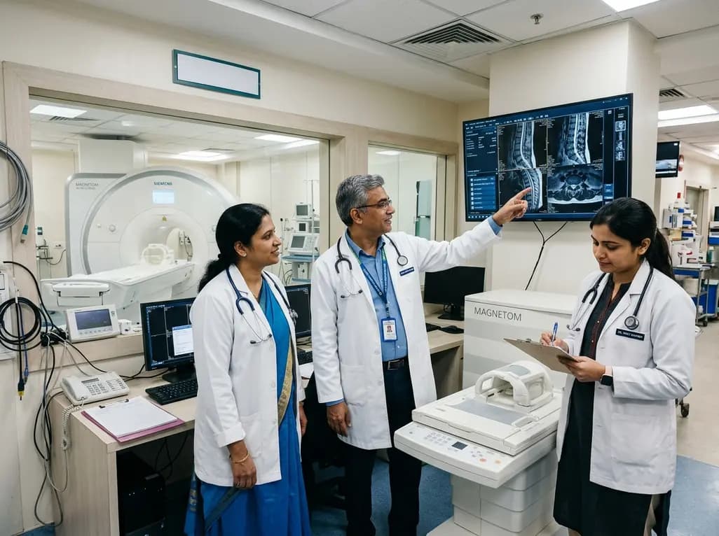
Under India's PLI initiative, domestic production of 57 critical medical devices has commenced, including MRI and CT scanners, cath labs, and heart valves. Global partnerships and local investments are expanding high-end technology, curbing import reliance, and strengthening clinical accessibility nationwide.
Today

A longitudinal study reveals that patients undergoing staged bilateral hip arthroscopy experience progressive contralateral labral deterioration during surgical intervals. However, structural degradation does not compromise 2-year postoperative clinical outcomes or patient-reported satisfaction scores.
Today

New immunological research reveals how PTPN22 forms a delayed inhibitory signaling complex to restrain T cell activation, explaining how the autoimmune-linked R620W variant disrupts feedback control and promotes persistent inflammation in conditions like rheumatoid arthritis and type 1 diabetes.
Today
