

Loading, please wait...
Loading, please wait...

Inherited retinal dystrophies present substantial diagnostic challenges because identical genetic variants can produce markedly divergent manifestations across affected family members. A recent clinical and molecular investigation has uncovered crucial insights into how a dominant CRX mutation drives cone-rod dystrophy with striking intrafamilial variability. The cone-rod homeobox gene encodes a key transcription factor regulating photoreceptor differentiation and survival. Consequently, mutations in this locus disrupt both rod and cone functions, causing visual impairment ranging from severe early blindness to subtle, adult-onset dysfunction.
The study evaluated a four-generation family segregating a Class III frameshift variant, c.590del or p.(Pro197Argfs*22). Six family members underwent detailed genetic testing with targeted dystrophy panels and Sanger confirmation. Remarkably, three confirmed carriers of this identical alteration exhibited starkly disparate clinical presentations. The proband experienced adult-onset cone-rod dystrophy characterized by progressive central vision loss, nyctalopia, and severe outer-retinal atrophy. In contrast, two other adult family members harboring the exact same mutation remained completely asymptomatic during longitudinal follow-up. These mildly affected relatives preserved normal visual acuity and standard full-field electroretinography responses. However, high-resolution optical coherence tomography and fundus autofluorescence demonstrated subclinical outer nuclear layer thinning and subtle retinal pigment epithelium alterations. Therefore, clinicians must recognize that identical pathogenic alleles do not guarantee uniform disease trajectories. Variable expressivity and incomplete penetrance can complicate pedigree tracking in routine clinical practice. Furthermore, these findings emphasize that asymptomatic carriers might escape timely diagnosis without comprehensive family screening.
Comprehensive phenotyping plays an indispensable role in detecting early photoreceptor compromise before functional visual loss emerges. In this pedigree, full-field electroretinography clearly documented combined rod and cone dysfunction in the symptomatic proband. Specifically, both scotopic and photopic amplitudes showed marked attenuation, correlating directly with patient complaints of nyctalopia and central blur. Conversely, electrophysiological studies remained completely normal in asymptomatic carriers, proving that global electrical responses can remain intact despite underlying structural alterations. Optical coherence tomography served as a pivotal modality by capturing microscopic thinning of the outer nuclear layer across all carriers. Fundus autofluorescence further revealed patchy hyperautofluorescence and subtle mottled hypoautofluorescence at the macula, reflecting ongoing metabolic stress within the retinal pigment epithelium. Thus, multimodal retinal imaging reliably detects occult structural deterioration even when conventional visual testing yields completely normal parameters. Clinicians should therefore integrate structural tomographic scans into regular screening protocols for at-risk first-degree relatives. This structured protocol enables timely baseline assessment and prevents diagnostic delay.
Understanding the functional impact of the CRX c.590del variant required rigorous ex vivo and in vitro biochemical characterization. Researchers performed dual-luciferase reporter assays using the rhodopsin promoter in both human embryonic kidney cells and mouse retinal explants. The experimental data revealed that the mutant protein completely lacked normal transactivation capability. Moreover, when co-expressed alongside wild-type CRX, the truncated protein actively suppressed wild-type transcriptional activity in a dose-dependent manner. Consequently, this variant functions as a classical dominant-negative allele rather than causing simple haploinsufficiency. The truncated transcription factor appears to retain its DNA-binding capacity while lacking the essential transactivation domain. Thus, the mutant protein effectively competes for promoter target sites and forms non-functional complexes, thereby blunting the transcription of critical downstream phototransduction genes. Because photoreceptors depend heavily on robust homeobox signaling for structural protein synthesis, this sustained transcriptional interference gradually triggers cellular apoptosis. These molecular findings convincingly elucidate the biological mechanism underlying progressive photoreceptor degeneration in affected individuals.
These research insights highlight the significant perils of relying purely on genotype-based prognostication in inherited retinal dystrophies. Because three family members with the identical sequence variant demonstrated widely divergent clinical outcomes, geneticists cannot predict visual loss severity solely from DNA sequencing reports. Genetic counselors must explain this biological nuance clearly to patients and their extended relatives. Specifically, counseling sessions should emphasize that possessing a pathogenic CRX allele establishes disease risk, but modifier genes or environmental factors might alter ultimate phenotypic severity. Furthermore, clinicians must counsel families about the autosomal dominant inheritance pattern, where each child carries a fifty percent probability of inheriting the altered gene. Identifying asymptomatic carriers through predictive screening allows ophthalmologists to establish regular surveillance intervals and initiate protective lifestyle interventions. Counselors should also prepare individuals for possible progression while providing reassurance that severe visual impairment is not an inevitable outcome for every mutation carrier. Consequently, comprehensive clinical staging through objective retinal imaging must always accompany genetic testing.
The identification of a dominant-negative disease mechanism has profound therapeutic ramifications for inherited retinal diseases. Standard gene augmentation strategies, which successfully treat recessive conditions like RPE65-associated retinal dystrophy, remain insufficient against dominant-negative alleles. Simply delivering supplemental wild-type CRX cDNA cannot overcome persistent transcriptional blockade imposed by the interfering mutant protein. Therefore, researchers are actively developing dual-action therapeutic platforms that combine allele-specific knockdown with gene augmentation. In this strategy, small interfering RNA or antisense oligonucleotides selectively degrade the mutant transcript, while an adeno-associated viral vector delivers healthy wild-type copies to restore physiological transcriptional equilibrium. Preclinical evaluations using retinal organoids and animal explants have shown remarkable promise in rescuing photoreceptor survival and restoring opsin synthesis. Furthermore, advances in targeted genome editing tools offer potential avenues to permanently silence the mutant allele in retinal tissue. These innovative molecular approaches signify a transformative paradigm shift in managing previously incurable autosomal dominant retinopathies.
A dominant CRX mutation produces a defective transcription factor that binds target retinal gene promoters but lacks normal transactivation capability. Consequently, the mutant protein outcompetes healthy wild-type proteins in a dose-dependent dominant-negative manner. This molecular disruption impairs the sustained transcription of vital structural proteins and phototransduction enzymes required for photoreceptor survival. Over time, both cone and rod photoreceptors undergo progressive degeneration, culminating in nyctalopia, macular atrophy, and severe central visual impairment.
Intrafamilial phenotypic variability occurs because disease manifestation depends on factors beyond the primary monogenic alteration. Specifically, genetic modifiers, epigenetic modifications, and variable transcriptional regulation across retinal cells influence how severely photoreceptors react to dominant-negative interference. Furthermore, differences in environmental exposures, oxidative stress levels, and individual cellular compensatory mechanisms can dictate whether a carrier develops symptomatic cone-rod dystrophy or remains subclinical with only subtle, asymptomatic outer nuclear layer thinning.
Treating dominant-negative retinopathies requires a coordinated dual-action strategy combining mutant allele knockdown with simultaneous gene augmentation. Because conventional gene replacement alone cannot overcome the toxic inhibition caused by mutant proteins, clinicians must first eliminate the abnormal transcript using allele-specific antisense oligonucleotides or RNA interference. Subsequently, an adeno-associated virus delivers functional wild-type CRX cDNA to restore proper photoreceptor transcription, effectively rescuing cellular architecture and preventing progressive visual decline.
Disclaimer: This content is for informational and educational purposes only. It is not intended to substitute for professional medical advice, diagnosis, or treatment. Always seek the advice of your physician or other qualified health provider with any questions you may have regarding a medical condition. Refer to the latest local and national guidelines for clinical practice.
References

Read summarized clinical updates, watch expert medical content, and earn CME certifications right from your smartphone.
Open your smartphone camera to scan and install.





A study on a dominant CRX frameshift mutation reveals marked intrafamilial phenotypic variability in cone-rod dystrophy and demonstrates a dominant-negative pathogenic mechanism, highlighting the need for knockdown plus gene augmentation strategies.
Today

Kenya's confirmation of its eighth Ebola infection highlights the acute risk of secondary transmission in hospital environments. Clinicians must maintain high vigilance, enforce strict infection prevention protocols, and implement rapid triage mechanisms for viral hemorrhagic fevers across healthcare settings.
Today
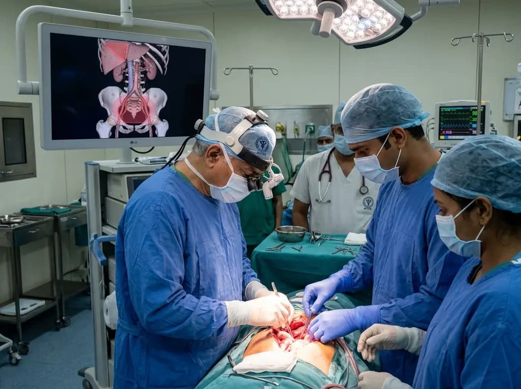
Surgical oncologists at AIIMS Delhi have mapped two critical anatomical landmarks: Ray's Zone in the deep pelvis and Ray's Hepatocaval Triangle near the liver. These predefined hazard boundaries guide dissection, dramatically reduce severe intraoperative bleeding, and preserve vital nerves and visceral organs.
Today

A study reveals that the MYLK c.4079G>A, p.(Gly1360Asp) variant is linked to high-penetrance heritable thoracic aortic disease and early-onset aortic dissection, showing a founder effect in Finnish cohorts. Findings emphasize early cascade screening and vigilant surveillance.
Today

A novel sacrificial-template technique combines FDM polymer 3D printing and micro-arc oxidation to fabricate orientation-controlled porous titanium scaffolds. These bioactive constructs offer tailored stiffness, exceptional multidirectional load resistance, and robust osseointegration for bone defect repair.
Today
A novel label-free electrochemical biosensor utilizing magnetic nanocomposites enables ultrasensitive TERT C228T mutation detection (LOD: 0.67 pM) for rapid risk stratification in papillary thyroid cancer, offering promising point-of-care utility for endocrine oncology.
Today
