
Loading, please wait...
Loading, please wait...

Digital microscopy blood films are becoming a cornerstone of modern diagnostic pathology. As laboratories seek to improve workflow efficiency, many are transitioning from traditional glass slides to digitized imaging. However, recent research suggests that while digital systems are efficient, they may have specific diagnostic blind spots compared to traditional optical methods.
A recent preliminary study evaluated the agreement between digital and optical cytology. Researchers tasking 22 clinical pathologists and residents to review canine, feline, and equine blood films found that digital slides generally perform well for common morphology. Pathologists could readily identify changes in erythrocyte shape, platelet counts, and leukocyte features on both digital and glass slides. However, the study revealed a significant concern regarding the detection of small intracellular organisms.
When using digital microscopy blood films, the identification of certain infectious agents proved challenging. For instance, organisms like Theileria haneyi were identified on significantly fewer digitized slides than on traditional glass slides. This discrepancy highlights a potential risk in clinical settings where blood-borne infectious diseases are suspected. Furthermore, the study noted low intra-observer consistency between the two methods, suggesting that the digital format may influence how a pathologist interprets fine cellular details.
While digital morphology is adequate for routine screening of red and white blood cell changes, it may not yet fully replace the high-resolution capabilities of 100x oil immersion optical microscopy. Clinicians and pathologists must exercise caution. If a patient presents with symptoms of a blood-borne parasite, manual optical review remains the gold standard to ensure diagnostic accuracy. Consequently, many experts recommend a hybrid approach where digital systems handle high-volume screening while complex or infectious cases undergo manual verification.
The transition to digital pathology offers undeniable benefits for remote consultation and archival storage. However, the current limitations in image resolution at higher magnifications can lead to missed diagnoses of intracellular pathogens. Therefore, ongoing technological advancements are necessary to improve the sensitivity of digital scanners. For now, integrating rigorous quality control and manual oversight is essential for any laboratory adopting digital hematology workflows.
Currently, no. While digital systems are excellent for general cell counts and morphology, they may lack the sensitivity required to detect rare cells or small intracellular parasites that are easily seen under a manual microscope.
The primary issue often relates to image resolution and focus. Some digital scanners may not capture the fine, multi-planar detail of an organism located inside a cell as effectively as a trained pathologist using a traditional 100x oil immersion lens.
Yes, it is highly effective for routine screening where the goal is to identify common abnormalities in red blood cells, white blood cells, and platelets, significantly speeding up laboratory turnaround times.
Disclaimer: This content is for informational and educational purposes only. It does not constitute professional medical advice, diagnosis, or treatment. Always seek the advice of your physician or other qualified healthcare provider with any questions you may have regarding a medical condition. Refer to the latest local and national guidelines for clinical practice.
References
Anderson S et al. Comparison of digital and optical microscopy for the evaluation of veterinary blood films: a preliminary study. J Vet Diagn Invest. 2026 Mar 21. doi: 10.1177/10406387261432653. PMID: 41863154.
Horiuchi Y et al. Real-World Application of Digital Morphology Analyzers: Practical Issues and Challenges in Clinical Laboratories. PMC. 2025 Mar 10.
Karner K et al. Comparative Analysis Reveals Potential Utility of Digital Microscopy in the Evaluation of Peripheral Blood Smears With Some Barriers to Implementation. Am J Clin Pathol. 2015 Jul 15.

Read summarized clinical updates, watch expert medical content, and earn CME certifications right from your smartphone.
Open your smartphone camera to scan and install.


A comparative study highlights the strengths and limitations of digital microscopy in blood film evaluation, particularly regarding infectious diseases....
4 months ago
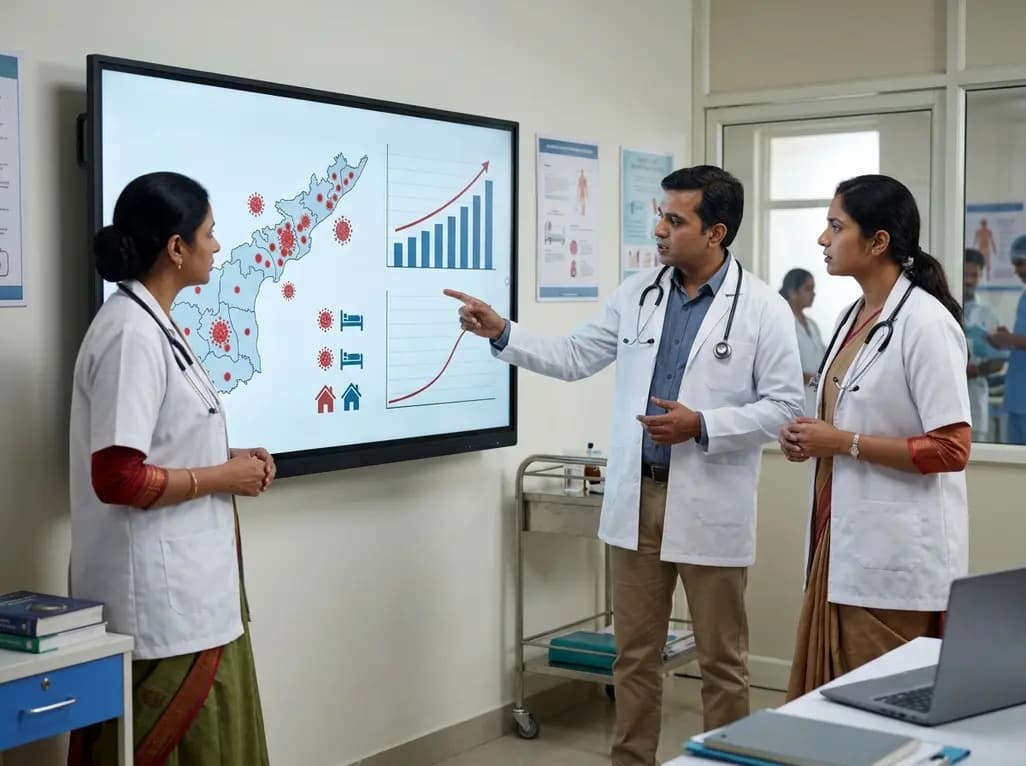
Andhra Pradesh reported 10 new Covid-19 cases, taking the state tally to 49 while deaths remain at four. With 24 patients hospitalized and 16 under home isolation, the Health Department has intensified monitoring. Medical professionals should review regional distribution, diagnostic protocols, and management plans.
Today

An 11-year Swedish registry study of 618 uterine sarcoma patients found that minimally invasive surgery yielded survival comparable to open surgery in early stages. However, adjuvant chemotherapy conferred no survival benefit in localized or advanced disease, highlighting stage and histology as key outcomes.
3 days back

A cross-sectional study evaluates post-intensive care syndrome in cardiac patients 2-4 weeks post-ICU discharge, highlighting cognitive, psychological, and functional impairments and the need for structured multidisciplinary rehabilitation.
3 days back

Anterior cruciate ligament reconstruction failure lacks uniform definition. A narrative review proposes an integrative framework incorporating objective and subjective instability, persistent pain, restricted motion, graft rupture, and secondary meniscal injury to standardize clinical reporting.
3 days back

With World Obesity Atlas data warning that over 41 million Indian children are overweight or obese, ICMR and NIN have unveiled a 10-point policy roadmap. The initiative calls for mandatory front-of-pack labeling, HFSS taxes, strict marketing bans, and healthier school environments to curb non-communicable diseases.
Today
