

Loading, please wait...
Loading, please wait...
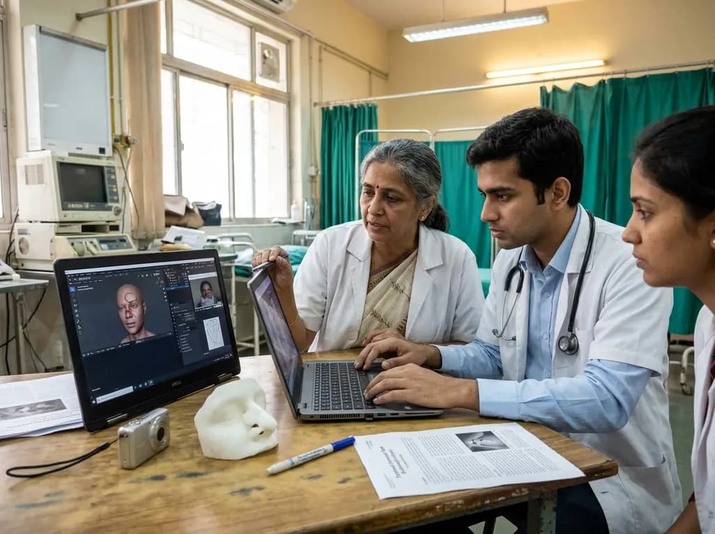
Reconstructing extensive nasal defects remains one of the most demanding tasks in facial plastic and reconstructive surgery. For over a century, the paramedian forehead flap has served as the gold standard for subtotal and multi-subunit nasal resurfacing. Traditionally, surgeons rely on freehand foil manipulation to transfer complex three-dimensional contours onto a two-dimensional forehead donor site. However, this manual technique introduces substantial geometric distortion and depends heavily on individual surgeon experience. Moreover, a groundbreaking preclinical validation study introduces an accessible, low-cost digital templating workflow that resolves these long-standing surgical limitations.
Nasal anatomy presents an intricate combination of convexities, concavities, and delicate free margins. When skin cancer excision or major facial trauma destroys these structures, reconstructive surgeons must restore both structural contour and patent airway mechanics. Historically, clinicians perform intraoperative templating using malleable materials like aluminum suture packaging or sterile foam sponges. However, flattened foil cannot easily replicate the compound three-dimensional curvature of the nasal tip, ala, and columella.
Furthermore, soft tissue collapse and post-excision wound edema frequently distort the true defect boundary during surgery. As a result, manual templates often produce dimensional discrepancies and unpredictable flap margins. If the harvested forehead tissue is too small, excessive closure tension can cause wound dehiscence or ischemic flap necrosis. Conversely, oversizing the flap requires extensive secondary debulking and increases forehead donor site morbidity. Therefore, modern head and neck surgeons require objective, standardized preoperative planning methods. By eliminating intraoperative guesswork, clinicians can design patient-specific cutaneous paddles with elevated precision, ensuring superior aesthetic outcomes and fewer revision surgeries.
To overcome the financial barriers of proprietary medical software, researchers developed an elegant, consumer-grade digital pipeline. The innovative workflow incorporates three widely accessible platforms: PhotoCatch, Blender, and Adobe Photoshop. First, the surgical team captures a continuous thirty-second video of the nasal defect using an ordinary smartphone camera. Next, PhotoCatch processes this short video sequence through advanced photogrammetry algorithms to construct a photorealistic three-dimensional polygonal mesh.
Subsequently, the digital asset imports into Blender, an open-source three-dimensional modeling suite. In Blender, software operators isolate the exact nasal defect boundaries and digitally unwrap the complex 3D surface into an accurate 2D planar template. Finally, clinicians import the flattened vector shape into Adobe Photoshop to calibrate the image against an absolute metric scale. Consequently, surgeons can print the final template onto sterile transparent film for precise intraoperative marking. This entire digital transformation requires neither expensive optical surface scanners nor specialized bioengineering hardware, making it immediately viable for resource-conscious clinics.
To validate this digital protocol, investigators conducted a prospective pilot evaluation involving twenty participants with simulated nasal defects. The research team generated digital flap designs and compared them directly with conventional manual reference forehead templates. Specifically, investigators measured three primary morphological parameters: maximum vertical height, maximum horizontal width, and total surface area.
The statistical findings demonstrated exceptional concordance between the digital reconstructions and physical defects. Mean maximum height measured 3.70 ± 1.39 cm physically versus 3.72 ± 1.46 cm digitally, showing no significant difference (P = 0.573). Similarly, maximum width demonstrated remarkable parity, registering 4.26 ± 1.80 cm physically versus 4.32 ± 1.77 cm digitally (P = 0.240). Furthermore, total surface area aligned closely at 10.59 ± 7.55 cm² compared to 10.73 ± 7.84 cm² (P = 0.270). Bland-Altman agreement analysis confirmed minimal bias and tight limits of agreement across all linear dimensions. Therefore, this digital method accurately reproduces true anatomical boundaries without clinically meaningful error, providing surgeons with reliable predictive metrics.
Beyond geometric precision, clinical workflows must exhibit reasonable time efficiency to achieve widespread surgical utility. In this preclinical study, the mean total template generation time was 24.95 ± 11.83 minutes. Importantly, capturing the baseline video data requires less than one minute during an initial clinic visit or preoperative consultation. The remaining digital steps occur asynchronously on standard office computers before the patient enters the operating suite.
Consequently, this pipeline completely eliminates the stressful manual template fabrication normally performed while the patient is anesthetized. By saving twenty to thirty minutes of operative time, surgical teams can decrease general anesthesia exposure and lower theater resource costs. Moreover, operating room teams can sterilize the pre-printed acetate templates alongside standard surgical instrumentation. In busy hospital centers across India and globally, saving theater time directly enhances operating room throughput and patient safety. Thus, this pragmatic digital strategy delivers substantial logistical and economic advantages for public and private healthcare facilities alike.
The integration of digital photogrammetry also offers tremendous educational benefits for resident surgeons and surgical fellows. Historically, trainees learned forehead flap design through subjective trial and error under senior scrubbed faculty. In contrast, digital modeling provides a reproducible, objective platform where trainees can visualize complex topological transformations in three dimensions. Residents can simulate flap rotation, evaluate aesthetic subunit boundaries, and predict soft-tissue closure tension before touching a scalpel.
Additionally, these high-resolution three-dimensional digital models enhance patient counseling and shared decision-making. Facial cancer resections cause severe anxiety regarding postoperative appearance and structural deformities. When surgeons present customized digital reconstructions during preoperative visits, patients comprehend the rationale for a staged paramedian forehead flap far more clearly. Furthermore, digital storage preserves an unalterable, objective record within the electronic medical system for prospective quality audits and longitudinal research. Ultimately, adopting low-cost digital workflows elevates surgical consistency, refines clinical education, and establishes a new benchmark for reconstructive rhinoplasty.
Digital templating replaces subjective freehand foil modeling with objective computer-assisted precision. By accurately unwrapping complex three-dimensional nasal defect topography into an exact planar geometry, this method avoids flap undersizing and tissue waste. Consequently, reconstructive surgeons can position donor flaps precisely over the supratrochlear axial pedicle. This meticulous alignment minimizes donor-site morbidity, preserves flap vascularity, reduces operative time, and ultimately improves aesthetic and structural symmetry for complex reconstructive patients.
The described protocol utilizes freely available and low-cost consumer software rather than expensive medical hardware. First, clinicians use PhotoCatch on an ordinary smartphone to generate three-dimensional photogrammetric models from simple video recordings. Next, Blender unwraps the digital model to flatten three-dimensional curvatures into planar patterns. Finally, Adobe Photoshop scales the graphic to life-size dimensions. Therefore, surgical teams can avoid high-end commercial scanning packages while maintaining high clinical precision.
In the preclinical validation study, the entire digital workflow required an average processing time of 24.95 minutes. Video acquisition takes merely thirty seconds at the bedside or clinic. Subsequently, computational rendering, digital surface unwrapping, and dimensional calibration take approximately twenty to twenty-five minutes. Furthermore, clinicians can perform these steps preoperatively outside operating theatre hours. Consequently, the digital process saves valuable intraoperative time, avoids prolonged patient anesthesia, and streamlines surgical execution.
Disclaimer: This content is for informational and educational purposes only and should not be considered medical advice. Always consult a qualified healthcare provider for diagnosis and treatment decisions. Refer to the latest local and national guidelines for clinical practice.
References

Read summarized clinical updates, watch expert medical content, and earn CME certifications right from your smartphone.
Open your smartphone camera to scan and install.





A preclinical validation study demonstrates that an accessible digital workflow using PhotoCatch, Blender, and Photoshop accurately templates 3D nasal defects for paramedian forehead flap reconstruction, matching manual accuracy while saving valuable operative time.
Today
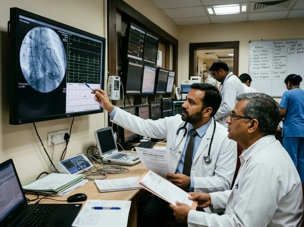
The randomized EARLY-RESYNC trial reveals that early left bundle branch pacing combined with guideline-directed medical therapy significantly improves LVEF and reduces LV end-systolic volume compared to medical therapy alone in patients with complete LBBB and mildly reduced ejection fraction (36%-50%).
Today
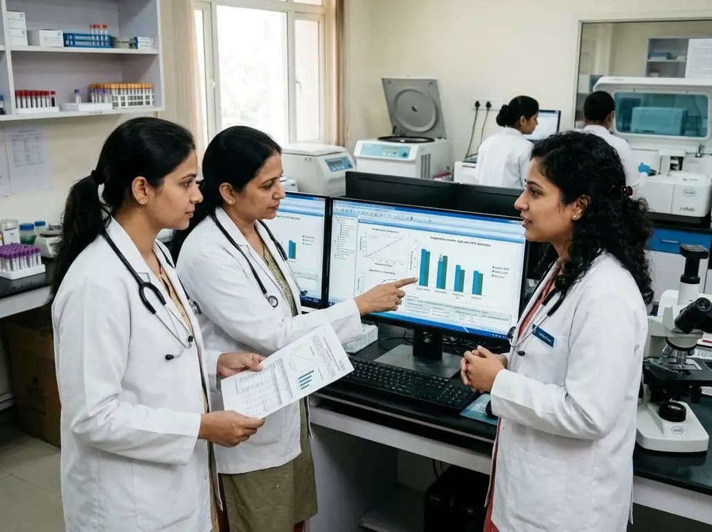
A comparative evaluation of INNO-LiPA, Cobas 4800, and Amplicor highlights significant assay-dependent variation in high-risk HPV detection, emphasizing the clinical distinction between analytical sensitivity and screening specificity.
Today
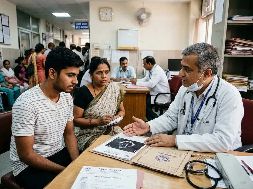
Recent data from PGIMER reveals that young adults diagnosed with type 2 diabetes face an aggressive disease trajectory. They encounter severe cardiovascular and microvascular damage within five years, occurring at higher rates than in type 1 diabetes despite lower mean blood glucose levels.
Today
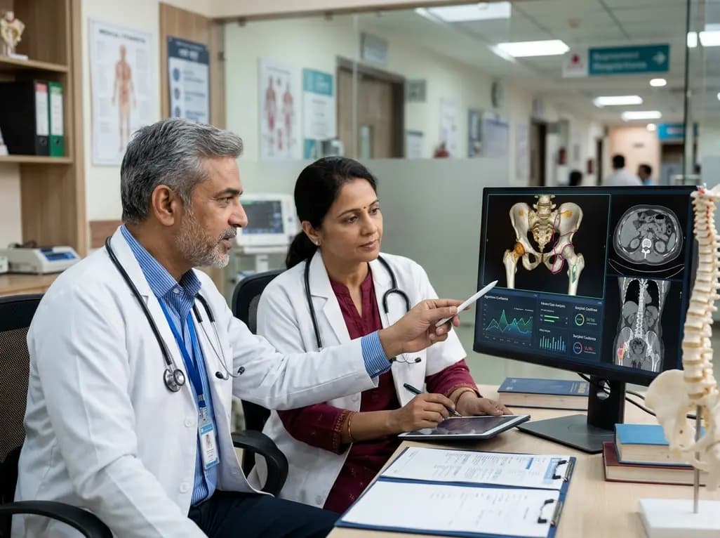
Discover the six core principles of responsible AI in orthopaedics, emphasizing algorithmic fairness, transparency, clinical reliability, and specialist oversight to safeguard patient trust and outcomes.
Today
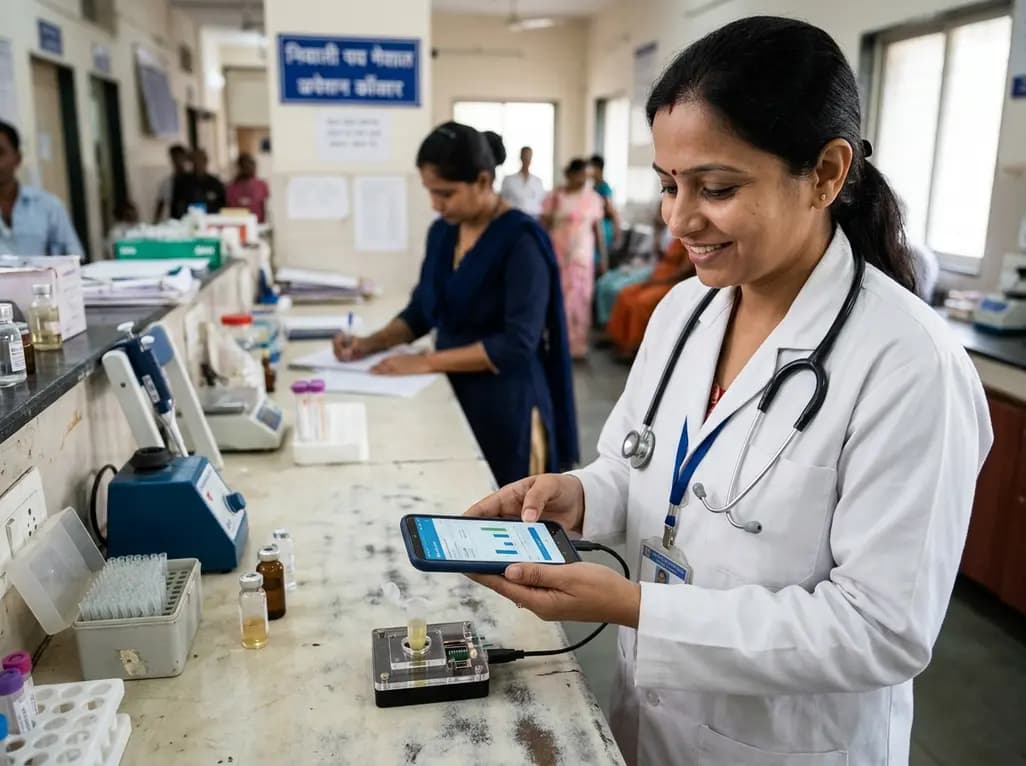
A novel smartphone-assisted biosensor utilizing molecularly imprinted polymers and catalytic nanozymes offers antibody-free, enzyme-free urinary microalbumin testing, enabling rapid, low-cost point-of-care screening for early chronic kidney disease.
Today
