

Loading, please wait...
Loading, please wait...

Phenotypic evaluation represents a cornerstone in medical genetics, especially during pediatric dysmorphology assessments. Clinicians recognize that one-third of syndromic disorders exhibit distinctive craniofacial anomalies. However, the diagnostic journey for affected families often spans several years of exhaustive testing. Recent advances in artificial intelligence offer hope for accelerating this evaluation. Specifically, researchers are testing computer vision algorithms for diagnosing rare genetic diseases from patient facial photographs. A landmark diagnostic accuracy study evaluated three open-weight multimodal large language models to assess their clinical precision and visual prompting utility.
Recognizing syndromic dysmorphism demands nuanced clinical training and extensive personal experience. In pediatric practice, atypical facial signs such as hypertelorism, epicanthal folds, or midface hypoplasia often indicate deeper systemic syndromes. Consequently, identifying subtle structural phenotypes early can prevent secondary complications and guide targeted molecular genetic testing. However, rare disorders remain exceptionally diverse, with thousands of unique molecular entities presenting overlapping phenotypic signatures. In resource-constrained settings, clinical geneticists remain scarce, leaving primary pediatricians to manage complex diagnostic dilemmas. Therefore, digital tools that parse facial images could provide critical decision support in underserved clinics. Dedicated deep learning architectures have historically demonstrated respectable classification abilities. Nevertheless, medical informatics has recently shifted toward general-purpose multimodal large language models. These foundation systems process clinical photography alongside unstructured descriptive language. Clinicians must now understand whether these broad systems provide dependable differential diagnoses across diverse patient populations.
To establish clear diagnostic benchmarks, investigators conducted a rigorous accuracy study using 4746 curated patient photographs from the GestaltMatcher Database. Specifically, the cohort comprised two distinct subsets: GMDB-Frequent, featuring 882 images across conditions with six or more photographs, and GMDB-Rare, containing 3864 images from ultra-rare conditions. The researchers locally deployed three open-weight multimodal large language models: Gemma-4-31B, Qwen3.6-35B-A3B, and Qwen3.8-27B. Each system generated 50 ranked candidate diagnoses mapped directly to Mondo Disease Ontology identifiers. The empirical findings demonstrated sobering performance boundaries. Within GMDB-Frequent, top-50 diagnostic accuracy ranged between 13.95% and 18.25% across the three architectures. More alarmingly, performance plummeted when evaluating GMDB-Rare cases, yielding top-50 accuracy figures between 0.57% and 1.35%. Consequently, the vast majority of rare genetic presentations eluded the diagnostic gaze of these models. These results indicate that raw multimodal systems cannot yet provide reliable automated syndromic identification in routine practice.
Because raw images presented substantial diagnostic hurdles, the study explored whether targeted computer vision pre-processing could enhance model accuracy. Researchers evaluated image restoration through CodeFormer super-resolution alongside automated visual prompting using Qwen-Image-Edit-2511 to place red circles around dysmorphic facial regions. Interestingly, red-circle prompting yielded numerical accuracy increases across five of the six model-subset evaluations. Specifically, Qwen3.8-27B achieved a statistically significant accuracy rise in GMDB-Rare, increasing from 0.57% to 1.27%. Furthermore, within the subset possessing high-quality visual prompts in GMDB-Frequent, Gemma-4-31B demonstrated notable improvements over super-resolution. The model exhibited accuracy increases of 5.76 and 4.46 percentage points across physician rating assessments. In addition, matched red-circle inputs decisively outperformed mismatched controls across all model configurations, proving that focused visual guidance genuinely directs model attention. Moreover, adopting a two-stage image-to-phenotype workflow yielded additional performance gains in selected subsets. Thus, structured visual prompting partially mitigates visual parsing failures in multimodal architectures.
Despite modest improvements from visual prompts, deeper mechanistic analyses revealed unsettling algorithmic behaviors. Most strikingly, a naive random baseline achieved 8.15% diagnostic accuracy in GMDB-Rare. This baseline comfortably exceeded all three multimodal architectures under both raw and red-circle input conditions. The explanation lies in severe prediction overlap and heavy text-frequency biases. When investigators performed image-replacement experiments, candidate lists remained remarkably consistent despite swapping different patient faces. Consequently, the models repeatedly outputted identical common syndromes regardless of unique dysmorphic features. Their top-tier predictions clustered heavily around a minute subset of well-documented syndromes. This indicates that model outputs were weakly anchored to actual facial geometry. Instead of extracting patient-specific facial biometrics, the models largely relied on internal text frequency priors from their training literature. Furthermore, models exhibited high diagnostic list perplexity and poor stability during repeated inference runs. These foundational flaws highlight severe vulnerabilities in relying on uncalibrated vision-language models for clinical diagnostics.
These critical findings carry profound clinical implications for practicing pediatricians, neonatologists, and medical geneticists. Across outpatient departments and neonatal intensive care units, clinicians frequently evaluate newborns presenting with unexplained dysmorphism. While artificial intelligence holds substantial promise, off-the-shelf multimodal large language models remain completely unsuited for autonomous clinical triage. Relying on their unverified outputs risks diagnostic distraction, inappropriate testing, and parental anxiety. Instead, clinicians must preserve rigorous clinical dysmorphology methodology. Pediatricians should meticulously document anatomical landmarks, measure craniofacial distances, and record developmental trajectories. When algorithmic assistance is desired, clinicians should utilize dedicated next-generation phenotyping platforms rather than general chat models. Specialized tools map geometric distances across standardized latent representations, minimizing the hallucination risks inherent in language models. In addition, future artificial intelligence development must focus on prospective multicenter validation and multimodal integration. Until then, careful clinical phenotyping by experienced physicians remains the indispensable gold standard in rare disease diagnosis.
No, current multimodal large language models cannot replace specialized dysmorphology platforms in clinical practice. The study proved that general models achieve top-50 accuracy rates below 19% in frequent conditions and barely 1% in rare syndromes. Furthermore, their predictions often rely on textual word frequencies rather than precise facial measurements. Consequently, clinicians must continue using validated, dedicated next-generation phenotyping algorithms and formal molecular genetic testing rather than general vision-language models for diagnostic decisions.
Visual prompting guides neural network attention toward specific regions of interest, such as dysmorphic eye shapes or nasal bridges. In the benchmark study, applying red circles around affected areas produced modest diagnostic improvements across several model comparisons. Moreover, matched visual prompts consistently outperformed mismatched controls, showing that targeted visual annotations deliver genuine informational value. However, prompting alone cannot overcome foundational reasoning deficits, meaning models still require structured phenotypic translation to generate clinically meaningful differential lists.
A random baseline attained 8.15% accuracy in ultra-rare diseases, exceeding all multimodal models evaluated on raw photographs. This occurred because multimodal models suffer from severe prediction collapse, repeatedly suggesting the same few common genetic disorders regardless of the input image. In contrast, the random baseline sampled uniformly across all possible rare entities in the ontology. Because ultra-rare diseases are numerous, uniform random sampling had a mathematically higher probability of hitting rare targets than heavily biased models.
Disclaimer: This content is for informational and educational purposes only and should not be taken as professional medical advice. Always consult with a qualified healthcare provider for personal health concerns or diagnosis. Never disregard professional medical advice or delay in seeking it because of something you have read here. In the event of a medical emergency, call your doctor or local emergency services immediately. Refer to the latest local and national guidelines for clinical practice.
References

Read summarized clinical updates, watch expert medical content, and earn CME certifications right from your smartphone.
Open your smartphone camera to scan and install.





A landmark diagnostic study evaluates multimodal large language models and visual prompting for identifying rare genetic diseases from facial images, highlighting major clinical limitations.
Today
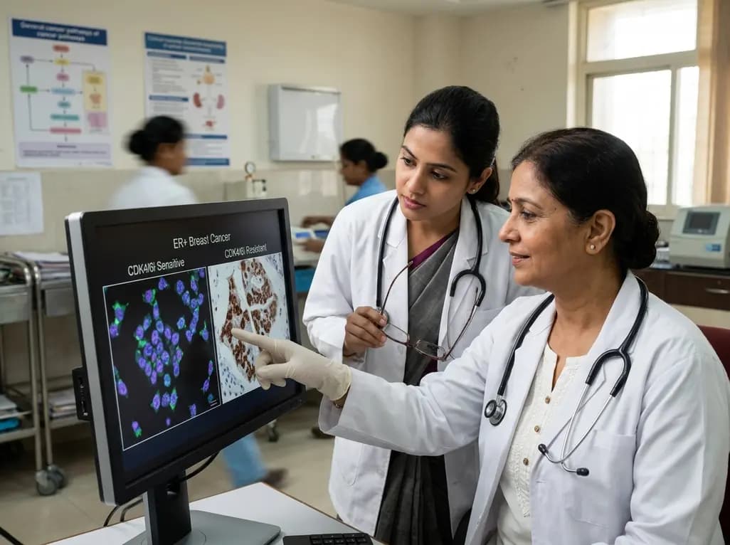
Overexpression of glycoprotein nonmetastatic melanoma protein B (GPNMB) mediates resistance to CDK4/6 inhibitors like abemaciclib and palbociclib in ER+ breast cancer through integrin-driven AKT/ERK signaling. Targeting GPNMB with antibody-drug conjugates like glembatumumab vedotin may overcome this resistance.
Today
A secondary analysis of the MOMS clinical trial reveals that infant sex does not influence neonatal opioid withdrawal syndrome severity in buprenorphine-exposed newborns. Maternal co-exposures and clinical factors drive outcomes, highlighting the need for objective assessment and supportive breastfeeding.
Today
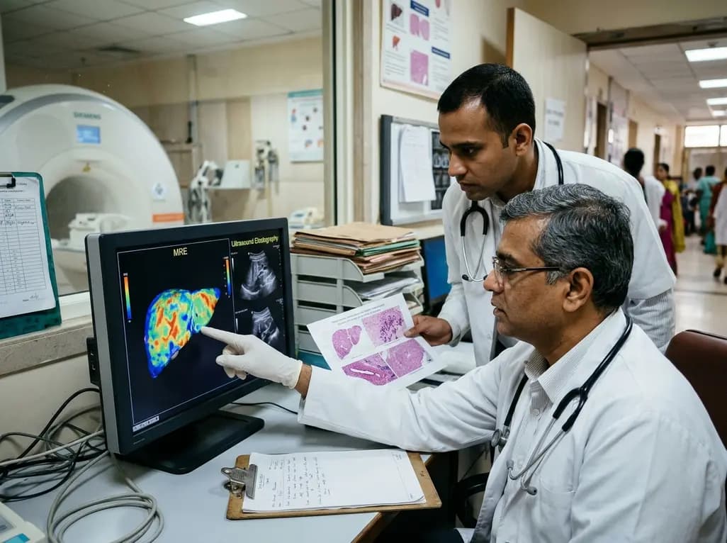
A comparative study reveals magnetic resonance elastography outperforms ultrasound elastography in assessing liver fibrosis in Fontan-associated liver disease, demonstrating stronger histological agreement and superior discrimination.
Today
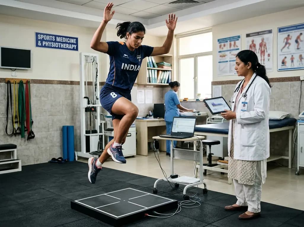
A systematic review and meta-analysis highlights the substantial impact of plyometric jump training on jump performance, reactive strength, and landing kinematics in female team-sport athletes, providing evidence-based programming guidelines for sports medicine and orthopedics.
Today
A comprehensive review of the role of growth hormone in diabetic kidney disease and podocytopathy. Learn how elevated GH activates ZEB2, Notch, and TGF-beta1, driving podocyte loss and glomerulosclerosis, and explore novel targeted therapeutic approaches.
Today
