

Loading, please wait...
Loading, please wait...

Neurosurgeons frequently utilize decompressive craniectomy as a life-saving intervention for malignant brain edema resulting from traumatic brain injury or ischemic stroke. While surgical survival rates have improved significantly, evaluating long-term post-craniectomy neuropsychiatric outcomes has become an urgent clinical priority. Patients often survive acute catastrophic intracranial hypertension, yet they face enduring psychiatric illness and cognitive deficits. Historically, clinical follow-ups focused almost exclusively on gross functional recovery through metrics like the Glasgow Outcome Scale. Consequently, clinicians frequently overlooked subtle yet debilitating neurobehavioral morbidities. A landmark systematic review and meta-analysis led by Bari and colleagues brings crucial clarity to this clinical dilemma. By synthesizing psychometric and cognitive data from multiple international cohorts, the study maps the psychiatric trajectory of craniectomy survivors. In nations like India, where road traffic accidents and acute neurovascular emergencies occur frequently, understanding these psychiatric consequences is particularly essential. Early surgical survival should represent the beginning, rather than the conclusion, of comprehensive neurological care. Therefore, neurosurgical teams must appreciate the extensive psychiatric footprint associated with cranial decompression to design targeted, proactive therapeutic pathways.
The meta-analysis delivers sobering quantitative figures regarding psychiatric morbidity in craniectomy cohorts. Specifically, the pooled prevalence of post-operative depression reached forty-seven percent across evaluated patient populations. Moreover, statistical assessments demonstrated remarkably low between-study heterogeneity, confirming that high depression rates represent a consistent global phenomenon rather than regional anomalies. In addition, several evaluated studies examined depressive severity in depth. Clinicians observed moderate depression rates ranging from eight to forty-five percent, while severe depressive states afflicted up to thirty-six percent of patients. Anxiety disorders similarly showed high prevalence rates across multiple validated psychometric assessments. These psychiatric disturbances do not merely reflect transient emotional adjustment disorders. Instead, they stem from profound organic disruption, altered cerebral hemodynamics, and significant psychological trauma. In everyday practice, however, these symptoms often go unrecognized because motor recovery dominates routine outpatient examinations. Consequently, persistent depressive disorders severely impair vocational reintegration, undermine home rehabilitation, and heighten caregiver distress. Recognizing this high epidemiological prevalence empowers healthcare teams to implement systematic psychometric screening protocols early during post-surgical convalescence.
Understanding the biological basis of emotional and cognitive deterioration requires examining post-craniectomy intracranial biomechanics. The surgical removal of a large cranial vault segment exposes the underlying brain to external atmospheric pressure. Consequently, this persistent pressure gradient distorts cerebral architecture, disrupts cerebrospinal fluid circulation, and impedes regional blood flow. Clinicians recognize this phenomenon as the syndrome of the trephined, or sinking skin flap syndrome. Furthermore, chronic exposure to atmospheric pressure gradients causes venous stasis and reduces metabolic glucose utilization across vulnerable cerebral cortex networks. The prefrontal cortex, temporal lobes, and limbic circuits suffer disproportionate functional disruption during this period of altered biomechanics. As a result, patients experience progressive apathy, severe emotional lability, psychomotor slowing, and executive dysfunction. In addition, secondary neuroinflammation and axonal shearing from the initial trauma exacerbate neurochemical imbalances in serotonergic and dopaminergic pathways. Therefore, post-craniectomy psychiatric morbidity reflects true neurobiological injury rather than purely psychological distress. Clinicians must address these physiological drivers promptly to prevent irreversible secondary neuronal damage and chronic neurocognitive deterioration.
Fortunately, surgical reconstruction of the cranial vault provides dramatic benefits beyond simple cosmetic repair and physical brain protection. Longitudinal investigations demonstrate that cranioplasty directly improves cerebral hemodynamics and normalizes intracranial pressure balance. By restoring a rigid, closed cranial vault, the reconstruction eliminates the negative atmospheric pressure gradient on cerebral tissue. Consequently, patients frequently display rapid cognitive and affective improvements following successful cranioplasty. The systematic review identified substantial longitudinal reductions in depression and anxiety scores at twelve months post-reconstruction. Furthermore, qualitative cognitive assessments consistently documented marked gains in attention, verbal fluency, memory retention, and processing speed. Restoring physiological cerebrospinal fluid pulsation appears to reactivate microvascular perfusion in previously hypoperfused perilesional cortical regions. However, surgeons must carefully balance the timing of cranioplasty against the risks of infection and persistent cerebral swelling. In typical Indian surgical centers, autologous bone flaps or customized alloplastic implants facilitate this structural normalization. Ultimately, cranioplasty serves as an active neurorestorative operation that establishes optimal physiological conditions for comprehensive neuropsychological recovery.
Given the high prevalence of mood disturbances, surgical teams must actively integrate structured psychometric evaluations into regular neurosurgical follow-ups. Relying exclusively on informal clinical impressions or spontaneous patient complaints frequently fails to detect severe internalizing disorders. Therefore, outpatient clinics should systematically deploy validated screening tools, such as the Patient Health Questionnaire and the Hospital Anxiety and Depression Scale. Early detection enables timely collaborative interventions involving consultation-liaison psychiatrists, clinical neuropsychologists, and occupational therapists. When initiating pharmacotherapy, clinicians should exercise caution because craniectomy survivors carry heightened vulnerability to post-traumatic seizures. Consequently, selective serotonin reuptake inhibitors with favorable neurological safety profiles and low proconvulsant potential represent suitable first-line agents. In contrast, physicians must avoid agents with strong seizure-inducing properties. In addition, physical and cognitive rehabilitation must proceed concurrently with psychiatric management. Family caregivers require dedicated counseling to recognize early signs of mood decline, social withdrawal, and executive apathy. By establishing formalized multidisciplinary pathways across outpatient settings, tertiary centers can significantly reduce disability, optimize neurocognitive recovery, and enhance long-term functional independence.
Clinicians should initiate baseline cognitive and psychological screening before hospital discharge once the patient regains cognitive responsiveness. In addition, outpatient protocols must include structured evaluations at one, three, and six months post-surgery. Early assessment identifies emerging depressive symptoms promptly, enabling rapid pharmacotherapy and psychotherapeutic support before severe functional decline occurs.
Yes, cranioplasty frequently improves cognitive performance by restoring physiological intracranial pressure and eliminating the atmospheric pressure gradient. Studies show significant post-operative gains in attention, executive functioning, and processing speed. Normalizing cerebral blood flow and cerebrospinal fluid dynamics resolves the syndrome of the trephined, which directly fosters meaningful neurocognitive recovery.
Clinicians must select psychotropic medications with low epileptogenic potential because craniectomy patients carry substantial seizure risks. Selective serotonin reuptake inhibitors, such as sertraline or escitalopram, represent ideal first-line options due to their favorable safety profiles. In contrast, providers should avoid tricyclic antidepressants and bupropion because they significantly lower the seizure threshold.
Disclaimer: This content is for informational and educational purposes only. It is not intended to be a substitute for professional medical advice, diagnosis, or treatment. Always seek the advice of your physician or other qualified health provider with any questions you may have regarding a medical condition. Refer to the latest local and national guidelines for clinical practice.
References

Read summarized clinical updates, watch expert medical content, and earn CME certifications right from your smartphone.
Open your smartphone camera to scan and install.





A comprehensive meta-analysis reveals a 47% pooled prevalence of depression following decompressive craniectomy, highlighting significant psychiatric morbidity and the neurorestorative potential of timely cranioplasty.
Today

A landmark study reveals that congenital DLK1 deficiency in women with a history of central precocious puberty causes visceral adiposity, mitochondrial dysfunction, oxidative stress, and neutrophil activation, highlighting the critical need for lifelong cardiometabolic surveillance.
Today

Estetrol (E4), a native fetal estrogen, offers a unique tissue-selective profile with minimal hepatic impact and lower thrombotic risk. This review highlights its clinical use in oral contraception, menopausal symptom management, and emerging applications across oncology and neurology.
Today

Explore revascularisation strategies in dialysis-dependent chronic limb-threatening ischaemia. This review examines surgical bypass vs endovascular therapy, BEST-CLI trial findings, procedural morbidity, conduit availability, and multidisciplinary decision-making for optimal limb salvage.
Today

A recent cross-sectional analysis reveals that over a quarter of randomized controlled trials for non-specific low back pain face discontinuation or unknown status, while more than 36% of completed trials remain unpublished, exposing notable reporting gaps in spine and musculoskeletal clinical research.
Today
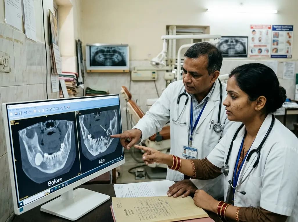
This case report examines the longitudinal CBCT evolution of Stage 2 MRONJ in a 70-year-old woman on denosumab at the site of a pre-existing mandibular dense bone island. It emphasizes the radiographic features, differential diagnosis, and multidisciplinary strategies required for high-risk bone lesions.
Today
