

Loading, please wait...
Loading, please wait...

Endoscopic third ventriculostomy has revolutionized the management of obstructive hydrocephalus across pediatric and adult populations. By establishing an internal bypass through the floor of the third ventricle, surgeons frequently eliminate the lifelong need for extracranial shunts. However, surgical teams still encounter distinct technical hurdles during the cranial entry and closure phases. Among these hurdles, postoperative cranial wound breakdown and fluid leakage pose grave risks to patient recovery. Reliable CSF leak prevention remains paramount because persistent external leakage dramatically increases the probability of retrograde bacterial inoculation, pseudomeningocele formation, and life-threatening intracranial infections. When cerebrospinal fluid escapes through the craniotomy or burr hole, it undermines soft tissue healing and prolongs inpatient hospital stays. Traditional neurosurgical interventions achieve closure through primary, watertight dural suturing. Unfortunately, endoscopic intraventricular procedures utilize narrow burr holes where direct dural suturing is technically impractical or unfeasible. As a result, surgeons require innovative, reproducible closure methods to secure the durotomy defect effectively. In a recent technical report, investigators introduced an elegant sutureless closure method combining autologous bone dust with an absorbable matrix. This multilayered technique bridges the critical barrier between intracranial pressure and scalp closure.
Postoperative fluid fistulas represent a formidable adversary following neuroendoscopic surgery. Although endoscopic third ventriculostomy restores physiological cerebrospinal dynamics, transient elevations in intracranial pressure frequently persist during early postoperative recovery. Furthermore, the surgeon creates a localized dural incision at the base of a narrow bony corridor. Because the endoscopic sheath dilates and tears the dura, re-approximating the edges with standard micro-sutures becomes nearly impossible. Consequently, high-pressure fluid escapes through the unsealed durotomy into the subgaleal space. This fluid accumulation can produce tense pseudomeningoceles, incite subgaleal collections, or extravasate directly through the cutaneous incision. In addition, persistent fistulas provide a direct conduit for external cutaneous pathogens to migrate into the subarachnoid space. Such retrograde contamination escalates the risk of catastrophic meningitis, ventriculitis, and systemic sepsis. Patients who develop external leaks frequently require prolonged antibiotic regimens, repeat lumbar punctures, or secondary revision surgeries. Therefore, neurosurgeons have explored numerous adjuncts, such as pericranial flaps, collagen matrices, and synthetic sealants. Nevertheless, many synthetic agents introduce substantial logistical costs, which severely burden healthcare centers in resource-constrained settings. Establishing an affordable, readily available, and technically straightforward closure protocol addresses a pressing worldwide clinical dilemma in contemporary pediatric and adult neurosurgery.
To overcome the limitations of narrow burr hole access, researchers designed a novel multilayered sandwich reconstruction. Specifically, this protocol integrates autologous bone debris with an absorbable hemostatic matrix to create a rigid, biological barrier. During the initial cranial entry, surgeons preserve bone shavings produced while drilling the precoronal burr hole. After completing the intraventricular fenestration and withdrawing the neuroendoscope, the surgical team begins the reconstruction. First, the surgeon places an initial layer of absorbable hemostatic matrix directly over the torn dural edges. This hemostatic substrate acts as an immediate scaffold, conforming smoothly across the irregular margins of the defect. Next, the surgeon packs the reserved autologous bone dust directly over this basal layer. By tamping the bone dust gently, the team reconstitutes the missing bony architecture within the burr hole aperture. Finally, the team applies a superficial capping layer of absorbable hemostatic matrix over the compacted bone debris. This second matrix layer anchors the bone particles firmly in position, preventing particle migration into adjacent subgaleal tissues. Consequently, this sandwich configuration constructs an impermeable, three-dimensional mechanical plug. The team then achieves anatomical closure of the overlying galea aponeurotica and skin using routine layered sutures.
The clinical success of this sandwich closure relies on the complementary mechanical and biological properties of its components. Absorbable cellulosic and hemostatic matrices, such as BloodSTOP, consist of etherified carboxymethylcellulose that rapidly hydrates upon contact with blood or exudate. When wet, the material transforms into a clear, adherent bio-gel. This dense gel immediately occludes microscopic fluid channels along the dural margins. Moreover, the gel exerts a gentle mechanical tamponade that arrests capillary bleeding without excessive tissue expansion. Meanwhile, the interspersed autologous bone debris provides structural rigidity. Unlike soft gelatin sponges that degrade quickly or compress under intracranial pulsations, compacted autologous bone resists physiological hydrostatic heads. In addition, autologous bone dust contains osteoinductive factors and viable osteoprogenitor elements that facilitate long-term osseous integration. By encasing the bone dust between two gel layers, the surgeon prevents accidental particulate dispersion into the subdural space or cerebral cortex. Historical literature cautioned that loose bone dust could potentially induce aseptic meningeal inflammation if particles migrated intraventricularly. However, the cohesive envelope formed by the gelatinous matrix securely encapsulates the bone fragments. Consequently, the construct promotes stable osteogenesis while maintaining immediate, durable hydrostatic resistance against persistent intracranial fluid pressures.
Neurosurgical centers traditionally rely on an array of commercial biomaterials to achieve dural closure when primary suturing fails. For example, surgeons frequently deploy collagen matrices, polyethylene glycol hydrogels, and fibrin sealants such as TachoSil. While these advanced sealants demonstrate robust efficacy across diverse cranial procedures, they also present distinct clinical and economic shortcomings. First, commercial fibrin sealants carry substantial financial costs, which significantly limits their regular adoption in resource-limited hospitals. Second, biological sealants derived from pooled human or equine plasma carry theoretical risks of allergic reactions, immunogenic responses, or transmission of infectious agents. Third, standalone synthetic hydrogels often lack rigid structural backing, making them vulnerable to displacement under fluctuating intraventricular pressures. In contrast, the autologous bone sandwich technique utilizes the patient’s own drilled bone debris, which entirely eliminates foreign body rejection and biological contamination risks. Furthermore, carboxymethylcellulose hemostatic matrices offer a cost-effective, shelf-stable, and hypoallergenic alternative to expensive biologic adhesives. Clinical observations across fourteen consecutive neuroendoscopic cases revealed zero incidences of incisional leaking, pseudomeningocele formation, or wound dehiscence. Thus, this accessible technique delivers surgical reliability comparable to premium commercial products while dramatically reducing per-procedure expenditures.
Adopting this sutureless closure requires minimal alterations to standard operative workflows, ensuring rapid learning curves for neurosurgical teams. During initial exposure, the operating team should meticulously collect bone dust using a slow-speed drill with irrigation or dedicated collector traps. Surgeons must store this autologous material in sterile saline to preserve cellular viability and prevent environmental contamination. Following neuroendoscopic fenestration, the operator should confirm stable ventriculostomy pulsation and achieve complete ventricular hemostasis before initiating closure. When packing the burr hole, surgeons must exercise caution to avoid pushing excessive bone dust deeply past the dural margins into the subdural space. Applying the inner matrix layer flat against the dura establishes a protective safety barrier that shields the underlying brain parenchyma. Subsequently, gentle compaction of bone fragments restores cranial contour and eliminates dead space beneath the scalp flap. Finally, surgeons must perform meticulous, tension-free closure of the galea aponeurotica using interrupted absorbable sutures. A well-vascularized, tightly approximated galea reinforces the sandwich construct and serves as the ultimate anatomical barrier against external pathogens. By integrating these straightforward precautions, surgical units can maximize procedural success, reduce complications, and accelerate postoperative discharge.
Primary dural suturing is technically impractical after neuroendoscopic surgery because surgeons access the ventricular system through a small burr hole. The tight bony corridor restricts needle maneuverability, while endoscopic instruments often fray the dural edges. Consequently, primary suturing fails to establish a reliable watertight barrier, necessitating sutureless multilayered closure methods.
Surgeons prevent bone dust migration by sandwiching the autologous fragments between two layers of absorbable hemostatic matrix. The deep matrix layer covers the dural tear, shielding the underlying brain parenchyma. When exposed to exudate, the matrix forms a dense, sticky gel that securely encapsulates bone particles and prevents intracranial dispersion.
The sandwich closure technique significantly reduces operative expenses by combining readily available autologous bone debris with affordable carboxymethylcellulose hemostatic matrices. In contrast, proprietary fibrin and polyethylene glycol sealants carry steep financial costs and cold-chain storage requirements, making the autologous sandwich method exceptionally advantageous for cost-conscious and resource-limited surgical centers worldwide.
Disclaimer: This content is for informational and educational purposes only... Refer to the latest local and national guidelines for clinical practice.
References

Read summarized clinical updates, watch expert medical content, and earn CME certifications right from your smartphone.
Open your smartphone camera to scan and install.





A novel sutureless closure using autologous bone dust and absorbable hemostatic matrix effectively achieves watertight burr hole sealing after endoscopic third ventriculostomy (ETV), providing an affordable, reproducible method for CSF leak prevention without requiring expensive synthetic sealants.
Today

A landmark study reveals that congenital DLK1 deficiency in women with a history of central precocious puberty causes visceral adiposity, mitochondrial dysfunction, oxidative stress, and neutrophil activation, highlighting the critical need for lifelong cardiometabolic surveillance.
Today

Estetrol (E4), a native fetal estrogen, offers a unique tissue-selective profile with minimal hepatic impact and lower thrombotic risk. This review highlights its clinical use in oral contraception, menopausal symptom management, and emerging applications across oncology and neurology.
Today
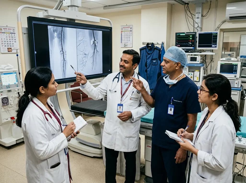
Explore revascularisation strategies in dialysis-dependent chronic limb-threatening ischaemia. This review examines surgical bypass vs endovascular therapy, BEST-CLI trial findings, procedural morbidity, conduit availability, and multidisciplinary decision-making for optimal limb salvage.
Today

A recent cross-sectional analysis reveals that over a quarter of randomized controlled trials for non-specific low back pain face discontinuation or unknown status, while more than 36% of completed trials remain unpublished, exposing notable reporting gaps in spine and musculoskeletal clinical research.
Today
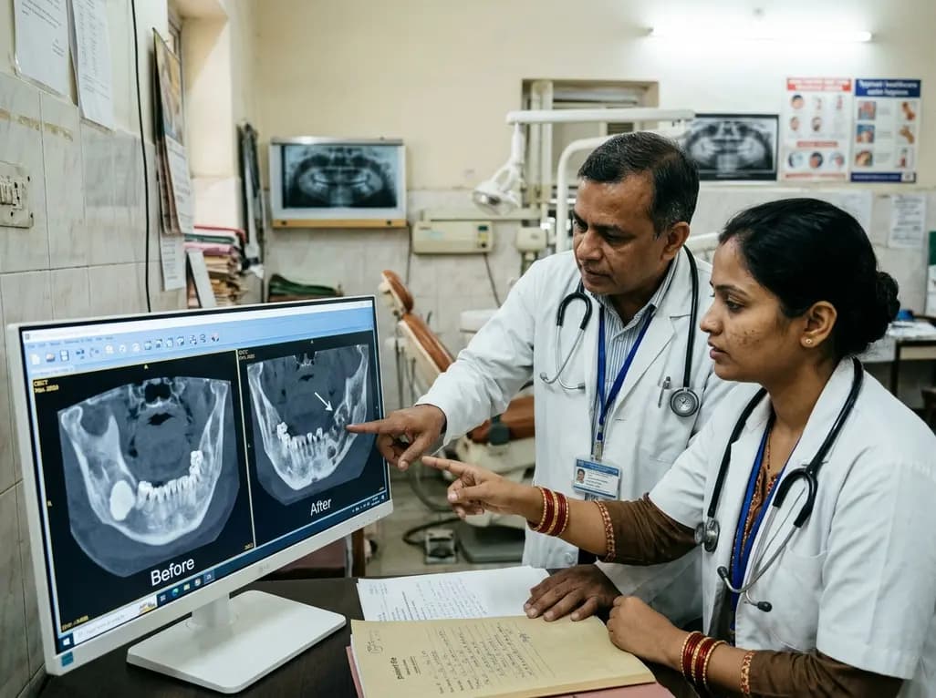
This case report examines the longitudinal CBCT evolution of Stage 2 MRONJ in a 70-year-old woman on denosumab at the site of a pre-existing mandibular dense bone island. It emphasizes the radiographic features, differential diagnosis, and multidisciplinary strategies required for high-risk bone lesions.
Today
