

Loading, please wait...
Loading, please wait...
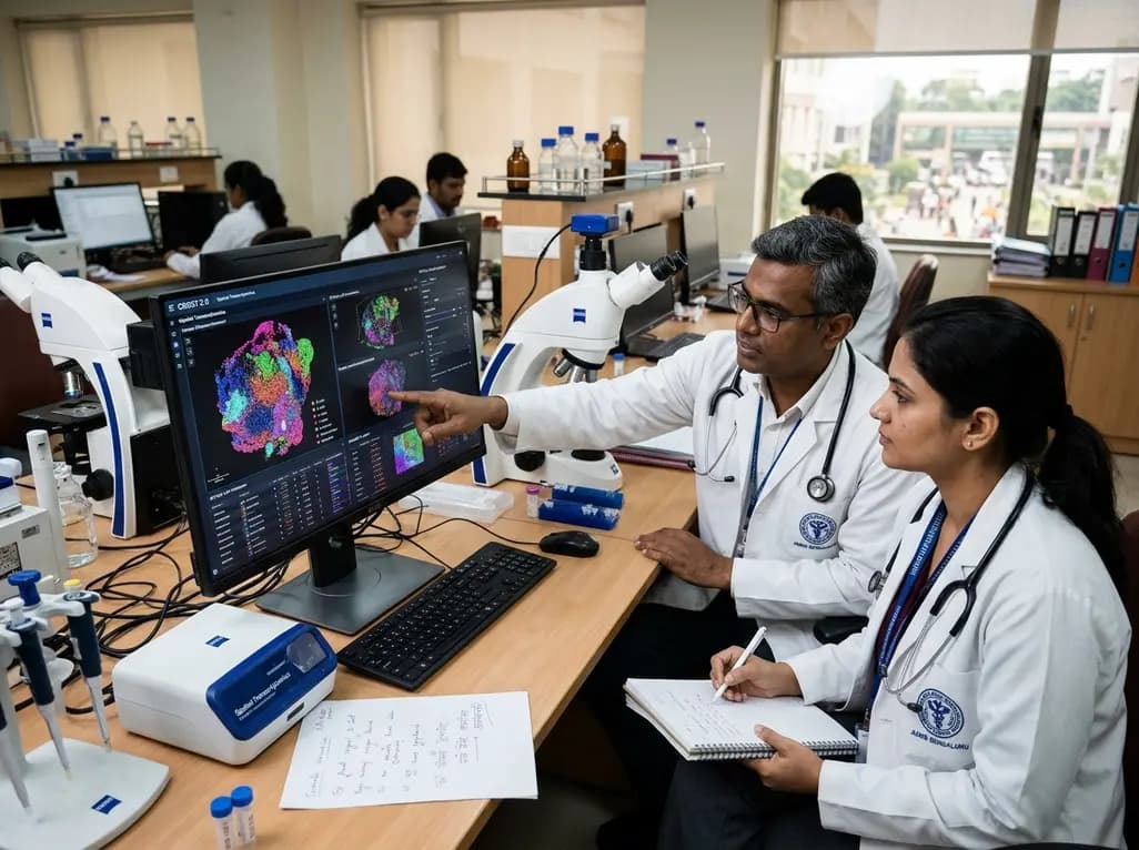
Translational oncology and cellular pathology increasingly rely on high-resolution tissue architecture to understand disease progression. In this context, a robust spatial transcriptomics repository provides clinicians and molecular researchers with indispensable reference datasets. CROST 2.0 represents a monumental milestone in this domain. Consequently, the updated platform bridges complex genomics with precise tissue microenvironment topography, accelerating cancer biomarker discovery across diverse malignancies.
The release of CROST 2.0 significantly expands data capacity for the medical oncology community. Initially, version 1.0 contained 182 projects and 1033 individual tissue samples. However, version 2.0 dramatically scales these holdings to 544 curated projects and 3538 biological samples. This nearly three-fold expansion encompasses diverse human organ systems and pathological conditions. Furthermore, the development team standardized all analytical pipelines to guarantee uniform quality across sequencing platforms. Researchers can easily query spatially resolved gene expression profiles across thousands of tumor specimens. In addition, the platform curates derived resources covering spatial colocalization and spatially variable cancer genes. Therefore, this expansive atlas provides robust statistical power to distinguish true biological signals from technical noise. Ultimately, CROST 2.0 centralizes heterogeneous spatial datasets into an accessible analytical hub.
Intra-tumoral heterogeneity presents a major clinical hurdle in modern oncology. Fortunately, CROST 2.0 introduces an advanced analytical framework designed for spatial microenvironment exploration. Specifically, the Cancer NicheDE module allows investigators to characterize niche-associated differential expression. Malignant clones actively manipulate adjacent fibroblasts, immune cells, and vascular networks within solid tumors. Consequently, this computational framework quantifies localized cell interactions and phenotypic shifts at invasive margins. In addition, users can execute spatially resolved gene-set activity analysis across distinct histological coordinates. For example, clinicians can track hypoxia pathways, immune exhaustion patterns, or metastatic signatures within microscopic tissue compartments. Moreover, spatial interaction hotspot analyses pinpoint coordinated cellular programs driving drug resistance. Thus, these analytical features convert raw sequencing reads into actionable biological insights.
Comparative tissue evaluation remains fundamental to anatomical pathology and oncology trials. To address this need, CROST 2.0 provides the innovative Spatial Compare utility. This interactive tool empowers researchers to compare spatial gene expression profiles directly across multiple patient specimens. Furthermore, investigators can evaluate shifts in cell-type distributions between normal tissues, pre-invasive lesions, and metastatic deposits. Clinicians frequently evaluate whether specific immune niches recur across independent cohorts. Therefore, Spatial Compare visualizes variations in niche composition with exceptional clarity. Additionally, the platform renders side-by-side topographic maps that highlight spatial discordance and shared molecular architecture. As a result, translational teams can correlate specific histological patterns with therapeutic responses. Ultimately, Spatial Compare accelerates our understanding of tissue remodeling during tumor progression.
Accurate cellular identification represents a cornerstone of spatial transcriptomic analysis. CROST 2.0 successfully integrates SCType, an automated online cell-type annotation module. Consequently, the platform seamlessly incorporates single-cell RNA sequencing reference information into spatial tissue sections. Many spatial technologies capture multi-cellular spots rather than single distinct cells. However, SCType resolves this limitation by deconvolving spot-level signals using curated cell marker databases. Moreover, automated annotation minimizes observer bias and standardizes classification across large patient cohorts. Pathologists can readily distinguish critical immune subsets, including cytotoxic T cells and tumor-associated macrophages. In addition, users can upload custom marker lists to identify rare cell phenotypes. Thus, SCType transforms complex expression patterns into clear cellular landscape maps.
Spatial biology is rapidly transitioning from basic laboratory research into translational oncology. Although routine clinical adoption in India faces infrastructure limitations, spatial datasets actively inform biomarker discovery and drug development. CROST 2.0 supports precision oncology by revealing microenvironmental vulnerabilities that bulk sequencing methods overlook. For instance, tertiary lymphoid structures within the tumor stroma often predict favorable responses to immune checkpoint inhibitors. By analyzing spatial interaction hotspots, clinical researchers can identify recurring cellular arrangements linked to treatment efficacy. Furthermore, academic pathology centers can cross-reference local biopsy specimens against thousands of curated spatial profiles. This comparative capability refines molecular risk stratification and sharpens therapeutic targets. Consequently, CROST 2.0 equips oncologists and pathologists with data-driven frameworks to study aggressive cancers.
While spatial transcriptomics offers unprecedented anatomical resolution, practical hurdles influence widespread clinical adoption. Currently, high sequencing costs, specialized imaging hardware, and substantial computational demands confine these technologies to tertiary academic centers. However, centralized databases like CROST 2.0 effectively democratize data access for clinicians and translational researchers. Investigators can test mechanistic hypotheses without incurring prohibitive laboratory expenses. In addition, future platform iterations will likely incorporate spatial proteomics and digital histology overlays. Pathologists can then correlate routine hematoxylin and eosin morphology with spatially resolved transcriptomic maps. Furthermore, standardized open databases foster international collaboration across developing oncology ecosystems. Therefore, CROST 2.0 represents an evolving resource driving spatial transcriptomics toward practical clinical utility.
CROST 2.0 expands data volume nearly threefold, offering 544 curated projects and 3538 biological samples compared to earlier versions. In addition, the update introduces sophisticated analytical frameworks such as the Cancer NicheDE module and spatial interaction hotspot analysis. The platform also integrates the SCType automated cell annotation tool and the Spatial Compare feature. Consequently, researchers can examine microenvironmental cellular interactions across multiple tumor cohorts with greater precision and statistical confidence.
The Cancer NicheDE module systematically evaluates niche-associated differential gene expression within heterogeneous tumor microenvironments. Tumors recruit stromal components, suppress localized immune responses, and alter adjacent normal architecture. By utilizing this module, oncologists can analyze how gene activity shifts across distinct cellular interfaces. Furthermore, this tool helps clinicians identify localized drug resistance mechanisms and evaluate spatially restricted biomarkers. Consequently, these findings guide the design of targeted combination therapies in advanced malignant diseases.
Many spatial sequencing techniques capture multiple cells within a single tissue spot, creating mixed molecular profiles. Therefore, integrating SCType allows researchers to deconvolute spot-level transcriptomic data using verified single-cell reference signatures. This automated annotation precisely classifies distinct cell types, including infiltrating lymphocytes and cancer-associated fibroblasts. Moreover, accurate annotation clarifies spatial interactions within the tumor stroma, providing pathologists with reliable topographical maps. Ultimately, these maps help clinicians understand immune evasion and therapeutic susceptibility across individual tissue samples.
Disclaimer: This content is for informational and educational purposes only... Refer to the latest local and national guidelines for clinical practice.
References

Read summarized clinical updates, watch expert medical content, and earn CME certifications right from your smartphone.
Open your smartphone camera to scan and install.





CROST 2.0 expands spatial transcriptomics resources to 544 projects and 3,538 samples. The database introduces the Cancer NicheDE module and SCType cell annotation, offering clinicians and researchers advanced tools to dissect tumor microenvironments and cross-sample spatial architectures.
Today
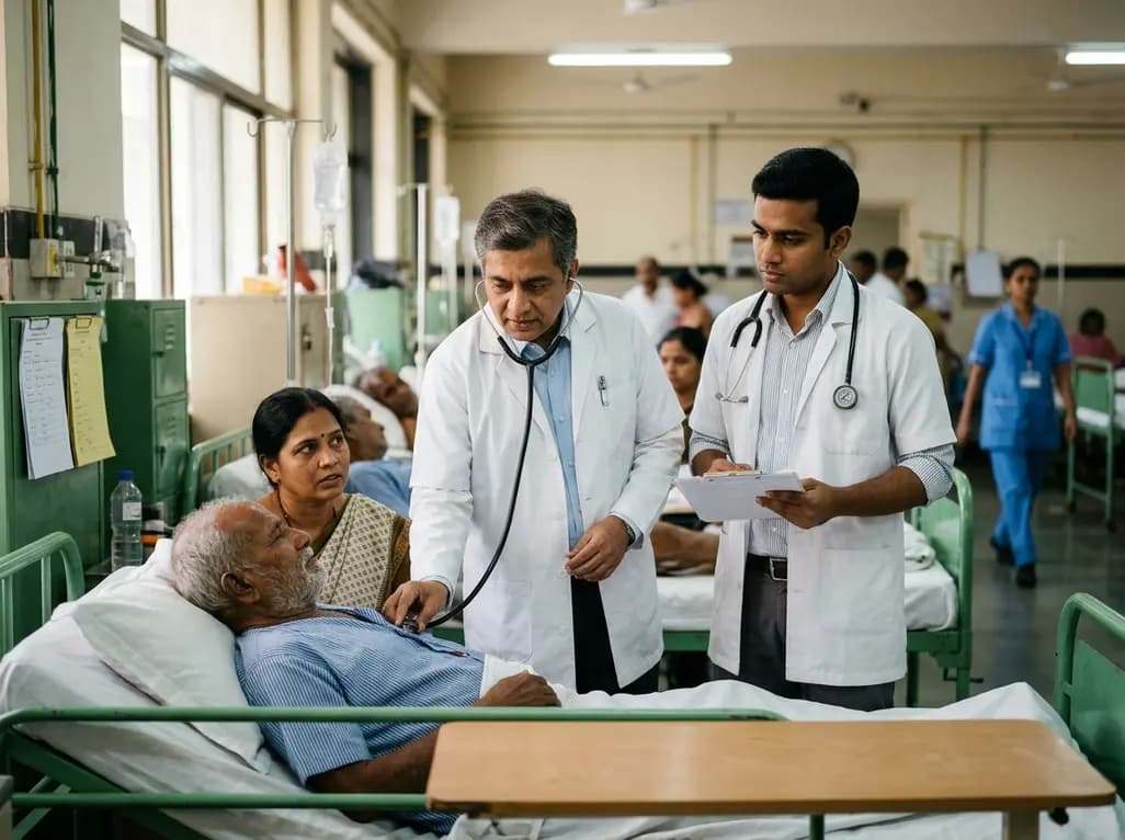
A qualitative study investigates whether traditional bedside clinical examinations still mirror contemporary physician practice. Findings support retaining long cases while reforming short cases toward hypothesis-driven assessments.
Today
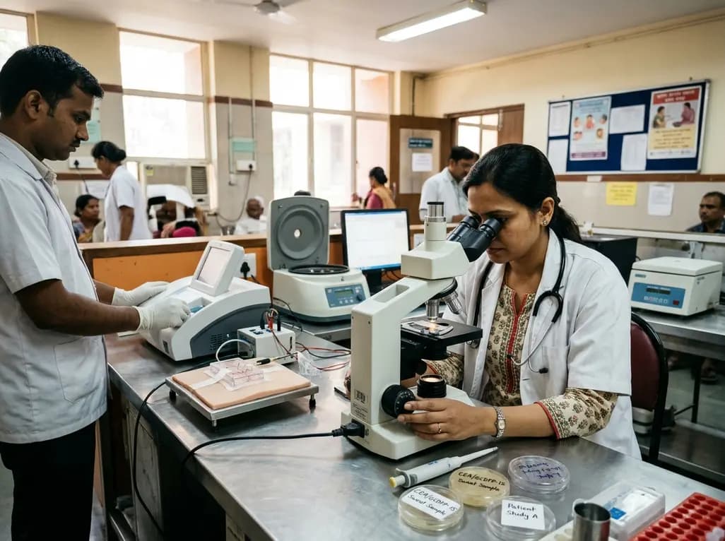
A pilot study evaluated WearDOXXN, a capillary microfluidic sweat patch, for noninvasive breast cancer monitoring by comparing sweat and serum CEA and PIP/GCDFP-15 levels.
Today
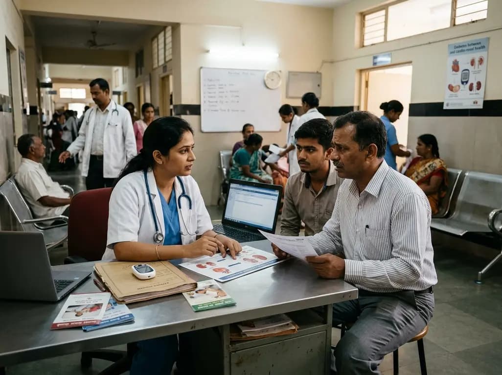
India faces a massive diabetes burden with over 89 million adults affected. Managing blood sugar in isolation fails to prevent severe cardiorenal complications. Clinicians must adopt an integrated metabolic ecosystem that prioritizes early detection, multifactorial risk assessment, and personalized therapies.
Today
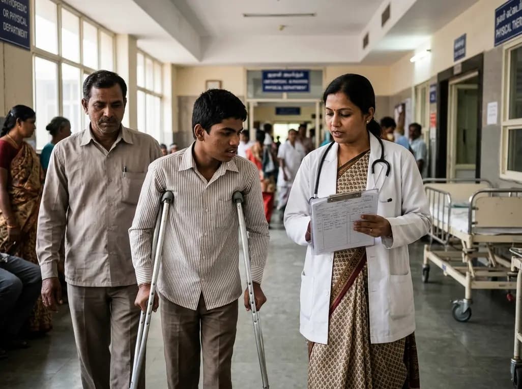
The Gross Motor Function Classification System (GMFCS) stops its validated age bands at 18. Recent evidence emphasizes that evaluating adult cerebral palsy requires purpose-sensitive documentation combining adolescent ratings with dynamic profiles of mobility, fatigue, pain, and community participation.
Today

A breakthrough 3D-printed piezoelectric collagen hydrogel activated by ultrasound accelerates diabetic wound repair, demonstrating 98.5% wound closure, robust antibacterial action, and enhanced vascularization without toxic chemical crosslinkers.
Today
