.webp&w=384&q=75)

Loading, please wait...
Loading, please wait...
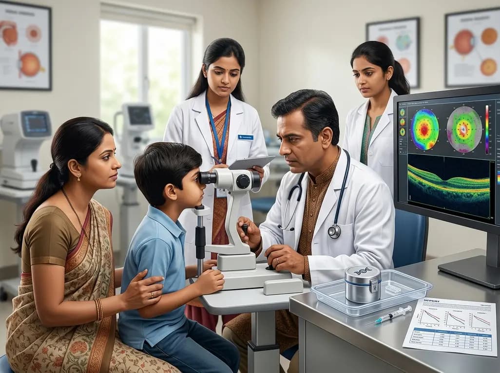
Inherited retinal dystrophies (IRDs) represent a heterogeneous group of genetic disorders that lead to progressive vision loss. Among these, mutations in the Crumbs homologue 1 (CRB1) gene are particularly significant. This gene encodes a protein vital for maintaining retinal cell polarity and adhesion. Consequently, defects in CRB1 lead to severe phenotypes such as Leber congenital amaurosis and retinitis pigmentosa. While the landscape of ocular medicine is shifting toward molecular interventions, the development of CRB1 gene therapy remains complex. Researchers face a substantial hurdle in identifying sensitive clinical endpoints that can accurately measure therapeutic efficacy over relatively short trial periods. Because the natural progression of these diseases is often slow or highly variable, standard metrics like best-corrected visual acuity may not capture subtle functional changes. Therefore, understanding the longitudinal natural history of CRB1-associated IRDs is an essential prerequisite for trial design. This understanding allows clinicians to distinguish between the natural decline of the disease and the potential benefits of a new treatment. Moreover, precise characterization of disease stages helps in selecting the optimal window for intervention, ensuring that patients receive therapy before irreversible structural damage occurs.
A recent prospective longitudinal cohort study has provided much-needed clarity on the natural progression of CRB1-associated IRDs. Conducted over a four-year period, this single-centre study assessed twenty patients who completed the full follow-up. The cohort primarily consisted of individuals with retinitis pigmentosa, although cone-rod dystrophy and macular dystrophy were also represented. By utilizing a comprehensive phenotyping protocol, investigators were able to track multiple functional and structural parameters simultaneously. This multifaceted approach included Goldmann perimetry, mesopic microperimetry, and full-field stimulus testing (FST). Furthermore, the study incorporated high-resolution imaging such as spectral-domain optical coherence tomography (OCT) and fundus autofluorescence. Such a rigorous assessment is crucial because CRB1 mutations often result in a thickened, disorganized retina that lacks clear lamination. These unique structural features make traditional imaging interpretation challenging. However, by following the same group of patients prospectively, the research team could observe the subtle yet significant changes that occur as the disease advances. These longitudinal data points serve as a vital baseline for any future CRB1 gene therapy, providing a clear picture of what the "untreated" path looks like for these patients.
The study results revealed several critical areas of functional decline that could serve as primary or secondary endpoints in clinical trials. Notably, there was a significant decrease in best-corrected visual acuity (BCVA), with patients losing an average of approximately 6.2 ETDRS letters over the four-year span. This finding is particularly relevant because BCVA is the standard regulatory endpoint for many ophthalmic trials. Nevertheless, visual acuity alone may not reflect the full impact of the disease on a patient's daily life. The research also highlighted a significant reduction in the seeing retinal area, as measured by Goldmann perimetry. Specifically, the V4e isopter showed a marked contraction, indicating a loss of peripheral and mid-peripheral vision. Perhaps most importantly, mean sensitivity on microperimetry showed a significant decline. Microperimetry is often considered more sensitive than BCVA because it maps the sensitivity of specific points within the macula. For patients being considered for CRB1 gene therapy, maintaining or improving macular sensitivity could be a more achievable and clinically meaningful goal. These functional losses correlate with the progressive nature of CRB1-related degeneration and suggest that these metrics are sensitive enough to detect changes within a reasonable timeframe for a clinical trial.
Structural integrity is another cornerstone of monitoring IRD progression. In CRB1-associated cases, the retina often exhibits a characteristic lack of normal layer differentiation, sometimes described as a "pachy-retina." Despite these baseline abnormalities, the four-year study sought to identify progressive structural markers. While some functional parameters showed clear declines, structural changes on OCT and fundus autofluorescence provided a more nuanced view of the disease. In many instances, the structural decay follows a predictable geographic pattern, even if the absolute retinal thickness remains high due to disorganized cell layers. Identifying these structural markers is essential because they can serve as surrogate endpoints. If a CRB1 gene therapy can demonstrate the preservation of specific retinal layers or the slowing of retinal thinning, it provides objective evidence of biological activity. Additionally, the study noted that full-field stimulus testing (FST) remained relatively stable in some patients, suggesting that FST might be more useful for assessing those with very advanced disease where microperimetry is no longer possible. The integration of structural and functional data allows for a more holistic assessment of retinal health, which is vital for the personalized management of these complex patients.
The primary objective of natural history studies is to pave the way for successful therapeutic interventions. By identifying sensitive clinical endpoints, this four-year study directly informs the design of CRB1 gene therapy trials. Specifically, the researchers suggested that BCVA, macular sensitivity, and Goldmann perimetry are the most sensitive measures for capturing disease progression in this population. Selecting the right endpoint is a delicate balance; it must be sensitive enough to show a difference but robust enough to withstand the variability of patient performance. Furthermore, the study highlights the importance of matching the endpoint to the patient's disease stage. For instance, microperimetry is excellent for early to mid-stage patients, whereas FST may be the only viable functional measure for those with profoundly low vision. As we move closer to clinical trials, these insights will help investigators determine the minimum sample size and the necessary duration of the study to achieve statistically significant results. Ultimately, the goal is to develop a treatment that can halt the progressive loss of sight, and having a validated roadmap of disease progression is the first step toward achieving that milestone for patients worldwide.
Looking ahead, the role of genetic testing in India and globally becomes even more critical. Since CRB1 mutations can lead to various clinical diagnoses, early and accurate molecular confirmation is necessary to identify candidates for CRB1 gene therapy. Clinicians should be aware that the window for effective treatment might be relatively narrow, especially in the more severe early-onset forms of the disease. Therefore, regular monitoring using the sensitive endpoints identified in this study is recommended. This proactive approach ensures that patients are "trial-ready" and that their baseline status is well-documented. Additionally, the development of these therapies underscores the need for a collaborative multidisciplinary approach, involving geneticists, retinal specialists, and low-vision rehabilitation experts. While the road to an approved treatment is long, the data provided by longitudinal studies offers a beacon of hope. It transforms our understanding of CRB1-associated IRDs from a collection of symptoms into a measurable biological process that can be targeted and, hopefully, slowed or reversed. As the scientific community continues to refine these genetic tools, the precision with which we monitor and treat these rare diseases will only continue to improve.
The CRB1 gene is essential for producing a protein that maintains the structural integrity of the retina. It specifically acts at the junctions between photoreceptors and Müller glia cells, helping to organize the retinal layers during development. When mutations occur, this organization is lost, leading to a thickened and poorly laminated retina. This structural disarray eventually causes progressive cell death, resulting in severe vision loss across various phenotypes like retinitis pigmentosa.
Best-corrected visual acuity (BCVA) only measures the eye's ability to see high-contrast detail at the very center of the macula. However, many inherited retinal diseases affect the surrounding macular area first. Macular sensitivity, measured through microperimetry, evaluates multiple points across the central retina. This makes it much more sensitive to subtle changes and early disease progression. For CRB1 gene therapy, capturing these early functional shifts is crucial for proving that a treatment is working.
The primary goals are to document how a disease progresses without treatment and to identify reliable clinical endpoints. For rare conditions like CRB1-associated IRDs, this information is vital for clinical trial design. It helps researchers determine which tests are most sensitive to change, how many patients are needed for a study, and how long the trial must last. Ultimately, these studies provide the necessary evidence to compare against the results of new genetic interventions.
Disclaimer: This content is for informational and educational purposes only. It is not intended as medical advice or a substitute for professional clinical judgment. Always seek the advice of a qualified healthcare provider regarding any medical condition or treatment. Refer to the latest local and national guidelines for clinical practice.
References
Karuntu JS et al. CRB1-Associated Inherited Retinal Dystrophies: Prospective Natural History Study With 4 Years of Follow-Up. Clin Exp Ophthalmol. 2026 Jul 11. doi: 10.1111/ceo.70142. PMID: 42433200.
den Hollander AI, et al. Mutations in the CRB1 gene cause Leber congenital amaurosis. Nat Genet. 1999;23(2):217-221. doi:10.1038/13847.
Talib M, et al. Genotypic and Phenotypic Characteristics of CRB1-Associated Retinal Dystrophies: A Long-Term Follow-up Study. Ophthalmology. 2017;124(6):884-895. doi:10.1016/j.ophtha.2017.01.031.
Boon CJF, et al. The Spectrum of Retinal Dystrophies Caused by Mutations in the CRB1 Gene. Genes (Basel). 2021;12(5):715. doi:10.3390/genes12050715.

Read summarized clinical updates, watch expert medical content, and earn CME certifications right from your smartphone.
Open your smartphone camera to scan and install.





A prospective 4-year study on CRB1-associated inherited retinal dystrophies identifies critical clinical endpoints for gene therapy. Significant declines in visual acuity and macular sensitivity provide a blueprint for evaluating the efficacy of emerging genetic interventions in these rare retinal diseases.
3 months ago

As monsoon seasons elevate arboviral disease threats, Tamil Nadu has initiated an ICMR-monitored evaluation of the tetravalent dengue vaccine in Tenkasi district. Clinicians emphasize clinical monitoring, public prevention, and high-quality early pediatric care to reduce disease morbidity across endemic regions.
Today

A comparative cohort study evaluates vNOTES vs TLH hysterectomy for benign conditions, demonstrating shorter operative duration, faster return to daily activities, and equivalent female sexual function scores without increased dyspareunia.
Today

Review how tailored exercise programs benefit frail older patients with heart failure. Explore pathophysiological mechanisms, individualized training prescriptions, and strategies to overcome adherence barriers in clinical practice.
Today

This comparative study evaluates posterior labral tears with and without glenohumeral instability, highlighting distinct demographic patterns, MRI underestimation in stable shoulders, and successful arthroscopic repair outcomes.
Today

A clinical study demonstrates that early intervention with tocilizumab, either as monotherapy or bridged with corticosteroids, achieves significant proptosis reduction and resolves active inflammation in treatment-naive thyroid eye disease.
Today
