
Loading, please wait...
Loading, please wait...

Recent research into craniosynostosis suture stem cells has unveiled a complex spatiotemporal atlas. This map explains why skull bones fuse prematurely in congenital disorders. By mapping these cells across developmental stages, scientists identified specific windows where stem cell fate diverges. Consequently, this study provides a new perspective on aberrant skull morphogenesis in Crouzon syndrome models.
To build this resource, researchers integrated single-cell RNA sequencing with high-resolution spatial transcriptomics. They focused on coronal sutures in Fgfr2 mice. Because of the high-resolution data, the team could observe the SuSC-to-osteoblast trajectory precisely. Furthermore, they discovered that pre-osteoblasts depleted earlier than the upstream SuSCs. This premature acquisition of osteogenic programs happens specifically near the suture midline.
Moreover, the research nominated Foxa3 as a primary candidate regulator in stem cell subsets. When the team performed siRNA knockdown of Foxa3, mineralization decreased significantly in the craniosynostosis background. Therefore, Foxa3 represents a promising intervention target for future molecular therapies. Additionally, communication analyses showed that signals from meningeal fibroblasts and immune cells converge on stem cell fate. This complex interaction suggests that the local niche plays a vital role in disease progression.
In conclusion, the results support a model where craniosynostosis involves disrupted temporal coordination. It is not merely a case of accelerated bone growth. However, this high-resolution resource provides a path toward therapeutic exploration. Ultimately, understanding these spatiotemporal dynamics allows clinicians and researchers to pinpoint better treatment timings.
Research indicates Foxa3 acts as a key regulator in suture stem cell subsets. Knockdown of this factor has been shown to reduce excessive mineralization in disease models, suggesting it is a potential therapeutic target.
The atlas showed that craniosynostosis involves a premature shift toward osteogenic programs. This suggests the disease is caused by disrupted timing of developmental stages rather than just simple bone growth acceleration.
Disclaimer: This content is for informational and educational purposes only and does not constitute medical advice. Always consult a qualified healthcare professional for diagnosis and treatment. Refer to the latest local and national guidelines for clinical practice.
References
1. Chen X et al. Spatiotemporal single-cell atlas of suture stem cell dynamics in craniosynostosis. Stem Cell Res Ther. 2026 May 03. doi: 10.1186/s13287-026-04987-6. PMID: 42071254.
2. Farmer DT et al. The developing mouse coronal suture at single-cell resolution. Nat Commun. 2021;12(1):4797. doi: 10.1038/s41467-021-24917-x.
3. Maruyama T et al. Stem cells of the suture mesenchyme in craniofacial development and repair. Dev Dyn. 2016;245(12):1186-1195. doi: 10.1002/dvdy.24443.
"
Read summarized clinical updates, watch expert medical content, and earn CME certifications right from your smartphone.
Open your smartphone camera to scan and install.


A high-resolution spatiotemporal atlas reveals how suture stem cells diverge in craniosynostosis, identifying Foxa3 as a potential therapeutic target....
2 months ago
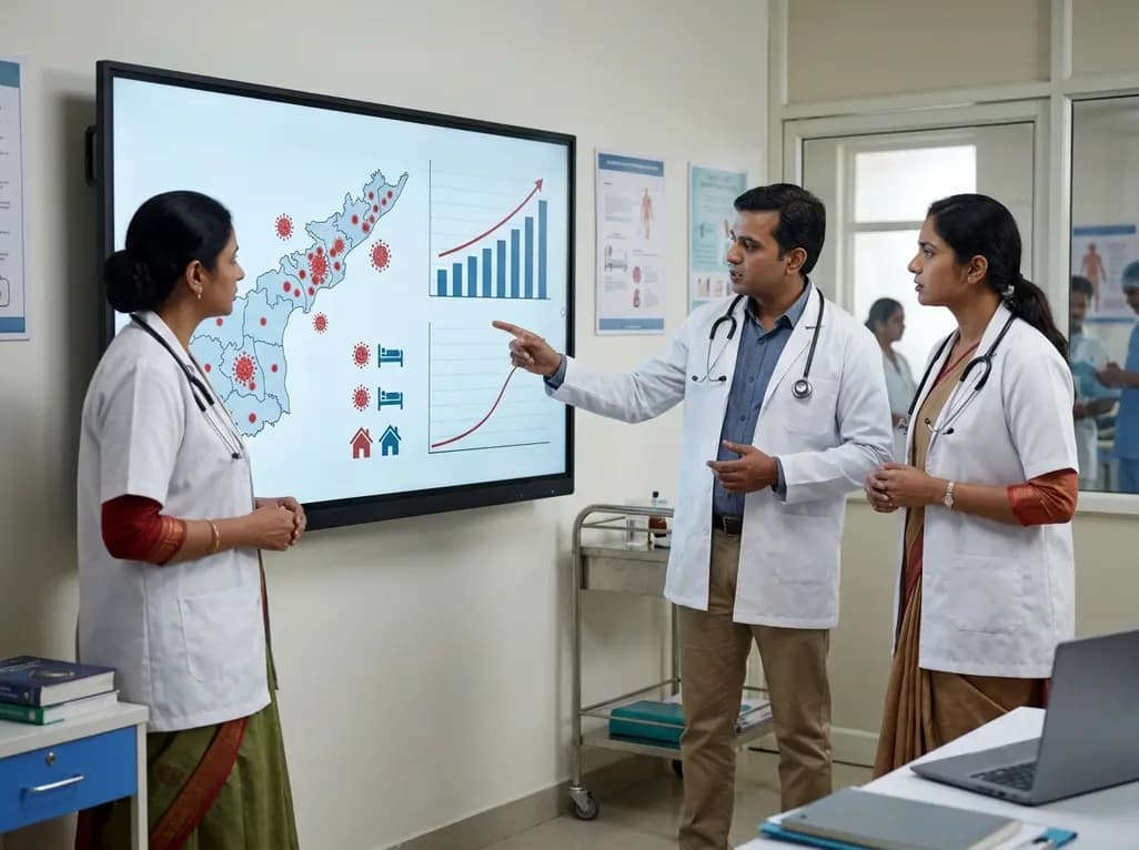
Andhra Pradesh reported 10 new Covid-19 cases, taking the state tally to 49 while deaths remain at four. With 24 patients hospitalized and 16 under home isolation, the Health Department has intensified monitoring. Medical professionals should review regional distribution, diagnostic protocols, and management plans.
Today

An 11-year Swedish registry study of 618 uterine sarcoma patients found that minimally invasive surgery yielded survival comparable to open surgery in early stages. However, adjuvant chemotherapy conferred no survival benefit in localized or advanced disease, highlighting stage and histology as key outcomes.
3 days back
A cross-sectional study evaluates post-intensive care syndrome in cardiac patients 2-4 weeks post-ICU discharge, highlighting cognitive, psychological, and functional impairments and the need for structured multidisciplinary rehabilitation.
3 days back
Anterior cruciate ligament reconstruction failure lacks uniform definition. A narrative review proposes an integrative framework incorporating objective and subjective instability, persistent pain, restricted motion, graft rupture, and secondary meniscal injury to standardize clinical reporting.
3 days back

With World Obesity Atlas data warning that over 41 million Indian children are overweight or obese, ICMR and NIN have unveiled a 10-point policy roadmap. The initiative calls for mandatory front-of-pack labeling, HFSS taxes, strict marketing bans, and healthier school environments to curb non-communicable diseases.
Today
