

Loading, please wait...
Loading, please wait...

Clinicians routinely monitor electrophysiological oscillations to evaluate cerebral function during diagnostic and therapeutic neurosurgical procedures. Specifically, low-frequency oscillations in the delta range of 1 to 4 Hz frequently reflect states of altered arousal, cortical deafferentation, and structural disruption. In surgical epileptology, clinicians place intracranial electrodes to localize the epileptogenic zone with spatial precision. However, surgical trauma and anesthetic agents alter immediate postoperative neurophysiology. A landmark study published in Epilepsia characterized the longitudinal trajectory of cortical delta power during chronic invasive epilepsy monitoring. The investigators evaluated 25 adult neurosurgical patients undergoing continuous intracranial monitoring. They recorded resting-state cortical electrophysiology across multiple consecutive sessions, beginning within 72 hours postoperatively. The team tracked spectral power changes using robust linear mixed-effects modeling. Consequently, the researchers discovered that delta power progressively declined across the entire brain throughout the chronic monitoring phase. Frontal, occipital, parietal, and temporal lobes demonstrated this continuous reduction, indicating a diffuse, global recovery process. In contrast, higher-frequency beta activity remained stable over time. Therefore, early low-frequency slowing represents a transient, resolving post-implantation phenomenon rather than permanent tissue dysfunction. Recognizing this trajectory helps neurophysiologists differentiate acute surgical artifact from true epileptogenic disturbances.
Surgical approach plays a pivotal role in governing the magnitude of postoperative cerebral slowing. Clinicians frequently choose between open craniotomy with subdural grid placement and minimally invasive stereoelectroencephalography (SEEG). The study revealed that patients undergoing open craniotomy exhibited markedly higher delta activity compared to patients undergoing SEEG. Open craniotomy requires extensive scalp incision, substantial bone flap removal, and direct mechanical manipulation of the dura and cortex. Furthermore, brain exposure causes atmospheric air entrainment, local microvascular shifts, and transient cortical surface irritation. These physical perturbations induce robust local neuroinflammatory responses and cerebral edema, which manifest electrophysiologically as amplified low-frequency slowing. Conversely, SEEG utilizes precise robotic or stereotactic guidance to insert multiple thin depth electrodes through small percutaneous twist drill holes. Consequently, stereotactic depth placement preserves cranial vault integrity, minimizes parenchymal compression, and limits cortical desiccation. Because tissue trauma remains localized along narrow electrode trajectories, SEEG elicits substantially less widespread baseline slowing. Therefore, neurosurgeons and clinical neurophysiologists must contextualize postoperative background activity relative to the specific surgical intervention. A patient who underwent extensive craniotomy naturally requires a longer physiological clearance window before baseline intracranial recordings stabilize for definitive surgical planning.
Beyond the surgical incision technique, operative duration and anesthetic clearance significantly affect intracranial electrophysiology. The study demonstrated clear associations between higher delta power and longer surgical procedures. Lengthy operations inevitably expose neuronal assemblies to sustained mechanical manipulation, hypothermia, fluid shifts, and systemic physiological stress. In addition, extended operative times necessitate prolonged administration of general volatile anesthetics and intravenous sedative infusions. These pharmacological agents depress thalamocortical networks and promote synchronized low-frequency oscillations. Furthermore, the analysis established that prolonged anesthesia emergence duration independently correlated with heightened delta activity during early invasive monitoring. Patients who took longer to regain full alertness maintained elevated slow-wave power within their resting-state intracranial recordings. Interestingly, other clinical variables showed no meaningful association with postoperative slow-wave power. The authors found no significant effects from patient age, biological sex, systemic white blood cell counts, or antiseizure medication dosages. Similarly, postoperative opioid analgesic dosing failed to explain differences in delta amplitude. Consequently, slow-wave excess predominantly reflects procedural trauma and recovery from deep anesthesia rather than routine perioperative medications. Neurologists should therefore account for intraoperative anesthesia logs when evaluating slow background activity during early telemetry days.
The specific hardware interface and recording depth also influence observed neurophysiological power spectra. Specifically, the researchers discovered that depth electrode contacts recorded significantly higher delta power than subdural surface electrodes. Depth electrodes penetrate deep cortical sulci, subcortical white matter tracts, and mesial structures such as the hippocampus and amygdala. Deep brain structures and white matter tracts intrinsically exhibit distinct baseline spectral dynamics compared to superficial neocortical gyri. Furthermore, subdural surface grids rest directly over neocortical gyral crowns and interface closely with cerebrospinal fluid within the subdural space. The presence of cerebrospinal fluid and postoperative subdural fluid collections can cause electrical shunting, which attenuates signal amplitude across frequency bands. Moreover, the surgical insertion of a depth wire generates micro-mechanical strain directly within three-dimensional neuronal parenchyma along the probe shaft. This focal tissue contact creates a micro-injury zone that transiently alters local membrane potentials and synaptic transmission. Consequently, intra-parenchymal contacts detect prominent local delta oscillations that superficial surface contacts do not capture. Neurophysiologists interpreting intracranial telemetry must recognize these technical nuances. They must not misattribute normal depth-derived slow oscillations to active pathological epileptogenicity or ischemic injury.
These findings deliver direct practical insights for comprehensive epilepsy centers performing surgical workups. Historically, clinical teams faced uncertainty when distinguishing postoperative surgical slowing from intrinsic epileptogenic encephalopathy. Because elevated slow activity naturally recedes over subsequent monitoring days, teams must interpret early electrophysiological findings with prudent caution. Specifically, clinicians should avoid premature seizure localization decisions based solely on interictal regional delta slowing recorded within the first 72 hours. In addition, surgical teams should leverage these physiological data when selecting diagnostic monitoring modalities. When clinical hypotheses permit, opting for SEEG over extensive craniotomy reduces baseline neurophysiological disruption and shortens recovery trajectories. Furthermore, tracking delta attenuation provides a quantitative physiological marker of brain recovery following electrode implantation. If delta power fails to resolve normally over time, clinicians should investigate potential complications, such as intracranial hemorrhage, infection, or focal edema. Moreover, neurocritical care units managing acute brain injuries can utilize this trajectory model to understand cerebral recovery dynamics. Ultimately, rigorous quantitative electrophysiology refines our understanding of the brain's response to surgery, improving diagnostic precision and clinical safety for patients with refractory epilepsy.
Postoperative elevated cortical delta power stems from acute mechanical tissue trauma, surgical manipulation, and prolonged general anesthesia exposure. Open craniotomy, extensive surgical duration, and delayed emergence disrupt thalamocortical networks, promoting low-frequency oscillations. This slowing represents a transient, resolving physiological recovery response rather than irreversible structural damage or new epileptogenic focus formation.
Open craniotomy induces substantially greater cortical delta power than stereoelectroencephalography because it involves extensive scalp incisions, bone flap removal, and direct cortical exposure. In contrast, stereoelectroencephalography uses minimally invasive percutaneous drill holes, preserving the cranial vault. Consequently, stereoelectroencephalography minimizes parenchymal edema and tissue inflammation, yielding significantly faster neurophysiological baseline stabilization.
Depth electrodes record higher delta power because their contacts directly penetrate deep parenchymal structures and subcortical white matter, where local micro-trauma occurs along the insertion tract. Conversely, subdural grid electrodes sit on the cortical surface, where surrounding cerebrospinal fluid and subdural fluid collections attenuate signal amplitude through electrical volume shunting.
Disclaimer: This content is for informational and educational purposes only and is not intended as medical advice. Healthcare professionals should make individual treatment decisions based on each patient’s clinical presentation. Refer to the latest local and national guidelines for clinical practice.
References

Read summarized clinical updates, watch expert medical content, and earn CME certifications right from your smartphone.
Open your smartphone camera to scan and install.





A study in Epilepsia demonstrates that cortical delta power rises significantly following open craniotomy and prolonged anesthesia before resolving globally during invasive epilepsy monitoring. The findings help clinicians differentiate acute post-surgical slowing from true epileptogenic zones.
Today

A Bayesian component network meta-analysis of 130 RCTs reveals that self-monitoring and gamification provide the clearest positive effects in mHealth physical activity interventions, while social comparison may have adverse psychological trade-offs.
Today

The GESS-AEEM consensus establishes a clear clinical implementation framework for testosterone therapy in postmenopausal women with hypoactive sexual desire disorder, focusing on transdermal dosing, precise laboratory monitoring, and safety communication.
Today
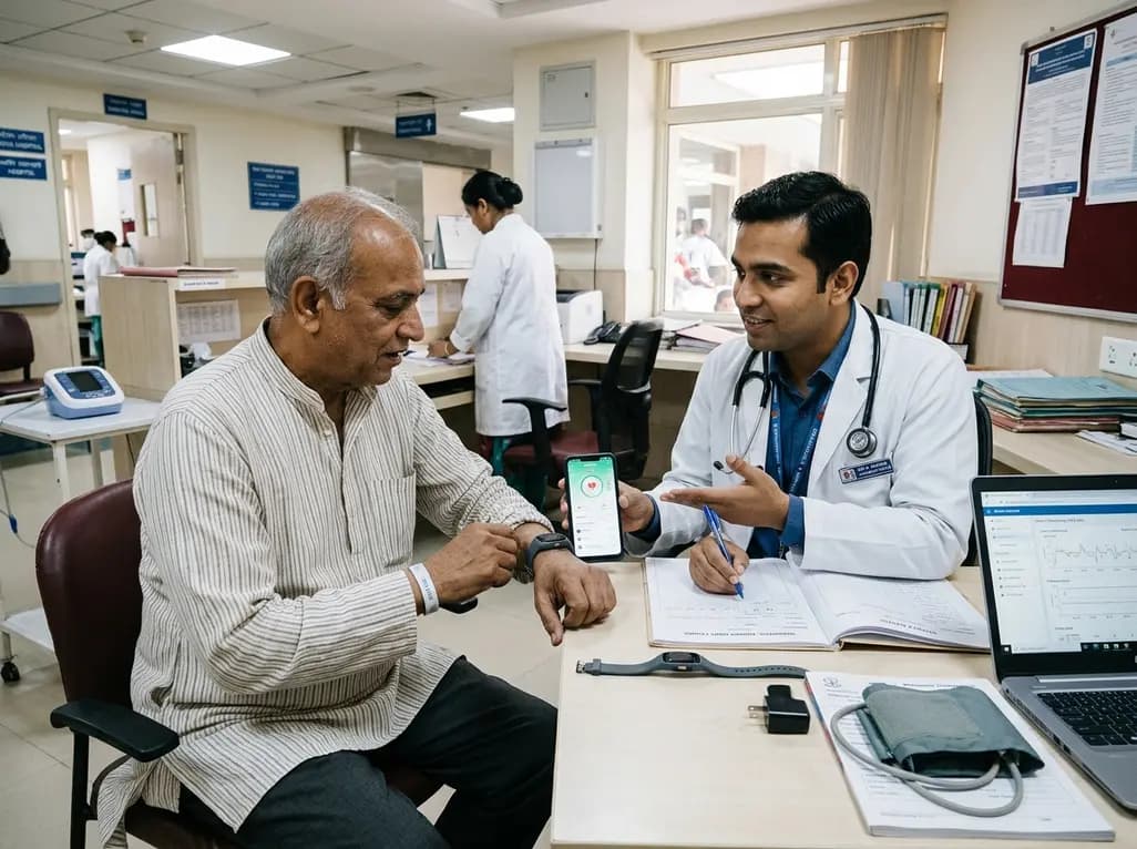
A multicenter observational study evaluated an 8-week digital health monitoring platform using wearable sensors in ambulatory heart failure. While participants achieved high daily wear-time of 11.7 hours and 80% completion, initial recruitment revealed significant technology-related onboarding barriers.
Today
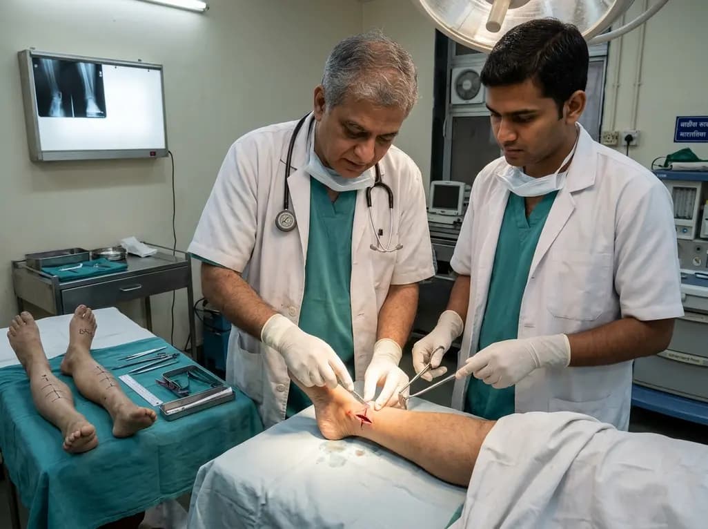
A paired cadaveric study compared triple and quintuple percutaneous Achilles tendon lengthening. While quintuple hemisection showed numerically greater elongation, differences were not statistically significant.
Yesterday

A clinical decision-support framework using health checkup data from 231,683 adults predicts incident obesity with high discrimination (AUROC 0.918). The system integrates temporal survival analysis and risk-factor explanations to prioritize individualized preventive interventions.
Today
