.webp&w=384&q=75)

Loading, please wait...
Loading, please wait...

Understanding knee osteoarthritis alignment represents a cornerstone in managing degenerative knee disorders effectively. Clinicians traditionally evaluate lower limb biomechanics through standard weight-bearing long-leg radiographs. However, conventional plain radiographs often fail to capture the complex soft-tissue changes occurring inside the joint space. Consequently, orthopedic surgeons frequently experience discrepancies when assessing static mechanical axes versus actual dynamic joint destruction. Yet, progressive cartilage thinning and meniscal extrusion fundamentally shift load distribution toward the medial compartment. Therefore, modern joint preservation surgery increasingly emphasizes subtle anatomical variations beyond simple bony geometry. Recent advances in three-dimensional imaging demonstrate that soft-tissue breakdown directly drives coronal plane deviation. For instance, meniscal subluxation diminishes functional tibiofemoral contact areas, which concentrates mechanical stress on the medial tibial plateau. As a result, subtle cartilage thinning accelerates coronal deformity and exacerbates varus malalignment. Recognizing these soft-tissue contributions allows clinicians to tailor corrective osteotomies and reconstructive strategies accurately. Moreover, comprehensive preoperative evaluations improve clinical outcomes by preventing unexpected postoperative overcorrection or undercorrection. Thus, evaluating both bony configuration and intra-articular soft-tissue architecture offers essential insights into progressive joint deterioration.
Orthopedic surgeons routinely utilize the arithmetic hip-knee-ankle angle to estimate native coronal alignment before joint wear began. Specifically, this mathematical calculation combines the medial proximal tibial angle and lateral distal femoral angle to recreate constitutional anatomy. However, the true weight-bearing mechanical axis also depends heavily on joint space alterations. Clinicians capture these intra-articular changes radiographically using the joint line convergence angle. Consequently, adding the joint line convergence angle to arithmetic alignment models markedly increases predictive precision for the overall mechanical axis. Nevertheless, weight-bearing radiographs represent a static two-dimensional projection of three-dimensional joint anatomy. Therefore, plain radiographs cannot fully separate the individual mechanical influence of meniscal extrusion from true articular cartilage loss. Furthermore, dynamic cartilage compression can alter joint line convergence unpredictably during plain radiographic acquisition. Because standing radiographs obscure subtle soft-tissue degradation, surgeons frequently miss early meniscal displacement that heralds joint instability. Additionally, patient positioning errors skew angular measurements, complicating longitudinal disease monitoring. Acknowledging the limitations of purely radiographic convergence metrics prompts practitioners to seek refined soft-tissue diagnostics. Understanding how soft tissues govern joint line convergence therefore clarifies the biomechanical progression of varus knee deformity.
Three-dimensional magnetic resonance imaging provides unmatched diagnostic clarity when visualizing intra-articular structures in degenerative joint disease. In particular, volumetric imaging accurately calculates the medial meniscus coverage ratio over the tibial plateau. When the medial meniscus extrudes beyond the tibial margin, it loses its essential hoop-stress capacity. Consequently, the uncovered tibial cartilage experiences unbuffered peak contact pressures during daily ambulation. A recent investigation evaluated ninety-four knees to examine how meniscal extrusion and cartilage loss influence overall mechanical alignment. Remarkably, researchers found that the medial proximal tibial angle significantly correlated with meniscal coverage in advanced osteoarthritis, but not in early-stage disease. Moreover, incorporating the medial meniscus coverage ratio into regression models predicted mechanical axis variance just as accurately as conventional joint line convergence angles. This key finding demonstrates that soft-tissue extrusion explains mechanical axis shifts with high mathematical fidelity. Therefore, advanced cross-sectional imaging directly uncovers the structural etiology behind joint space narrowing. In addition, three-dimensional assessments eliminate projection artifacts inherent to standard plain radiography. By precisely mapping meniscal coverage, clinicians can detect impending joint collapse before catastrophic bone-on-bone contact develops. Subsequently, sports physicians and joint preservation surgeons can select appropriate restorative interventions promptly.
Articular cartilage morphology plays an equally vital role alongside meniscal integrity in maintaining balanced joint biomechanics. Specifically, asymmetric cartilage wear alters the coronal tilt of the femoral condyles relative to the tibia. Researchers calculate the height-normalized medial-to-lateral cartilage thickness difference on three-dimensional magnetic resonance sequences to capture this imbalance. In a comprehensive statistical analysis, combining this cartilage thickness differential with meniscal coverage explained eighty-four percent of mechanical axis variance. Furthermore, all individual soft-tissue parameters achieved statistical significance within the predictive multivariable model. This outcome underscores that progressive cartilage attrition directly dictates limb axis deviation alongside bony anatomy. When cartilage wears down medially while remaining preserved laterally, the joint line tilts into an obligatory varus orientation. Consequently, mechanical load shifts further toward the medial compartment, creating an aggressive cycle of wear and deformity. However, conventional radiographs only approximate this loss through indirect joint space narrowing measurements. In contrast, magnetic resonance imaging directly maps topographical cartilage volume loss with millimeter accuracy. Thus, cross-sectional chondral thickness quantification provides objective, reproducible data for clinical decision-making. Orthopedic surgeons can harness these precise volumetric measurements to optimize alignment goals during corrective osteotomy or arthroplasty procedures.
Integrating advanced soft-tissue mapping into clinical workflows carries substantial implications for modern knee preservation surgery. For example, high tibial osteotomy candidates require meticulous evaluation to determine whether soft-tissue collapse will exacerbate postoperative malalignment. If severe medial meniscal extrusion and chondral loss already compromise the joint, standard bony corrections may fail to unload damaged compartments adequately. Therefore, surgeons must account for anticipated soft-tissue laxity and joint space restitution when calculating correction angles. Moreover, contemporary kinematic and functional alignment techniques in total knee arthroplasty demand precise knowledge of native joint lines. By evaluating magnetic resonance imaging before surgery, surgeons can distinguish between constitutional bony varus and acquired soft-tissue loss. Consequently, this detailed distinction prevents unnecessary over-resection of femoral condyles or tibial plateaus during implant placement. In addition, sports medicine clinicians managing traumatic meniscal root tears can recognize patients at elevated risk of accelerated mechanical deviation. Early surgical repair of displaced meniscal roots restores coverage, thereby preserving normal load distribution across the tibiofemoral joint. Ultimately, adopting comprehensive imaging paradigms bridges the divide between radiographic bony metrics and living soft-tissue biomechanics.
Medial meniscal extrusion reduces the shock-absorbing capacity of the fibrocartilage ring by compromising circumferential hoop tension. Consequently, the medial femoral condyle directly compresses the unshielded tibial plateau cartilage during weight-bearing activities. This concentrated compressive stress accelerates cartilage wear and progressively narrows the medial joint space. Therefore, the tibia tilts into progressive varus alignment, shifting the overall mechanical axis medially and compounding asymmetrical degenerative joint changes over time.
The arithmetic hip-knee-ankle angle accurately reflects constitutional bony alignment by combining femoral and tibial joint angles. However, it completely ignores soft-tissue wear and intra-articular joint space loss occurring during osteoarthritis progression. When cartilage erodes or meniscal tissue extrudes, the joint line convergence angle widens significantly. Therefore, arithmetic models alone underestimate the actual functional varus deformity without incorporating cross-sectional soft-tissue metrics or radiographic joint line convergence assessments during surgical planning.
Three-dimensional magnetic resonance imaging provides objective volumetric quantification of meniscal subluxation and regional cartilage loss across compartments. Consequently, surgeons can evaluate whether medial compartment narrowing stems from reversible meniscal extrusion or irreversible full-thickness bone exposure. This vital distinction enables clinicians to plan high tibial osteotomies with superior precision. Furthermore, understanding precise chondral thickness differentials helps surgeons set accurate realignment targets, avoiding postoperative overcorrection and ensuring long-term joint survival in active patients.
Disclaimer: This content is for informational and educational purposes only and should not be considered medical advice. Always consult a qualified healthcare professional regarding any medical condition or treatment. Refer to the latest local and national guidelines for clinical practice.
References
Sasabe A et al. Magnetic resonance imaging-based meniscal coverage and cartilage thickness are associated with mechanical axis alignment in knee osteoarthritis. J Exp Orthop. 2026 Oct undefined. doi: 10.1002/jeo2.70927. PMID: 42829635.
Hunter DJ, Sharma L, Skaife T. Alignment and osteoarthritis of the knee. J Bone Joint Surg Am. 2009 Feb;91 Suppl 1:85-89. doi: 10.2106/JBJS.H.01409. PMID: 19182031.
Bax EA, Kerkhof JAJ, van Egmond N, et al. The impact of varus and valgus alignment on knee cartilage quality assessed by magnetic resonance imaging: insights from the IMI-APPROACH cohort. Knee. 2025 Oct;57:477-487. doi: 10.1016/j.knee.2025.10.005. PMID: 39423719.

Read summarized clinical updates, watch expert medical content, and earn CME certifications right from your smartphone.
Open your smartphone camera to scan and install.





A new study reveals that MRI-based meniscal coverage and cartilage thickness explain mechanical axis alignment in knee osteoarthritis comparably to joint line convergence angle. These soft-tissue insights improve preoperative planning for osteotomies and arthroplasty.
Today
Psychometric validation of the WHO BeSD survey identifies vaccine importance and social responsibility as primary drivers of adult influenza vaccination uptake, offering clinicians actionable strategies to overcome hesitancy.
Today
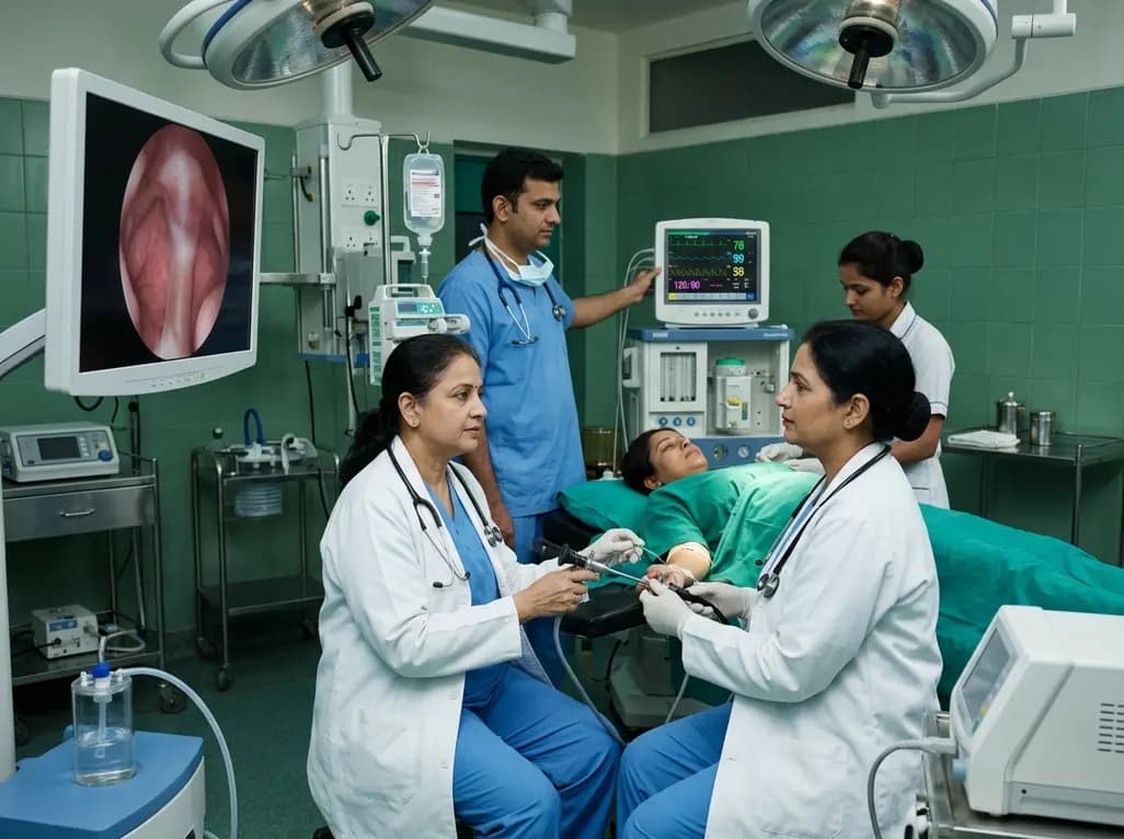
A meta-analysis of nine RCTs comparing remimazolam versus propofol in hysteroscopy reveals remimazolam significantly reduces injection pain, hypotension, and respiratory depression while maintaining comparable sedation success, recovery times, and procedural duration.
Today
Radiofrequency catheter ablation of the cavotricuspid isthmus successfully terminates typical atrial flutter. This article reviews the mechanisms driving coronary sinus electrogram changes, wavefront activation pathways, and clinical criteria to verify bidirectional block during electrophysiology studies.
Today

A breakthrough study leverages natural language processing to accurately identify incident medication-related osteonecrosis of the jaw and track antiresorptive drug holidays from clinical narratives in electronic health records, advancing osteoporosis pharmacovigilance and dental safety.
Today
