.webp&w=384&q=75)

Loading, please wait...
Loading, please wait...
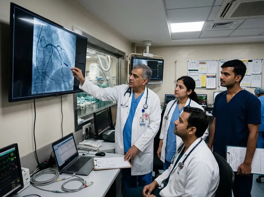
When patients present with sudden uneasiness or atypical chest discomfort, clinical teams must act promptly. Recently, Union Health Minister JP Nadda underwent evaluation at AIIMS Delhi following acute symptoms. Physicians performed a diagnostic coronary angiography to evaluate his coronary vasculature and ruled out immediate instability. Consequently, he remained under observation in the cardiology ward in stable condition. For clinicians, this clinical scenario highlights the vital role of invasive diagnostic pathways in ruling out acute coronary syndromes.
Prompt risk stratification remains the cornerstone of modern emergency cardiovascular care. Therefore, clinicians must evaluate acute chest symptoms, unexplained diaphoresis, or sudden uneasiness with high vigilance. Initial emergency workups require a standard twelve-lead electrocardiogram within ten minutes of arrival. Furthermore, serial high-sensitivity cardiac troponin assays help identify subtle myocardial injury rapidly. When non-invasive findings show equivocal results or when high clinical suspicion persists, diagnostic coronary angiography provides definitive anatomical visualization. Moreover, contemporary guidelines recommend early invasive evaluation for patients exhibiting intermediate or high cardiovascular risk profiles. Clinicians assess coronary artery luminal diameter, detect atheromatous plaques, and identify flow-limiting stenoses. In addition, prompt angiography helps exclude life-threatening plaque rupture, severe multivessel disease, or acute vessel occlusion. Consequently, emergency physicians and cardiologists can make swift, evidence-based decisions regarding medical therapy or mechanical revascularization.
Interventional cardiologists perform coronary angiography using standardized catheterization techniques to achieve optimal visualization. Today, operators predominantly select the transradial arterial approach over the transfemoral route. Consequently, transradial access significantly reduces access-site bleeding complications and vascular pseudoaneurysms. Furthermore, radial access allows earlier patient ambulation, thereby improving overall post-procedural comfort. During the procedure, the cardiologist introduces specialized diagnostic catheters into the coronary ostia under fluoroscopic guidance. Subsequently, non-ionic iodinated contrast media allows detailed cineangiographic recording of the coronary tree. Operators systematically visualize the left main, left anterior descending, left circumflex, and right coronary arteries across multiple orthogonal projections. In addition, fractional flow reserve or intravascular imaging may supplement diagnostic angiography when anatomical lesions show intermediate stenosis. Therefore, the diagnostic procedure provides an exceptional balance of safety, high diagnostic accuracy, and low procedural morbidity.
Following diagnostic cardiac catheterization, structured inpatient observation remains essential to ensure patient stability. Therefore, dedicated cardiology nursing teams monitor vital signs, access-site integrity, and distal perfusion at regular intervals. Clinicians maintain continuous telemetry monitoring to detect transient arrhythmias, conduction blocks, or recurrent ischemic episodes. Furthermore, adequate post-procedure oral or intravenous hydration promotes contrast clearance and mitigates contrast-induced nephropathy risks. This preventive measure is particularly important for patients with baseline renal impairment or diabetes mellitus. In addition, healthcare providers monitor radial compression bands closely to achieve complete hemostasis without compromising radial artery patency. Fortunately, serious post-procedure complications remain rare in modern catheterization laboratories. However, vigilant observation during the initial twelve to twenty-four hours ensures prompt recognition of hematomas, delayed allergic reactions, or hemodynamic shifts. Consequently, timely observation protects patient safety and confirms clinical stabilization.
Patients presenting with acute thoracic uneasiness often present significant diagnostic challenges to clinicians. Therefore, physicians must maintain a broad differential diagnosis beyond classic acute coronary syndromes. Non-cardiac etiologies frequently mimic ischemic discomfort, including gastroesophageal reflux, esophageal spasms, musculoskeletal inflammation, and acute pulmonary conditions. Furthermore, conditions such as pulmonary embolism, acute pericarditis, and aortic dissection require rapid exclusion through targeted testing. Clinicians utilize validated risk scores, such as the HEART score, to guide downstream testing. When diagnostic angiography demonstrates unobstructed coronary arteries, clinicians investigate non-obstructive causes of cardiac ischemia. For example, microvascular dysfunction, coronary vasospasm, and stress-induced cardiomyopathy can cause acute symptoms despite normal epicardial vessels. Consequently, thorough diagnostic evaluation ensures accurate phenotyping and prevents both underdiagnosis of cardiac pathology and unnecessary interventional procedures.
Comprehensive discharge planning consolidates the diagnostic findings and establishes long-term cardiovascular risk reduction. Therefore, clinicians must provide clear communication regarding diagnostic results, physical activity guidance, and prescribed medications. When angiography demonstrates non-obstructive coronary atherosclerosis, aggressive preventive therapy remains vital to halt plaque progression. Consequently, cardiologists initiate guideline-directed medical therapy, including statins, antiplatelet agents, and blood pressure control. Furthermore, clinicians emphasize lifestyle modifications such as dietary changes, structured exercise programs, weight management, and stress reduction. Patients receive explicit instructions regarding red-flag symptoms that warrant immediate re-evaluation. Additionally, clinicians schedule early outpatient follow-up visits to assess functional capacity and monitor medication adherence. Through systematic secondary prevention and patient education, healthcare providers significantly reduce future major adverse cardiovascular events.
Q1: Why is coronary angiography performed after a patient reports acute uneasiness?
Clinicians perform diagnostic angiography when acute uneasiness, atypical symptoms, or abnormal cardiac biomarkers suggest potential myocardial ischemia. The procedure provides definitive anatomical visualization of the coronary arteries. Consequently, it allows cardiologists to confirm or exclude significant coronary artery stenosis, acute plaque rupture, or flow-limiting obstructions, thereby guiding immediate clinical management.
Q2: What are the primary advantages of transradial coronary angiography?
Transradial catheterization offers significant clinical advantages over the traditional transfemoral approach. Specifically, radial access markedly reduces major bleeding complications, hematomas, and vascular access-site injuries. Furthermore, patients experience greater procedural comfort and can mobilize much earlier. Consequently, radial access shortens hospital stays and lowers overall post-procedural morbidity.
Q3: How long should patients remain under observation following diagnostic angiography?
Observation duration typically ranges between four and twenty-four hours depending on procedural access, underlying clinical stability, and patient comorbidities. During this period, clinical teams monitor vital signs, evaluate puncture site hemostasis, and maintain adequate hydration to facilitate contrast clearance. This observation ensures complete cardiovascular stability before safe hospital discharge.
Disclaimer: This content is for informational and educational purposes only. It does not constitute medical advice or replace professional judgment. Refer to the latest local and national guidelines for clinical practice.
References

Read summarized clinical updates, watch expert medical content, and earn CME certifications right from your smartphone.
Open your smartphone camera to scan and install.





Following the admission of Union Health Minister JP Nadda to AIIMS Delhi for acute uneasiness, clinical teams utilized diagnostic coronary angiography and inpatient observation. This clinical review outlines acute triage pathways, procedural protocols, post-procedure observation, and long-term risk management.
last month

Evaluating concordance between patient-reported and clinician-assessed modified Rankin Scale scores reveals how nonmotor symptoms, caregiver reliance, and cultural factors affect functional disability ratings in chronic neurologic disorders.
Today

A scoping review evaluates 116 studies on AI in women's reproductive health, finding that 83.6% carry high risk of bias. While ensemble models show promise, disparities and lack of validation hinder LMIC deployment.
Today

Differential oxygenation during peripheral TA-NRP can cause undetected coronary hypoxemia due to arch vessel clamping. A novel circuit with continuous aortic root monitoring detects desaturation early, facilitating left ventricular unloading and preventing allograft dysfunction and unnecessary heart discard.
Today

A longitudinal study reveals that patients undergoing staged bilateral hip arthroscopy experience progressive contralateral labral deterioration during surgical intervals. However, structural degradation does not compromise 2-year postoperative clinical outcomes or patient-reported satisfaction scores.
Today

A post hoc analysis of the PARM-T2D study shows pemafibrate exerts baseline-dependent effects on LDL-C and particle size in type 2 diabetes and hypertriglyceridemia. It lowers triglycerides and remnants while favorably remodeling atherogenic LDL particles based on initial lipid control status.
Today
