

Loading, please wait...
Loading, please wait...
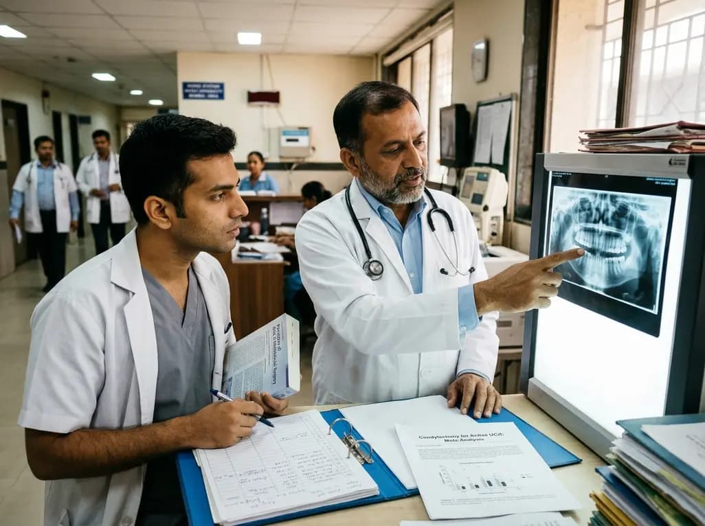
Unilateral condylar hyperplasia presents a major surgical challenge characterized by progressive, asymmetrical overgrowth of the mandibular condyle. Clinicians often struggle to determine whether surgical arrest through condylectomy sufficiently halts disease progression or if secondary corrective osteotomies remain inevitable. Recently, a systematic review and single-arm meta-analysis synthesized data from seventeen studies involving 476 patients. The landmark findings provide crucial evidence on surgical stability and secondary intervention rates.
Active unilateral condylar hyperplasia involves uncontrolled, non-neoplastic proliferation of the condylar head cartilage. Consequently, patients develop progressive facial asymmetry, occlusal canting, and lateral crossbites that degrade functional chewing and facial harmony. The pathological process predominantly accelerates during late adolescence and early adulthood. Nuclear imaging, specifically single-photon emission computed tomography, confirms active cellular growth by demonstrating increased radionuclide uptake. Without timely surgical intervention, persistent asymmetry induces secondary compensatory deformities in the maxilla, dentoalveolar complex, and soft tissues. Furthermore, progressive articular changes can lead to severe temporomandibular joint dysfunction and chronic myofascial pain. Therefore, stopping abnormal osteogenesis represents the immediate priority in modern maxillofacial treatment algorithms. Early condylectomy directly removes the active proliferative cartilage layer, immediately halting progressive skeletal distortion. However, clinicians traditionally debated whether arresting the growth center alone permits spontaneous functional compensation or leaves residual deformities requiring extensive secondary orthognathic correction. Synthesizing multicenter international evidence provides oral and maxillofacial surgeons with reliable prognostic data, enabling clearer clinical counseling during initial surgical evaluations.
The primary aim of the recent meta-analysis was quantifying the proportion of patients needing secondary jaw repositioning after condylectomy. Across 476 patients from seventeen studies, the pooled random-effects estimate for secondary orthognathic surgery was 15.4 percent. Thus, approximately 85 percent of treated patients avoided subsequent osteotomies after initial condyle resection. This compelling statistic demonstrates that condylectomy functions as an extraordinarily effective growth-stabilizing procedure. In many patients, early growth arrest coupled with targeted post-surgical orthodontic mechanics successfully aligns the dentition without extra skeletal surgeries. Nevertheless, the wide 95 percent confidence interval spanning 4.7 to 29.7 percent reflects notable clinical variability between surgical centers. Differences in orthodontic camouflage capabilities, baseline deformity severity, and surgeon threshold for secondary intervention drive this variance. For instance, severe vertical discrepancies and preexisting maxillary plane canting often demand secondary bimaxillary procedures regardless of condylar growth arrest. Consequently, maxillofacial teams must inform patients that while condylectomy frequently provides definitive surgical control, secondary bimaxillary correction remains possible for full aesthetic harmony.
Surgical execution varies widely regarding the exact volume of condylar tissue resected during surgery. Maxillofacial surgeons typically select between high condylectomy, proportional condylectomy, and adaptable condylectomy techniques. High condylectomy resects the superior three to five millimeters of the condylar head, directly eliminating the hyperactive prechondroblastic cell layers. In contrast, proportional condylectomy removes an anatomical portion equal to the measured discrepancy between the affected and healthy contralateral condyle. Meanwhile, adaptable condylectomy tailors resection boundaries according to preoperative three-dimensional virtual surgical planning and real-time intraoperative occlusal assessments. Notably, the meta-analysis revealed no statistically significant differences in secondary orthognathic surgery rates among these three approaches. Subgroup comparative analysis yielded a non-significant p-value of 0.41 across the different resection strategies. Therefore, current literature indicates that thorough removal of the pathological growth zone matters far more than the specific resection design. Surgeons can confidently choose the resection methodology that best preserves articular disc stability and anatomical joint function.
Demographic pooling within the meta-analysis highlighted distinct patient characteristics that assist clinicians with early identification. Female patients constituted 63 percent of all analyzed cases, confirming a distinct female predilection in condylar hyperplasia. Although hormonal factors and estrogen receptor expression in articular cartilage may influence this gender distribution, definitive biological pathways require deeper investigation. Additionally, the pooled mean age at surgical intervention was 23.47 years. However, marked statistical heterogeneity characterized this finding, with the clinical age range spanning 19.72 to 27.22 years. This distribution underscores that condylar hyperplasia frequently persists or presents beyond typical physiological pubertal growth completion. Young adults frequently notice late-onset facial asymmetry, worsening dental crossbite, or shifting dental midlines during college years. Because active hyperplasia in adulthood mimics temporomandibular joint disorders, primary care clinicians may misdiagnose early manifestations. Recognizing these distinct demographic markers ensures prompt referral to maxillofacial specialists before dentoalveolar compensations become severely entrenched.
In the Indian clinical context, managing unilateral condylar hyperplasia requires standardized multidisciplinary collaboration between orthodontists and maxillofacial surgeons. Patients often present late due to delayed diagnostic suspicion and limited access to nuclear imaging facilities in smaller towns. Therefore, tertiary centers in India must implement accessible diagnostic pathways combining serial cone-beam computed tomography with SPECT bone scintigraphy. When active condylar hyperactivity is confirmed, clinicians should recommend early condylectomy to stabilize facial growth and prevent progressive maxillary canting. Performing condylectomy preserves the natural mandibular framework and significantly simplifies subsequent orthodontic decompensation. Moreover, digital virtual surgical planning enables precise execution, reducing operative morbidity and temporomandibular joint complications. Because secondary orthognathic surgery is required in only 15 percent of cases, early intervention offers substantial economic advantages for Indian families by avoiding complex bimaxillary reconstructions. Collaborative postoperative monitoring ensures timely orthodontic guiding elastics, restoring excellent masticatory function and symmetrical aesthetics.
The exact etiology of unilateral condylar hyperplasia remains incompletely understood, but researchers consider it a multifactorial disorder. Genetic predisposition, local microtrauma, vascular hypervascularity, and hormonal influences likely contribute to prolonged condylar cartilage growth. Specifically, increased expression of growth factors and estrogen receptors within the condylar fibrocartilage stimulates persistent cellular proliferation. Consequently, this persistent overgrowth continues well beyond typical pubertal development, resulting in progressive facial asymmetry, crossbite, and functional joint impairment.
Surgeons determine disease activity by combining sequential clinical examinations, serial three-dimensional radiographic imaging, and nuclear scintigraphy. Single-photon emission computed tomography bone scanning with technetium-99m serves as the diagnostic gold standard for assessing metabolic turnover. A condylar uptake discrepancy exceeding ten percent relative to the contralateral healthy side indicates active pathology. In addition, serial superimposition of cone-beam computed tomography scans demonstrates ongoing anatomical elongation, confirming the necessity of surgical condylectomy to halt further progression.
Orthodontic treatment alone cannot resolve active unilateral condylar hyperplasia because braces cannot arrest pathological cartilage growth within the temporomandibular joint. Attempting dental camouflage while the condyle remains active usually causes severe periodontal compromise and eventual treatment relapse. However, orthodontic therapy plays a crucial collaborative role alongside surgery. Orthodontists decompensate the dental arches before or after condylectomy, facilitating proper occlusal settling and ensuring long-term skeletal and dental stability following definitive growth arrest.
Disclaimer: This content is for informational and educational purposes only and does not constitute formal medical or surgical advice. Clinicians should assess each individual patient case carefully and refer to the latest local and national guidelines for clinical practice.
References

Read summarized clinical updates, watch expert medical content, and earn CME certifications right from your smartphone.
Open your smartphone camera to scan and install.





A single-arm meta-analysis of 476 patients shows condylectomy effectively halts active unilateral condylar hyperplasia, with only 15.4% requiring secondary orthognathic surgery. Techniques showed comparable outcomes in predominantly young adult females.
Today
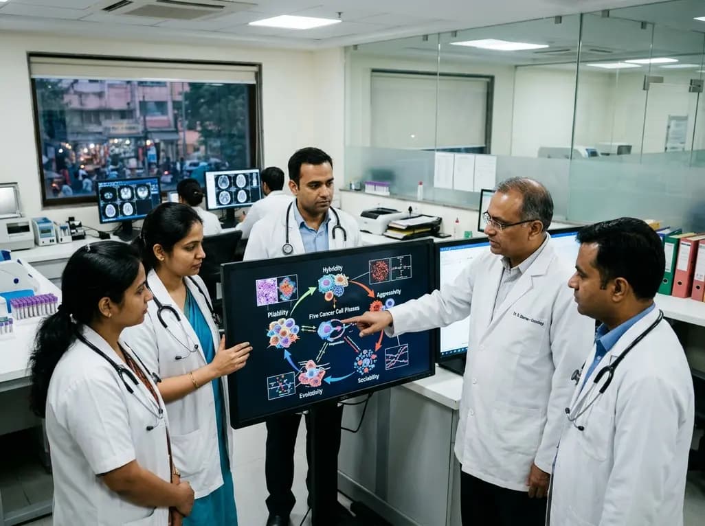
The PHASE framework conceptualizes cancer biology through five interacting processes: Pliability, Hybridity, Aggressivity, Sociability, and Evolutivity. This regulatory circuit offers a systems-level perspective on tumor heterogeneity, disease progression, and interaction-guided therapeutic strategies.
Today
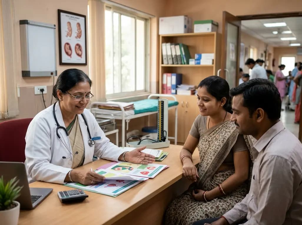
Maternal cardiovascular risk factors such as hypertension, obesity, and diabetes significantly elevate offspring cardiovascular disease risk through placental dysfunction and fetal programming. Enhancing preconception screening and antenatal care offers clinicians a powerful strategy to protect long-term heart health.
Yesterday
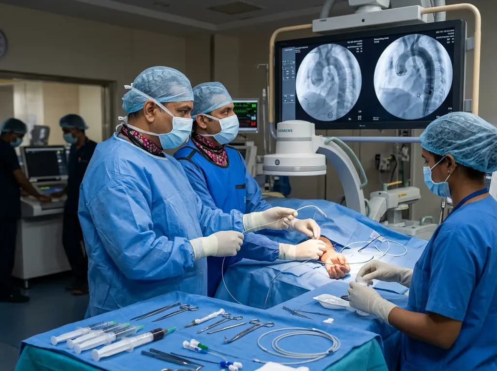
The novel CAST technique utilizes the indwelling Safari XS guidewire as a tracking rail to reposition a radial pigtail catheter into the descending aorta during minimalist TAVI, overcoming challenging vascular anatomy without requiring secondary femoral access or specialized equipment.
Today
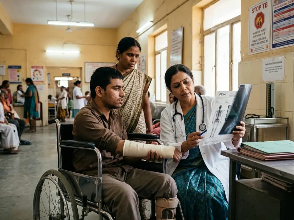
A retrospective cohort study reveals that lumbar spine bone mineral density does not distinguish extremity long bone fracture status in adults with severe motor and intellectual disabilities. Because age drove fracture risk rather than spinal density, clinicians must adopt comprehensive assessment tools.
Yesterday
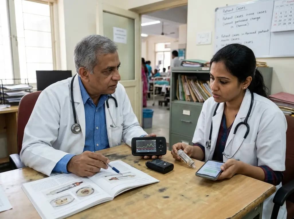
A case report details how reusing single-use insulin cartridges caused device failure and diabetic ketoacidosis in a type 1 diabetes patient using an advanced hybrid closed-loop system. The report highlights technological blindspots, mechanical degradation, and safety protocols during supply shortages.
Today
