.webp&w=384&q=75)

Loading, please wait...
Loading, please wait...
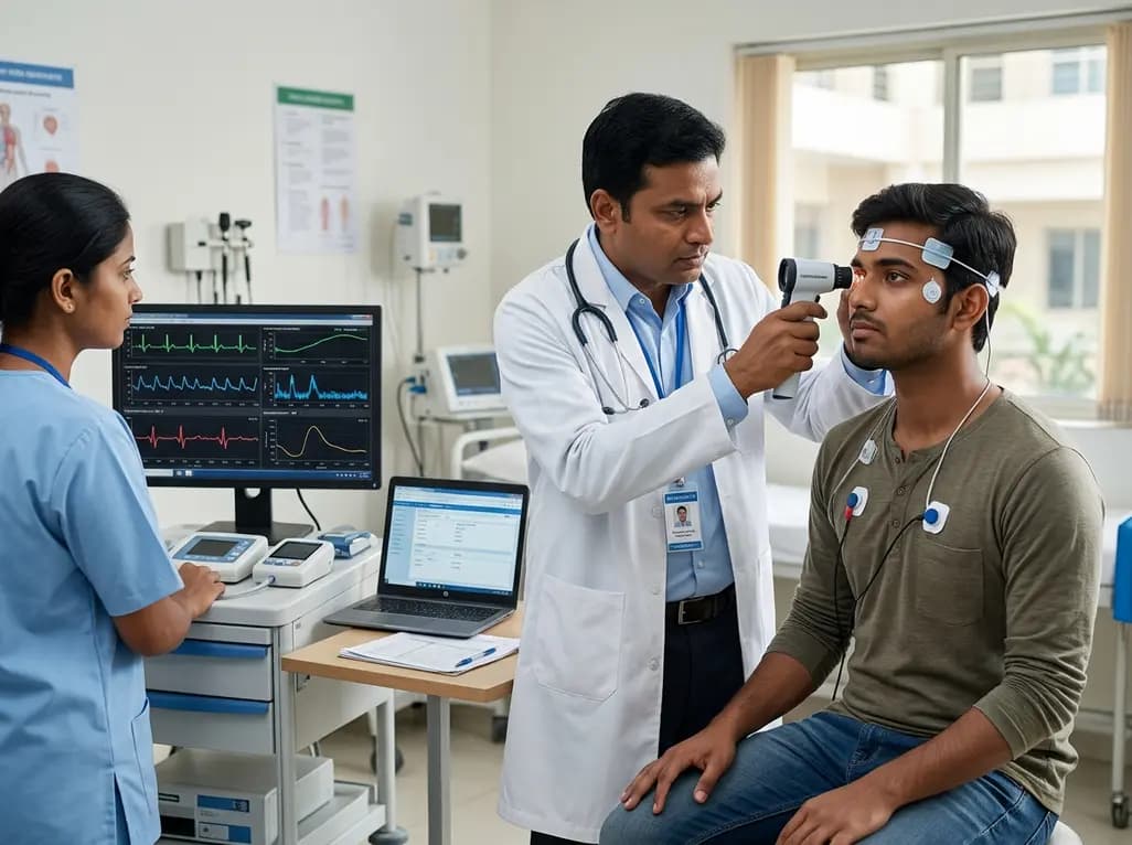
Traumatic brain injuries alter neural networks and disrupt systemic physiological balance. Following mild traumatic brain injury (mTBI), many patients experience prolonged symptoms that interfere with physical activity, academic pursuits, and professional performance. Emerging clinical evidence indicates that autonomic nervous system dysregulation plays a fundamental role in post-concussion morbidity. Disruption of autonomic pathways frequently causes orthostatic intolerance, exercise intolerance, persistent headaches, and emotional lability. However, clinicians historically lacked standardized, reproducible physiological markers to track autonomic recovery systematically. Most return-to-activity protocols continue to depend on subjective patient reports, which creates significant vulnerability to premature clearance or prolonged disability.
The central autonomic network spans interconnected regions within the cerebral cortex, insula, amygdala, hypothalamus, and brainstem. Consequently, biomechanical forces during a concussive impact can stretch delicate axonal projections across these networks. This mechanical strain precipitates neurometabolic cascades, ionic shifts, and altered neurovascular coupling. As a result, the body struggles to maintain equilibrium between sympathetic excitation and parasympathetic regulation.
In healthy individuals, the autonomic branches constantly adjust cardiac output, vascular resistance, and respiratory patterns to match environmental and physical stressors. Following head trauma, patients frequently exhibit uncoupled sympathetic overdrive and depressed vagal modulation. Therefore, simple daily tasks or mild physical exertion can trigger disproportionate tachycardia, lightheadedness, and systemic fatigue. Moreover, autonomic instability alters cerebral blood flow autoregulation, worsening cognitive fatigue and headache severity during recovery. Identifying these physiological impairments allows clinicians to address the root causes of persistent post-concussive symptoms rather than managing isolated subjective complaints.
Although prior studies linked head trauma to dysautonomia, several methodological limitations restricted the clinical translation of physiological biomarkers. Past investigations often suffered from small sample sizes, heterogeneous testing protocols, and inadequate control for confounding factors such as pre-injury conditioning, sleep quality, and psychological distress. To overcome these barriers, researchers developed the Measuring Autonomic Nervous System in Warfighters Following Exertion and Resting Stressors (Measuring ANSWERS) study.
This comprehensive protocol establishes an observational framework evaluating both military personnel and collegiate club athletes. Specifically, the investigation incorporates a cross-sectional arm and a longitudinal tracking arm. In the cross-sectional cohort, investigators evaluate 150 participants, comprising 50 military service members and 100 collegiate athletes stratified by concussion history. Meanwhile, the longitudinal cohort prospectively monitors 50 concussed athletes alongside 50 matched healthy controls. By employing standardized autonomic stress testing at rest and during physical exertion, the study directly links objective autonomic parameters with functional clinical outcomes.
The Measuring ANSWERS protocol deploys objective physiological markers to quantify both resting and exertional autonomic control. Among these, heart rate variability (HRV) serves as a primary noninvasive measure of cardiovagal and sympathetic interaction. Healthy autonomic networks produce dynamic beat-to-beat variations in response to respiration and baroreceptor feedback. Conversely, reduced HRV parameters, such as root mean square of successive differences (RMSSD) and high-frequency spectral power, reflect compromised vagal tone and sympathetic hyperarousal.
Additionally, the study utilizes quantitative infrared pupillometry to capture subtle disruptions in brainstem autonomic circuitry. The pupillary light reflex (PLR) reflects rapid parasympathetic constriction and subsequent sympathetic redilation in response to calibrated light stimuli. Following concussion, patients frequently demonstrate delayed constriction latencies, reduced constriction velocity, and prolonged dilation recovery. By capturing both HRV and PLR at baseline, during physical exertion, and throughout post-exercise recovery, clinicians can obtain a high-resolution physiological profile of autonomic integrity across multiple organ systems.
Subjective symptom resolution does not always coincide with complete physiological brain recovery. In many cases, athletes report feeling fully recovered while latent neurovascular and autonomic deficits persist under physiological stress. If an athlete returns to contact sports before autonomic autoregulation normalizes, they face heightened risks of secondary injury, prolonged post-concussion syndrome, and severe neurovascular complications.
To address this clinical dilemma, the longitudinal arm of the Measuring ANSWERS study evaluates participants across three critical recovery milestones: within 7 days of injury, at participant-reported symptom resolution, and 1 to 2 weeks after clinical clearance for return to activity. This serial evaluation clarifies whether exertional autonomic abnormalities outlast self-reported symptom resolution. Furthermore, integrating physiological monitoring with standardized vestibular, oculomotor, cognitive, and dynamic balance assessments ensures a comprehensive evaluation of neuromuscular and autonomic readiness prior to full unrestricted activity.
The outcomes of the Measuring ANSWERS study promise substantial improvements in clinical management across sports medicine, neurology, and military healthcare. By validating standardized autonomic testing protocols, clinicians can implement targeted sub-symptom threshold aerobic exercise regimens with greater confidence. Active rehabilitation approaches, such as the Buffalo Concussion Treadmill Test protocol, safely restore autonomic balance and cerebral blood flow when guided by reliable physiological thresholds.
Moreover, objective autonomic metrics can help clinicians stratify concussion severity, predict protracted recovery trajectories, and identify patients who require tailored autonomic rehabilitation. As enrollment progresses toward the targeted 250 participants, the study establishes a reproducible foundation for modern neurotrauma care. Ultimately, shifting clinical paradigms from subjective symptom tracking to objective autonomic monitoring will protect active populations and optimize long-term neurological recovery.
Concussive biomechanical forces disrupt axonal integrity across the central autonomic network, including the brainstem, hypothalamus, and cerebral cortex. This traumatic insult triggers neurometabolic cascades and neurovascular uncoupling, which impairs the delicate balance between sympathetic and parasympathetic signaling. Consequently, patients develop heightened sympathetic reactivity, blunted vagal modulation, and dysregulated blood pressure and heart rate responses during physical exertion and postural transitions.
Clinicians assess autonomic dysfunction using noninvasive physiological tools, including resting and exertional heart rate variability (HRV), quantitative dynamic pupillometry, and orthostatic vital sign tracking. Additionally, graded exertion protocols, such as the Buffalo Concussion Treadmill Test, safely identify exercise intolerance thresholds. Combining these objective physiological markers with comprehensive vestibular, oculomotor, and cognitive testing batteries provides an accurate assessment of post-injury autonomic health.
Persistent autonomic dysregulation frequently outlasts subjective symptom resolution, leaving patients vulnerable if they resume strenuous activity prematurely. When autonomic balance remains impaired, exertional stress can compromise cerebral blood flow regulation, increase reinjury risks, and prolong post-concussive morbidity. Integrating objective physiological biomarkers into return-to-play assessments ensures athletes achieve genuine neuromuscular and cardiovascular recovery before engaging in unrestricted physical exertion.
Disclaimer: This content is for informational and educational purposes only and should not be considered medical advice. Always consult a qualified healthcare professional regarding any medical condition or before making healthcare decisions. Refer to the latest local and national guidelines for clinical practice.
References

Read summarized clinical updates, watch expert medical content, and earn CME certifications right from your smartphone.
Open your smartphone camera to scan and install.





Mild traumatic brain injury frequently disrupts autonomic control. The Measuring ANSWERS study protocol establishes a standardized framework combining heart rate variability, pupillary light reflex, and multimodal clinical testing to track autonomic nervous system dysregulation after concussion.
last month

Ceftriaxone therapy can lead to biliary sludge and gallstone formation due to calcium-ceftriaxone precipitation. A recent cohort study shows that the noninvasive Fibrosis-4 (FIB-4) index, alongside treatment duration, independently predicts lithogenesis, enabling proactive risk stratification.
Today

A multiparametric MRI approach combining quantitative T2 shading, apparent diffusion coefficient (ADC) measurements, and morphological signs provides superior diagnostic accuracy over subjective evaluation to differentiate ovarian endometriomas from benign hemorrhagic cysts.
Today

A landmark case report reveals a donor heart beating for a century in a 63-year-old recipient, challenging conventional age limits in cardiac transplantation and offering critical lessons on organ longevity, donor selection, and long-term graft survival.
Today
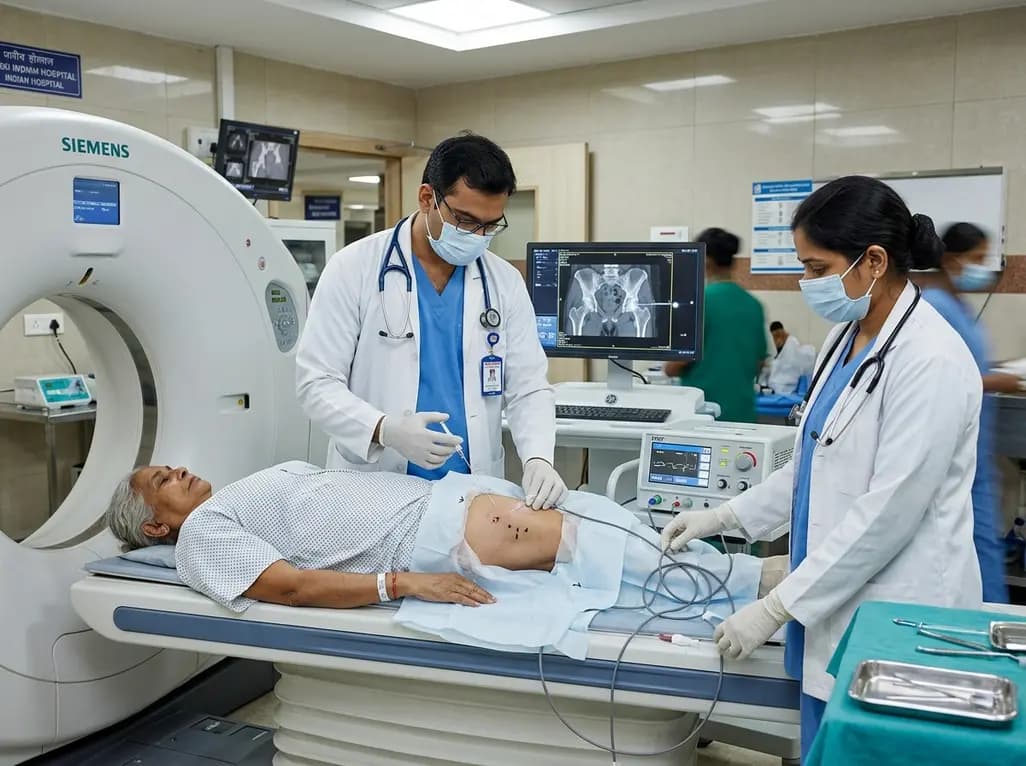
CT-guided pulsed radiofrequency ablation of the obturator nerve delivers durable pain relief up to 12 months in mild-to-moderate hip osteoarthritis, while offering a safe short-term palliative bridge for severe grade 4 disease without procedural complications.
Today

New research reveals that M1 macrophage-derived exosomes aggravate diabetic nephropathy by transferring WTAP to stabilize S1PR2 mRNA. Silencing WTAP in these vesicles attenuates endothelial injury and renal fibrosis, pointing toward innovative nanomedicine therapies.
Yesterday
