

Loading, please wait...
Loading, please wait...

Surgical decompression and clot evacuation remain critical interventions for acute supratentorial intracerebral hemorrhage. However, acute postoperative rebleeding frequently offsets surgical success and triggers devastating secondary neural injury. Clinicians often observe that preexisting vascular, metabolic, and systemic disorders exacerbate early hemorrhagic recurrence. Despite this clinical intuition, the precise link between documented comorbidity burden and radiologically significant hematoma growth requires systematic evaluation. In neurosurgical practice, determining which patients harbor the highest risk of postoperative rebleeding allows surgical teams to individualize perioperative protocols. Chronic vasculopathies, hypertension, renal impairment, and anticoagulant exposure consistently disrupt normal vascular autoregulation. Consequently, delicate capillary beds within the friable resection cavity fail to maintain adequate hemostatic control. When surgical teams relieve intracranial tamponade during craniotomy or minimally invasive evacuation, fragile microvessels experience abrupt hemodynamic shifts. Therefore, identifying patient-specific risk profiles beforehand provides crucial opportunities for aggressive surveillance. Modern neurocritical care units must continually refine stratification tools to anticipate catastrophic hemorrhage before clinical deterioration emerges. This study evaluates how documented multimorbidity interacts with specific neuroimaging definitions to dictate clinical trajectories after hematoma evacuation.
Investigating surgical outcomes requires standardized and reproducible imaging endpoints. The single-center cohort study analyzed 111 consecutive adult patients who underwent supratentorial hematoma evacuation between 2016 and 2025. Researchers systematically grouped patients into two clinical categories based on documented baseline medical conditions. Specifically, the team compared individuals with 0 to 1 comorbidity against those presenting with 2 or more systemic comorbidities. The primary imaging endpoint demanded strict radiographic criteria between early and second postoperative computed tomography scans. To establish definitive primary rebleeding, investigators required a visually confirmed new or expanding hemorrhagic component. Additionally, the lesion had to show an absolute interval increase exceeding 20 mL and 25% of baseline volume. The researchers also examined alternative volumetric definitions to test whether diagnostic thresholds modified observed associations. For example, secondary definitions assessed simple volume expansion above 25% of baseline or an interval growth exceeding 6 mL. By employing rigorous imaging parameters, the researchers effectively distinguished minor surgical oozing from clinically meaningful, space-occupying rehemorrhage. Furthermore, prospective data collection combined with blinded radiographic reassessments ensured high methodological validity throughout the study period.
The clinical results demonstrated a striking disparity in hemorrhagic recurrence across comorbidity cohorts. Among patients with low comorbidity, primary rebleeding developed in only 2 of 78 patients, representing a 2.6% incidence. In contrast, patients with 2 or more comorbidity categories suffered primary rebleeding in 9 out of 33 cases, reaching 27.3%. Statistical analysis revealed a conditional odds ratio of 13.84, with an exact 95% confidence interval spanning from 2.62 to 140.31. This dramatic elevation carried high statistical significance and reflected an absolute risk increase of 24.7 percentage points. Furthermore, secondary surgical rescue became necessary for 8 of the 11 patients who experienced primary rebleeding. Sensitivity analyses specifically excluding 5 patients who received intervening revisions for residual clot showed persistent risks. Within that refined subcohort of 106 individuals, multimorbidity retained an odds ratio of 14.47 for major recurrent hemorrhage. Patients experiencing these substantial hematomas also required significantly longer neurointensive care management. Specifically, median intensive care unit stays rose from 6 days in stable cohorts to 10 days among rebleeding cases. These striking metrics underline how systemic medical fragility translates directly into catastrophic surgical complications.
Intriguingly, the study illustrated that the strength of the statistical association depended heavily on the chosen imaging definition. When researchers evaluated less stringent definitions of rebleeding, the apparent risk magnification attenuated considerably. For instance, defining rebleeding simply as an interval volume increase greater than 25% yielded an odds ratio of only 2.84. Similarly, adopting an alternative threshold of 6 mL or 33% relative volume growth produced an odds ratio of 1.54. Neither of these secondary comparisons achieved formal statistical significance. Consequently, these findings indicate that systemic multimorbidity specifically predisposes patients to large, high-volume hemorrhage rather than minor radiographic petechiae. In addition, anticoagulant therapy represented an important clinical confounder in this surgical cohort. Crude analyses demonstrated that preadmission anticoagulant exposure was strongly linked to postoperative hemorrhagic expansion. However, anticoagulant use heavily overlapped with systemic cardiovascular multimorbidity, including atrial fibrillation, ischemic heart disease, and peripheral vascular disorders. Therefore, separating the mechanical vascular friability of chronic disease from pharmacologically induced coagulopathy remains challenging. Clinicians must recognize that multimorbid patients frequently harbor dual threats of compromised microvascular integrity and altered clotting cascades.
The identification of high-risk surgical patients demands heightened vigilance during both operative intervention and subsequent intensive care. Surgeons operating on multimorbid individuals should consider adopting meticulous hemostatic techniques to secure fragile bed margins. Furthermore, neuroanesthetists must prevent sharp blood pressure surges during emergence and extubation, as acute hypertension provokes bleeding into freshly decompressed cavities. Following surgery, neurocritical care teams should enforce stringent hemodynamic targets and avoid excessive blood pressure variability. Continuous invasive arterial monitoring provides the precision required to maintain stable cerebral perfusion without inducing hyperperfusion injury. Moreover, clinicians must prioritize rapid reversal of all antiplatelet and anticoagulant agents before and immediately after surgical evacuation. Point-of-care coagulation assays, including viscoelastic testing, assist in identifying subtle platelet dysfunction and clotting factor deficiencies. Routine surveillance imaging also plays a pivotal role in post-evacuation management. Obtaining an early baseline scan within 6 hours, followed by a scheduled follow-up scan, detects silent expansion early. Timely radiological identification allows prompt intervention before irreversible secondary brainstem compression and herniation occur. Multidisciplinary collaboration between neurosurgeons and intensivists optimizes intracranial pressure control and systemic stability. Consequently, individualized post-craniotomy pathways significantly reduce secondary neurological decline in vulnerable patients.
In India, the burden of spontaneous intracerebral hemorrhage continues to rise alongside increasing life expectancy and non-communicable diseases. Indian tertiary centers frequently admit elderly patients presenting with uncontrolled hypertension, poorly regulated type 2 diabetes, and chronic kidney disease. Concurrently, the expanding use of oral anticoagulants for atrial fibrillation and coronary stents amplifies perioperative complexity across Indian intensive care units. In community and district hospital setups, limited access to serial advanced neuroimaging can delay the recognition of rebleeding. Therefore, Indian neurosurgical centers must establish standardized local protocols for postoperative monitoring in high-risk patients. When resources permit, adopting minimally invasive parafascicular evacuation or neuroendoscopy may minimize surgical tract trauma in friable vascular beds. However, maintaining strict blood pressure targets in resource-constrained wards requires dedicated nursing protocols and affordable titratable infusions like labetalol. Clinicians should also educate families regarding the substantial risks of reoperation in elderly patients with multiple coexisting morbidities. Furthermore, establishing regional stroke networks ensures prompt referral of complicated surgical cases to comprehensive neurocritical centers. Ultimately, proactive risk scoring and rigorous postoperative surveillance will elevate the safety and success of neurosurgical care nationwide.
Coexisting conditions like uncontrolled hypertension, diabetes, and chronic kidney disease induce widespread microvascular fragility and endothelial dysfunction. When surgical evacuation rapidly decompresses intracranial tissues, these damaged parenchymal microvessels experience acute shear stresses. Furthermore, underlying metabolic derangements impair normal platelet function, predisposing patients to large recurrent hematomas within the friable resection cavity.
Stringent volumetric thresholds, such as requiring an increase exceeding 20 mL, isolate clinically catastrophic recurrent hematomas that cause mass effect and require surgical revision. In contrast, smaller thresholds capture minor, asymptomatic postoperative oozing. Consequently, multimorbidity strongly predicts major structural rebleeding, whereas smaller parenchymal volume fluctuations correlate more with local surgical manipulation than systemic disease.
Preventing hematoma recurrence requires continuous arterial line monitoring to maintain systolic blood pressure strictly between 130 and 140 mmHg. Clinicians must promptly correct coagulopathy using prothrombin complex concentrates or specific reversal agents. Additionally, performing scheduled baseline and follow-up computed tomography scans within 24 hours ensures early identification of interval expansion before clinical deterioration ensues.
Disclaimer: This content is for informational and educational purposes only. It is not intended to be a substitute for professional medical advice, diagnosis, or treatment. Always seek the advice of your physician or other qualified health provider with any questions you may have regarding a medical condition. Refer to the latest local and national guidelines for clinical practice.
References

Read summarized clinical updates, watch expert medical content, and earn CME certifications right from your smartphone.
Open your smartphone camera to scan and install.





A single-center cohort study shows that having two or more comorbidity categories dramatically increases the risk of large-volume interval rebleeding (OR 13.84) after supratentorial intracerebral hematoma evacuation, emphasizing the need for tailored neurointensive surveillance.
Today

A landmark study reveals that congenital DLK1 deficiency in women with a history of central precocious puberty causes visceral adiposity, mitochondrial dysfunction, oxidative stress, and neutrophil activation, highlighting the critical need for lifelong cardiometabolic surveillance.
Today

Estetrol (E4), a native fetal estrogen, offers a unique tissue-selective profile with minimal hepatic impact and lower thrombotic risk. This review highlights its clinical use in oral contraception, menopausal symptom management, and emerging applications across oncology and neurology.
Today

Explore revascularisation strategies in dialysis-dependent chronic limb-threatening ischaemia. This review examines surgical bypass vs endovascular therapy, BEST-CLI trial findings, procedural morbidity, conduit availability, and multidisciplinary decision-making for optimal limb salvage.
Today

A recent cross-sectional analysis reveals that over a quarter of randomized controlled trials for non-specific low back pain face discontinuation or unknown status, while more than 36% of completed trials remain unpublished, exposing notable reporting gaps in spine and musculoskeletal clinical research.
Today
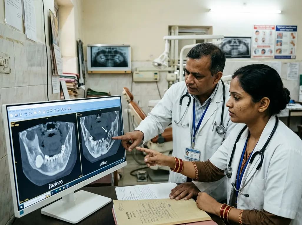
This case report examines the longitudinal CBCT evolution of Stage 2 MRONJ in a 70-year-old woman on denosumab at the site of a pre-existing mandibular dense bone island. It emphasizes the radiographic features, differential diagnosis, and multidisciplinary strategies required for high-risk bone lesions.
Today
