

Loading, please wait...
Loading, please wait...

Clinical neurotology increasingly emphasizes the relationship between sensory inputs and cortical processing. Modern balance assessment extends beyond simple reflex arcs, as emerging evidence proves that cognitive image recognition fundamentally shapes visuo-vestibular interaction. When clinicians evaluate patients suffering from balance impairments, isolated reflex measurements often fail to reflect subjective disability. Consequently, understanding how working memory and visual attention modulate oculomotor responses provides vital diagnostic clarity. By examining the brain under dual-task cognitive demands, clinicians can resolve persistent discrepancies between objective laboratory gains and daily functional impairment.
Clinicians traditionally relied on bedside head impulse testing to evaluate vestibulo-ocular reflex function. However, the video head impulse test revolutionized balance medicine by quantifying angular vestibulo-ocular reflex gain. Furthermore, the newer suppression head impulse paradigm, or SHIMP, introduced an anti-compensatory tracking task. In conventional testing, healthy individuals suppress corrective saccades because reflexes stabilize visual targets. In contrast, SHIMP requires patients to fixate on a target moving synchronously with their head. Therefore, healthy individuals generate large, predictable anti-compensatory saccades after head rotation stops. Conversely, patients with severe vestibular loss show absent SHIMP saccades. As recovery progresses, however, the central nervous system remodulates these saccades. Clinicians observe that the reappearance and timing of these movements signal meaningful neurological compensation. In addition, researchers designed the functional head impulse test to measure dynamic visual acuity during passive thrusts. Combining these innovative tools allows neurotologists to study gaze stabilization under controlled conditions.
The cerebral cortex coordinates visual processing and spatial orientation through interconnected neural networks. Specifically, shared cortical substrates within the posterior parietal cortex oversee spatial attention and gaze stabilization. When individuals encounter rapid head movements, the brain integrates vestibular signals with visual input to maintain clear retinal images. However, when individuals simultaneously recognize complex visual symbols, cognitive load increases substantially. Consequently, this cognitive demand challenges cortical resource allocation. Visual recognition requires working memory, rapid feature discrimination, and precise decision-making. Therefore, when the brain performs image recognition and vestibulo-ocular modulation together, central processing pathways compete for finite attention. Researchers observe that this simultaneous demand modifies saccadic latency and clusters corrective eye movements differently. Moreover, these neurophysiological shifts confirm that higher cognitive centers directly influence reflexive oculomotor pathways. Thus, visuo-vestibular interaction represents an integrated cortical-subcortical network rather than an isolated brainstem loop.
To quantify these neurological dynamics, investigators evaluated healthy cohorts alongside patients experiencing unilateral or bilateral vestibular hypofunction. The research protocol paired video head impulse testing in standard and suppression modes with the functional head impulse test. Initially, participants completed the tests separately to establish baseline performance. Subsequently, subjects performed the tests simultaneously under a unified dual-task protocol. During the combined challenge, patients identified the orientation of transient Landolt optotypes while undergoing rapid head impulses. Consequently, researchers analyzed Landolt optotype recognition rates, saccadic latency, and saccade clustering metrics across both protocols. The data revealed striking differences between isolated testing and joint dual-task execution. Notably, simultaneous cognitive recognition altered saccadic programming and response latencies across patient groups. In addition, the cognitive burden unmasked subtle functional limitations that single-modality assessments consistently missed. Therefore, integrating cognitive challenges into routine vestibular protocols provides an accurate representation of dynamic gaze stability in real-world environments.
Subjective dizziness often correlates poorly with objective canal paresis measured during standard caloric or impulse testing. However, the degree of saccadic remodulation observed during dual-task testing demonstrates a robust relationship with Dizziness Handicap Inventory scores. Specifically, patients who organize their corrective saccades into tight, predictable clusters experience substantially fewer functional difficulties during daily activities. When the central nervous system successfully remodulates SHIMP saccades, patients regain visual confidence during rapid head rotations. Conversely, scattered, poorly organized saccades prolong retinal slippage and exacerbate chronic motion intolerance. Furthermore, dual-task experiments revealed that differences between disjointed and combined protocols directly reflect symptom severity on clinical questionnaires. As cognitive demands rise, patients with incomplete central compensation struggle to maintain target recognition. Consequently, measuring saccadic latency shifts under cognitive load offers a quantifiable biomarker for functional recovery. Clinicians can harness these insights to identify individuals requiring targeted cognitive-motor balance rehabilitation rather than standard repetitive adaptation exercises.
In India, vestibular disorders represent a major clinical burden across busy neurology and otolaryngology outpatient clinics. Practicing clinicians frequently manage patients reporting disabling vertigo despite normal vestibular test metrics. Therefore, incorporating cognitive dual-task principles into Indian clinical workflows offers immediate diagnostic value. Clinicians can integrate optotype recognition tasks or mental arithmetic challenges into dynamic visual acuity testing and bedside examinations. Furthermore, Indian neurotologists can utilize functional head impulse protocols to formulate personalized vestibular rehabilitation therapy. For instance, physiotherapists can train patients using combined dual-task exercises that simulate bustling Indian market environments or dense traffic conditions. As patients learn to resolve visual targets while moving their heads under cognitive strain, central compensatory networks strengthen rapidly. In addition, tracking Dizziness Handicap Inventory improvements alongside saccadic reorganization ensures reliable treatment monitoring. Ultimately, adopting these multi-domain assessment frameworks bridges the gap between laboratory reflex testing and genuine patient well-being across Indian healthcare settings.
Clinicians assess suppression head impulse testing because it offers an intuitive measure of residual vestibular function and central compensation. Unlike standard testing where covert saccades complicate analysis, SHIMP prompts healthy individuals to generate large compensatory saccades at head movement termination. Consequently, tracking whether these saccades reappear, cluster, and shorten in latency helps physicians monitor neurological adaptation. Furthermore, this dynamic parameter correlates closely with functional recovery and symptom relief in vestibular patients.
Cognitive image recognition interferes with vestibulo-ocular reflexes because both processes share parietal cortical substrates and attentional resources. When a patient identifies visual optotypes during sudden head movement, working memory and pattern recognition create competing neurological demands. Consequently, central processing networks prioritize information processing, which alters compensatory saccade latency and spatial clustering. Furthermore, this cognitive interference explains why balance-impaired individuals frequently experience symptom worsening when navigating visually complex, highly demanding everyday environments.
Clinicians can implement dual-task assessments by combining standard dynamic visual acuity protocols with structured cognitive tasks during routine evaluations. For example, neurotologists can instruct patients to read fluctuating optotypes or perform mental arithmetic while executing active or passive head rotations. Additionally, computerized functional head impulse testing provides automated, objective measurements of dynamic image recognition under rotational stress. These combined protocols help specialists identify functional deficits that standard single-modality reflex testing consistently misses.
Disclaimer: This content is for informational and educational purposes only. It is not intended to substitute for professional medical advice, diagnosis, or treatment. Seek the advice of your physician or another qualified healthcare provider with any questions you may have regarding a medical condition. Refer to the latest local and national guidelines for clinical practice.
References

Read summarized clinical updates, watch expert medical content, and earn CME certifications right from your smartphone.
Open your smartphone camera to scan and install.





New research demonstrates that cognitive image recognition during functional head impulse testing directly influences visuo-vestibular interaction and saccadic compensation, providing a practical framework for ecological balance assessment in neurotology.
Today

A comprehensive review of malnutrition in older adults, addressing multifactorial drivers, screening tools, and targeted nutritional and exercise interventions to foster healthy ageing.
Today

A prospective study reveals significant first-trimester trace-element alterations in pregnant women with epilepsy. Lower serum selenium independently predicted subsequent seizure recurrence despite prior remission, highlighting the vital clinical role of micronutrient profiling during antenatal care.
Today
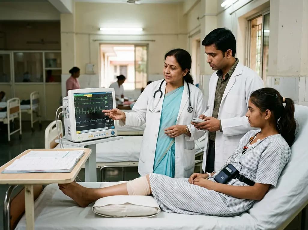
A 15-year-old patient presented with knee arthralgia and acute rheumatic fever, exhibiting complex arrhythmias including AV blocks and non-sustained ventricular tachycardia despite lacking severe valvulitis. This highlights the importance of extended Holter monitoring in pediatric rheumatic carditis.
Today

A biomechanical study shows that removing a stripped nonlocking screw during metacarpal bridge plate fixation safely avoids hardware migration without reducing construct strength or creating a stress riser, resolving a common intraoperative dilemma in hand trauma surgery.
Today
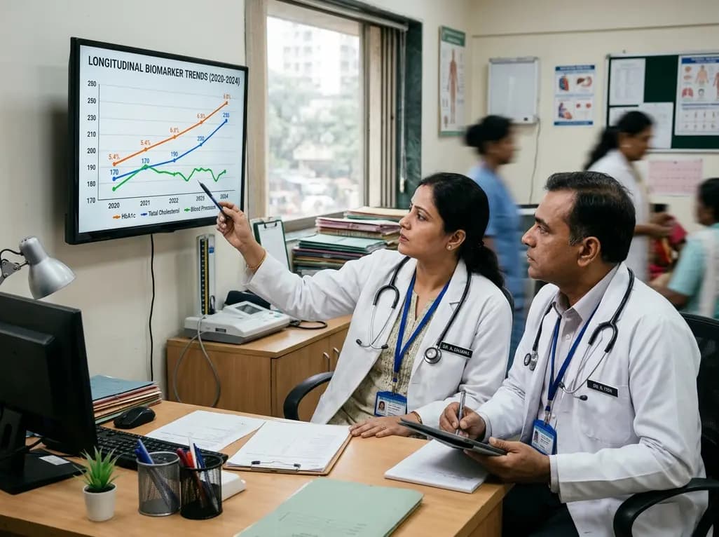
Annual health checks only offer static snapshots. Real-world evidence from 2.1 million insured Indians reveals that tracking dynamic biomarker trends over time enables clinicians to spot evolving cardiometabolic risks early, reverse borderline pathology, and deliver tailored preventive interventions.
Yesterday
