

Loading, please wait...
Loading, please wait...

Clinical teams frequently encounter central nervous system neoplasms that present with classic anatomical features. However, metastatic renal cell carcinoma can occasionally develop within cranial and spinal nerves, closely mimicking benign nerve sheath tumors. Because renal malignancies exhibit an unpredictable and delayed biology, secondary deposits often manifest nearly a decade after radical nephrectomy. Therefore, clinicians must maintain a heightened index of suspicion when evaluating neural lesions in oncology patients.
Clear cell renal cell carcinoma notoriously displays protracted dormancy followed by aggressive distant dissemination. Historically, clinicians expect metastases to involve familiar sites such as the lungs, bone, liver, and cerebral parenchyma. Nevertheless, tumor emboli can colonize peripheral nerve sheaths, spinal nerve roots, and cranial nerves through hematogenous routes or retrograde venous plexuses. In illustrative clinical series, patients developed neurogenic lesions between six and nine years following initial nephrectomy. For example, lesions have emerged within the cervical dorsal root ganglion, the cerebellopontine angle, and the cerebellomedullary junction. Because decades may elapse after surgical cure of the primary renal tumor, clinicians often attribute newly discovered solitary neural masses to primary nerve sheath neoplasms like schwannomas. Consequently, patients risk diagnostic delay when clinicians overlook distant surgical history. Medical practitioners should therefore routinely review comprehensive urological records whenever atypical neurogenic masses arise.
Conventional magnetic resonance imaging frequently misleads clinicians because neural metastases can adopt benign morphologic traits. On standard T1-weighted and T2-weighted sequences, metastatic deposits along nerve pathways often present as sharply circumscribed, well-demarcated masses. Moreover, these lesions cause adjacent bony remodeling and neural foraminal expansion, which radiographically suggests an indolent, slow-growing schwannoma. Routine contrast-enhanced scans demonstrate intense, homogeneous enhancement, thereby reinforcing the erroneous diagnosis of a benign peripheral nerve sheath tumor. In the cerebellopontine angle, an avidly enhancing mass along the fifth cranial nerve strongly mimics a trigeminal schwannoma. Similarly, an expanding foramen in the cervical spine typically leads radiologists to report a benign neurofibroma or schwannoma. Consequently, surgeons may schedule non-urgent surveillance or plan conservative resections under false diagnostic assumptions. Such pitfalls emphasize that conventional anatomical imaging alone cannot reliably exclude renal metastases in high-risk patients.
Fortunately, advanced neuroimaging modalities provide crucial physiologic biomarkers that unmask underlying malignancy. Although conventional sequences show deceptive margins, arterial spin labeling perfusion MRI exposes profound tumor hyperperfusion characteristic of hypervascular renal cell deposits. In contrast, typical benign schwannomas exhibit significantly lower cerebral blood flow and modest perfusion values. Furthermore, susceptibility-weighted imaging often reveals an irregular hemosiderin rim, reflecting microhemorrhage and internal neoangiogenesis within the metastatic matrix. Significant peritumoral vasogenic edema also signals aggressive parenchymal infiltration, which benign sheath lesions rarely produce. Additionally, fluorodeoxyglucose positron emission tomography or DOTATATE imaging can pinpoint hypermetabolic systemic disease and detect silent extraneural deposits. When radiologists combine multi-parametric MRI sequences with metabolic nuclear imaging, they can effectively differentiate hypervascular metastases from common cranial nerve sheath neoplasms.
Managing metastatic neural lesions requires aggressive, customized multimodality therapy tailored to patient performance and tumor anatomy. Because renal cell carcinoma exhibits relative radioresistance to traditional fractionated radiation, stereotactic radiosurgery serves as a cornerstone for local control. Radiosurgeons deliver highly focused, ablative radiation doses that effectively disrupt tumor microvasculature while sparing adjacent cranial or spinal nerves. However, when large masses compress the brainstem or cause progressive motor deficits, neurosurgeons must pursue microsurgical decompression. Modern systemic therapy further transforms long-term disease management. Oncologists frequently administer combination regimens featuring tyrosine kinase inhibitors, such as cabozantinib or lenvatinib, alongside immune checkpoint inhibitors targeting PD-1 and CTLA-4 pathways. Thus, integrating targeted radiosurgery, meticulous surgical resection, and modern immunotherapy provides durable local disease stabilization and prolonged systemic survival.
Obtaining an accurate histological diagnosis remains imperative before initiating definitive oncological therapy. While imaging biomarkers strongly suggest secondary disease, tissue confirmation establishes true tumor biology and guides molecular therapies. Therefore, multidisciplinary teams should evaluate whether a safe biopsy or surgical debulking is feasible. If a patient presents with multiple metastases, interventional radiologists can safely target an accessible visceral site to avoid high-risk cranial nerve exploration. Conversely, when an isolated neurogenic lesion causes neurological deterioration, open surgical exploration achieves both histological verification and neural decompression. Clinicians must recognize that assuming a benign etiology without histopathology can result in underdosing radiosurgery or withholding lifesaving systemic therapies. Through diligent multidisciplinary collaboration among neurosurgeons, oncologists, and radiologists, clinical teams can avoid catastrophic diagnostic delays and optimize functional outcomes.
Long-term surveillance remains essential for all individuals who undergo curative-intent nephrectomy for clear cell carcinoma. Because malignant renal cells can remain dormant in distant neural capillary beds for years, surveillance protocols must extend beyond standard five-year windows. Oncologists should educate cancer survivors about subtle neurological symptoms, including localized radicular pain, facial numbness, unilateral hearing loss, and unexplained cranial nerve palsies. When neurological deficits appear, clinicians should immediately order targeted contrast-enhanced magnetic resonance neurography rather than attributing symptoms to degenerative spine disease or benign neuropathies. Moreover, routine multidisciplinary tumor boards facilitate prompt reassessment when imaging demonstrates unusual interval lesion growth. By maintaining prolonged clinical vigilance and recognizing unusual patterns of tumor relapse, healthcare providers can detect metastatic neural lesions early, preserve critical neurologic function, and significantly improve patient survival.
Yes, metastatic renal cell carcinoma frequently exhibits delayed recurrence, occasionally presenting 6 to 10 years after nephrectomy. Renal clear cell tumors possess an indolent dormancy phase, allowing circulating malignant cells to survive within distant capillary beds before proliferating. Consequently, clinicians must evaluate any new neurological mass in a cancer survivor with heightened suspicion, regardless of how many years have passed since their initial curative cancer surgery.
Although both entities demonstrate contrast enhancement, advanced magnetic resonance sequences reveal critical differences. Renal metastases typically exhibit marked arterial spin labeling hyperperfusion, reflecting rich neoangiogenesis, whereas benign schwannomas show low perfusion. Furthermore, metastatic lesions frequently present with susceptibility-weighted hemosiderin rims from microhemorrhages, substantial peritumoral vasogenic edema, and rapid interval growth on sequential neuroimaging studies, which benign nerve sheath tumors rarely display.
Histopathological confirmation is essential because metastatic lesions require vastly different radiation dosing and systemic regimens compared to benign schwannomas. While clinicians treat benign schwannomas with moderate radiation doses or observation alone, renal metastases mandate higher ablative stereotactic radiosurgical doses to overcome radioresistance. Additionally, confirming clear cell histology immediately qualifies patients for targeted kinase inhibitors and modern immune checkpoint therapy, which significantly alters overall prognosis and survival.
Disclaimer: This content is for informational and educational purposes only and should not be taken as professional medical advice. Always consult with a qualified healthcare provider for diagnosis and treatment of any health condition. Clinicians should exercise independent clinical judgment. Refer to the latest local and national guidelines for clinical practice.
References
Nageeb G et al. CNS metastatic renal cell carcinoma radiologically mimicking nerve sheath tumors: illustrative cases. J Neurosurg Case Lessons. 2026 Nov 09. doi: undefined. PMID: 43002766.
Patel A et al. Renal cell carcinoma intraspinal metastases: a peripheral nerve sheath tumor mimic. Illustrative case. J Neurosurg Case Lessons. 2026;7(26):CASE26244.
Voss MH et al. Incidence and clinical outcomes of asymptomatic central nervous system metastases in patients with renal cell carcinoma. J Natl Compr Canc Netw. 2021;19(4):421-428.

Read summarized clinical updates, watch expert medical content, and earn CME certifications right from your smartphone.
Open your smartphone camera to scan and install.





Metastatic renal cell carcinoma can mimic benign nerve sheath tumors along cranial and spinal nerves years after nephrectomy. This article reviews diagnostic pitfalls on MRI, advanced perfusion biomarkers, and multimodal management strategies combining surgery, stereotactic radiosurgery, and immunotherapy.
Today

A multicenter evaluation reveals moderate and heterogeneous implementation of safety practices for high-risk medications across hospitals. While oncology drugs achieved high compliance, critical vulnerabilities persist in concentrated potassium, opioids, barcode verification, and clinical decision support.
Today

An intracranial EWSR1::CREM fusion sarcoma in a 31-year-old female manifested as a paraneoplastic inflammatory syndrome with anemia and secondary infertility. The dural mass mimicked meningioma, but gross-total resection resolved her systemic inflammation and restored natural fertility within one month.
Today

The DOMINO study protocol presents a multidisciplinary implementation mapping approach to overcome provider, patient, and payer barriers to antiobesity medications, offering vital insights for Indian clinicians managing the growing diabesity burden.
Today
An age-matched cross-sectional study reveals that lower muscle quality and impaired lower-extremity function strongly correlate with an increased fear of falling in older women, highlighting the necessity of targeted resistance training and balance interventions in geriatric care.
Today
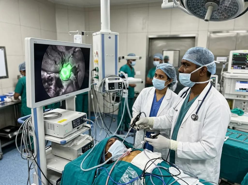
Near-infrared autofluorescence provides real-time label-free optical guidance during endoscopic transsphenoidal pituitary surgery. By highlighting stark signal contrasts between normal pituitary gland and adenomas, this technique aids gross-total tumor resection while safeguarding critical endocrine function.
Today
