.webp&w=384&q=75)

Loading, please wait...
Loading, please wait...
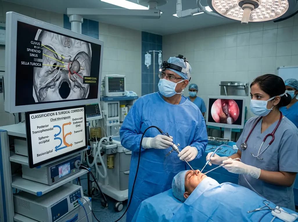
Congenital or primary clival meningoceles represent an exceptionally rare subset of skull base dysraphisms. These lesions occur due to disrupted embryonic fusion between the sphenoid and occipital bones. During normal cranial development, complex chondrification centers join to form the clivus. However, incomplete fusion along the spheno-occipital synchondrosis creates focal bony defects. Consequently, intracranial meninges herniate into the nasopharyngeal or prepontine space, creating a fluid-filled cystic mass. Effective clival meningocele surgical management relies on understanding these precise developmental origins and anatomical boundaries.
Unlike secondary skull base defects caused by trauma or elevated intracranial pressure, primary clival meningoceles stem strictly from congenital dysgenesis. Patients often present during late childhood or early adulthood with insidious symptoms. Clinical features frequently include persistent cerebrospinal fluid rhinorrhea, chronic headaches, or unexplained bacterial meningitis. Furthermore, large lesions can exert direct mass effect on the brainstem or cranial nerves. Therefore, early detection through high-resolution computed tomography and magnetic resonance imaging remains vital. Precise neuroimaging clearly depicts the bony defect and surrounding vascular structures, paving the way for targeted surgical planning.
Standardized classifications historically lacked specific subcategory definitions for rare transclival meningoceles. To address this clinical gap, investigators synthesized systematic review data with detailed skull base anatomical dissections. Consequently, a preliminary anatomical classification framework divides clival defects into three distinct subtypes. These categories comprise posterior transsphenoidal defects, spheno-occipital defects, and basioccipital defects. This tripartite system directly mirrors the embryonic origin of the lesion while providing clear guidance for surgical corridor selection.
Posterior transsphenoidal defects involve the upper portion of the clivus near the sphenoid body. Conversely, basioccipital defects affect the inferior third of the clivus near the foramen magnum. Spheno-occipital defects occupy the transitional middle region along the synchondrosis. In systematic analysis, posterior transsphenoidal cases represented the majority of documented presentations, followed by basioccipital defects. Spheno-occipital presentations occurred least frequently. Furthermore, this classification scheme allows skull base surgeons to accurately predict exposure limitations, ensuring surgical teams select the most direct trajectory with minimal soft tissue disruption.
Selecting the optimal surgical corridor is paramount in clival meningocele surgical management to ensure durable dural closure. Anatomical dissection studies confirm that the endoscopic endonasal approach offers unmatched visualization for upper and middle clival regions. Specifically, this corridor provides a direct line-of-sight trajectory to posterior transsphenoidal and superior spheno-occipital defects. Surgeons navigate through the nasal cavity and ethmoids, performing a sphenoidotomy to fully expose the sellar and clival dura. Consequently, surgical teams avoid extensive external incisions or facial bone disruption.
However, reaching the lower third of the clivus via a pure endonasal route presents technical challenges. The hard palate acts as an anatomical barrier restricting downward instrument maneuverability. As a result, attempting to reach basioccipital defects through the nose requires aggressive bony drilling and extensive mucosal resection. Moreover, steep working angles increase the risk of vascular injury to paraclival internal carotid arteries. Therefore, skull base specialists reserve the endoscopic endonasal corridor primarily for upper clival pathology, where high rates of defect obliteration are routinely achieved.
For defects located in the lower third of the clivus, the transoral endoscopic approach serves as the preferred surgical corridor. Basioccipital defects lie close to the craniocervical junction and anterior arch of the atlas. Therefore, entering through the oral cavity provides a perpendicular trajectory to the lower clivus. Endoscopic equipment inserted through the mouth offers magnified, wide-angle visualization without requiring soft palate splitting in most patients. Retracting the uvula superiorly reveals the posterior pharyngeal wall, allowing direct access to the basiocciput.
Recent surgical advancements highlight the feasibility of primary clival meningocele repair using a pure transoral endoscopic technique. This corridor bypasses the hard palate restriction inherent to endonasal routes. Consequently, surgeons achieve optimal instrument articulation when dissecting the meningocele sac and preparing bony margins for reconstruction. Furthermore, transoral endoscopic repair reduces the risk of postoperative velopharyngeal insufficiency compared to open transpalatal operations. Additionally, soft tissue healing along the posterior pharyngeal wall proceeds reliably, making the transoral route an effective strategy for inferior clival defects.
Synthesized data from systematic literature reviews provide key insights into the demographic and clinical profiles of primary clival meningocele patients. Analysis demonstrates a clear female predominance, with female patients accounting for over 70 percent of identified cases. The mean age at clinical diagnosis is approximately 30.7 years. This delayed presentation underscores the slow progression of congenital clival defects, which may remain asymptomatic until adult life or until trigger factors elevate intracranial pressure.
Common presenting complaints include clear nasal drainage, chronic headaches, and recurrent episodes of bacterial meningitis. In addition, some patients experience cranial nerve palsies due to localized traction or brainstem compression. Systematic review findings reveal that endoscopic endonasal approaches achieved excellent repair outcomes for upper clival cases. Similarly, transoral endoscopic repair successfully obliterated lower clival lesions without major neurological complications. Overall, systematic evidence confirms that choosing a targeted endoscopic corridor based on anatomical classification leads to high surgical success rates and low complication profiles.
Achieving a watertight seal is the cornerstone of successful skull base reconstruction in clival meningocele surgical management. Because clival defects lie adjacent to high-flow prepontine cisterns, cerebrospinal fluid leaks pose a major postoperative risk. Surgical techniques focus on meticulous sac reduction or resection, followed by multi-layered defect repair. Surgeons routinely employ inlay dural substitutes combined with autologous fascia lata, fat grafts, or rigid bone flaps. Furthermore, vascularized pedicled flaps provide robust tissue coverage that accelerates healing and prevents recurrent fistulas.
In endonasal repairs, the nasoseptal flap based on the posterior nasoseptal artery serves as the gold standard for defect coverage. Conversely, for transoral repairs, posterior pharyngeal mucosal flaps provide durable vascularized tissue layers. Additionally, precise intraoperative neuronavigation and high-definition endoscopes allow safe dissection around vital structures, including the basilar artery and lower cranial nerves. Consequently, comprehensive reconstruction protocols combined with rigorous postoperative care minimize complications such as meningitis, flap breakdown, or persistent CSF rhinorrhea.
A primary clival meningocele is a rare congenital condition caused by incomplete fusion of the embryonic sphenoid and occipital bones during skull base development. This developmental defect creates a bony opening along the clivus, allowing intracranial meninges to herniate into the nasopharynx or prepontine space over time.
Surgeons determine the optimal approach by classifying the defect's anatomical location. The endoscopic endonasal approach is preferred for upper clival (posterior transsphenoidal) lesions because it offers direct line-of-sight access. Conversely, the transoral endoscopic corridor is ideal for lower clival (basioccipital) defects, bypassing hard palate obstruction.
Patients with a primary clival meningocele often present with persistent cerebrospinal fluid rhinorrhea, recurrent unexplained bacterial meningitis, and chronic positional headaches. Larger lesions may also exert mass effect on the brainstem or adjacent cranial nerves, causing localized neurological deficits, facial numbness, or swallowing difficulties in severe cases.
Disclaimer: This content is for informational and educational purposes only and does not constitute medical advice. Refer to the latest local and national guidelines for clinical practice.
References

Read summarized clinical updates, watch expert medical content, and earn CME certifications right from your smartphone.
Open your smartphone camera to scan and install.





A systematic review and anatomical study introduce a new classification for primary clival meningoceles, mapping posterior transsphenoidal, spheno-occipital, and basioccipital defects to endoscopic endonasal and transoral surgical corridors for optimal reconstruction.
last month

Groundbreaking research reveals that naive CD8 T cells are not functionally uniform. Over time, prolonged homeostatic tonic TCR signaling primes long-lived naive cells into superior effectors with enhanced survival and memory potential, providing new opportunities for immunotherapy and vaccine design.
Today

Explore the crucial link between dietary protein and immunocompetence. This review examines how low-protein nutrition shifts metabolic resources away from pathogen defense toward early reproduction, compromising host survival and immunity against severe bacterial infections.
Today
Under India's PLI initiative, domestic production of 57 critical medical devices has commenced, including MRI and CT scanners, cath labs, and heart valves. Global partnerships and local investments are expanding high-end technology, curbing import reliance, and strengthening clinical accessibility nationwide.
Today

A longitudinal study reveals that patients undergoing staged bilateral hip arthroscopy experience progressive contralateral labral deterioration during surgical intervals. However, structural degradation does not compromise 2-year postoperative clinical outcomes or patient-reported satisfaction scores.
Today

New immunological research reveals how PTPN22 forms a delayed inhibitory signaling complex to restrain T cell activation, explaining how the autoimmune-linked R620W variant disrupts feedback control and promotes persistent inflammation in conditions like rheumatoid arthritis and type 1 diabetes.
Today
