.webp&w=384&q=75)

Loading, please wait...
Loading, please wait...

Cerebral venous thrombosis is an uncommon yet severe cerebrovascular disorder that accounts for a notable fraction of non-arterial strokes. Although it represents less than one percent of overall stroke presentations, the condition primarily affects young adults and women of childbearing age. Furthermore, early diagnosis remains notoriously difficult because symptoms frequently mimic common primary headaches or systemic disorders. Consequently, clinicians must maintain high diagnostic vigilance to ensure prompt therapeutic intervention and prevent irreversible tissue damage.
Clinicians encounter diverse clinical syndromes when assessing suspected cerebral venous thrombosis. Headache represents the most frequent presenting symptom, occurring in nearly ninety percent of confirmed patients. Typically, this headache develops progressively, though acute thunderclap presentations also occur. In addition, elevated intracranial pressure frequently produces papilledema, transient visual obscurations, and lateral rectus palsy. Focal deficits like hemiparesis and aphasia emerge when venous congestion precipitates localized tissue injury. Moreover, seizures occur in over one-third of acute presentations, manifesting as focal motor events with or without secondary generalization. Progressive encephalopathy indicates severe intracranial hypertension or deep venous involvement. Therefore, physicians must maintain heightened suspicion when young adults present with uncharacteristic neurological complaints. Specifically, medical teams should evaluate women using hormonal contraceptives or those in the puerperium. Systemic prothrombotic conditions, inherited thrombophilias, autoimmune disorders, and localized cranial infections also contribute substantially to disease pathogenesis. Rapid recognition enables timely emergency stabilization and prevents irreversible neurological harm.
Accurate neuroimaging remains vital for prompt confirmation in cerebral venous thrombosis. Although non-contrast computed tomography represents the standard initial emergency study, it lacks sufficient sensitivity to exclude dural sinus occlusion. Clinicians detect the classic dense triangle or cord sign in only a minority of presentations. Consequently, emergency protocols require dedicated vascular imaging whenever suspicion persists. Magnetic resonance imaging paired with magnetic resonance venography provides the definitive noninvasive standard. Specifically, T2-weighted and susceptibility-weighted sequences reliably visualize intraluminal thrombi and secondary parenchymal changes. Furthermore, contrast-enhanced computed tomographic venography provides an excellent rapid alternative when magnetic resonance imaging is unavailable or contraindicated. Modern imaging modalities also delineate critical parenchymal phenotypes that guide clinical therapy. For example, neuroimaging distinguishes reversible vasogenic edema from irreversible cytotoxic infarction. In addition, imaging identifies venous hemorrhagic transformation, which results from venous congestion rather than arterial disruption. Characterizing these patterns allows clinicians to tailor monitoring strategies and anticipate neurological deterioration.
Immediate therapeutic anticoagulation represents the cornerstone therapy for cerebral venous thrombosis. Clinicians should initiate anticoagulation promptly upon diagnosis, even when imaging shows hemorrhagic transformation. Clinical evidence confirms that anticoagulation halts thrombus propagation, facilitates recanalization, and prevents pulmonary embolism without worsening bleeding. In acute settings, physicians typically choose weight-adjusted low-molecular-weight heparin over unfractionated heparin because of superior bioavailability and predictable pharmacokinetics. However, clinical teams utilize unfractionated heparin if patients face imminent surgical interventions requiring rapid anticoagulation reversal. Following clinical stabilization, clinicians transition patients to oral anticoagulant therapy. Historically, vitamin K antagonists such as warfarin served as standard oral treatment, requiring frequent international normalized ratio monitoring. Nevertheless, recent randomized trials and prospective registries show that direct oral anticoagulants deliver equivalent safety and therapeutic efficacy. Specifically, agents like dabigatran, rivaroxaban, and apixaban achieve similar recanalization rates while significantly lowering major hemorrhagic risks. Consequently, contemporary international guidelines endorse direct oral anticoagulants as preferred first-line options for non-pregnant patients lacking antiphospholipid syndrome.
Although anticoagulation facilitates recovery in most individuals, a subset of patients suffers rapid clinical decline. For these severely affected patients, teams evaluate advanced endovascular or surgical interventions. Historically, interventional radiologists utilized endovascular mechanical thrombectomy and local chemical thrombolysis to restore venous patency. However, randomized trial evidence demonstrates that routine endovascular therapy does not confer superior functional outcomes compared with medical management. Therefore, clinicians reserve endovascular procedures strictly for individuals exhibiting progressive deterioration despite optimal parenteral heparinization. In contrast, surgical decompressive craniectomy represents a life-saving intervention for malignant cerebral venous thrombosis. Venous infarction and massive hematomas frequently cause severe mass effect and impending brainstem herniation. Consequently, emergent surgical decompression relieves refractory intracranial hypertension, restores cerebral perfusion, and prevents fatal herniation syndromes. Observational registries show that early decompressive craniectomy achieves favorable functional survival in over seventy percent of operated patients. Thus, neurointensive care teams must maintain rigorous monitoring to identify surgical candidates before irreversible injury occurs.
Overall functional survival following cerebral venous thrombosis has improved dramatically, with most patients achieving functional independence. However, long-term recovery extends far beyond basic neurological scores. Many surviving patients experience persistent neurocognitive deficits, chronic headache syndromes, mood disturbances, and profound fatigue. These lingering symptoms frequently hinder occupational reintegration and reduce quality of life. Furthermore, secondary prevention requires meticulous identification and management of underlying thrombophilic risk factors. For patients with transient provoked triggers, such as oral contraceptive use or localized infection, clinicians typically recommend anticoagulation for three to six months. In contrast, individuals with unprovoked thrombosis, recurrent venous events, or high-risk thrombophilias require extended or indefinite anticoagulation. Despite major therapeutic progress, important gaps persist across global healthcare environments. In particular, limited access to specialized neuroimaging and modern anticoagulants challenges standard clinical care in low-resource settings. Moreover, future clinical trials must develop validated prognostic scores integrating circulating biomarkers, clinical signs, and neuroimaging parameters. Resolving these challenges will ensure equitable, individualized patient care worldwide.
Recent randomized clinical trials and prospective observational studies confirm that direct oral anticoagulants demonstrate comparable safety and efficacy to vitamin K antagonists. These agents achieve similar rates of venous sinus recanalization and recurrent thrombosis while significantly reducing major intracranial bleeding complications. Furthermore, direct oral anticoagulants eliminate routine coagulation monitoring, improving patient compliance. However, clinicians should avoid these drugs during pregnancy, lactation, or in patients diagnosed with active antiphospholipid syndrome.
Surgeons consider decompressive craniectomy when acute cerebral venous thrombosis causes massive venous infarction, severe lobar hemorrhage, and refractory intracranial hypertension. Furthermore, this emergency intervention is critical when space-occupying lesions trigger progressive neurological decline, uncal herniation, or imminent brainstem compression despite aggressive medical therapy. Robust multicenter registry data indicate that early surgical decompression significantly reduces overall mortality and fosters favorable functional recovery in over seventy percent of these malignant cases.
Although most patients achieve functional independence, many experience subtle but disabling cognitive impairments. Commonly reported long-term symptoms include executive dysfunction, processing speed delays, impaired working memory, and sustained concentration difficulties. In addition, chronic headache, sleep disturbances, and debilitating mental fatigue frequently persist for months or years after the acute episode. Consequently, comprehensive outpatient follow-up should incorporate cognitive screening, structured neuropsychological rehabilitation, and mental health resources to support complete societal and vocational reintegration.
Disclaimer: This content is for informational and educational purposes only... Refer to the latest local and national guidelines for clinical practice.
References

Read summarized clinical updates, watch expert medical content, and earn CME certifications right from your smartphone.
Open your smartphone camera to scan and install.





A comprehensive clinical review of cerebral venous thrombosis, detailing advanced neuroimaging diagnosis, evidence-based anticoagulation, endovascular intervention, surgical decompression, and long-term neurocognitive prognosis.
Today

With measles cases surpassing 3,400 across the United States, public health agencies face mounting scrutiny over mortality reporting. The US CDC and CSTE are now formulating a standardized surveillance case definition to address discrepancies between state-level reports and federal vital statistics registers.
Today

A cross-sectional study of 700 college students highlights significant contraceptive knowledge disparities between sexually active and inactive young adults. The findings demonstrate a critical need for proactive, comprehensive sexual health counseling before sexual debut.
Yesterday
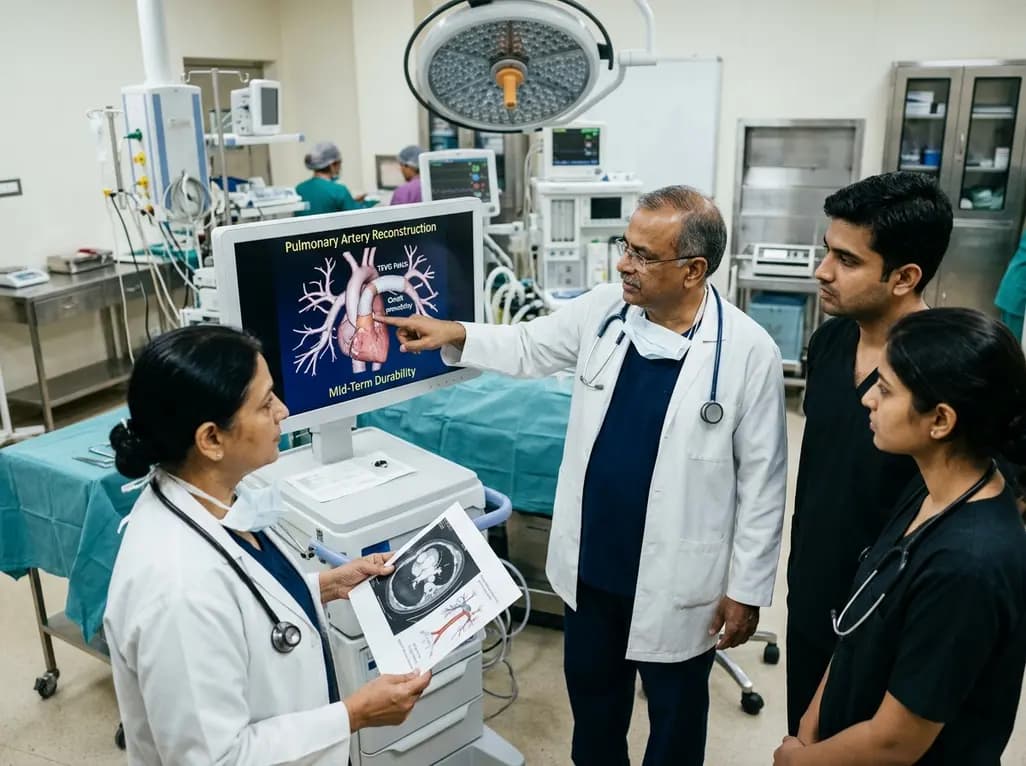
Autologous tissue-engineered vascular graft patches created via in-body tissue architecture show excellent mid-term durability and safety for pulmonary artery reconstruction in congenital heart surgery, particularly for peripheral branch augmentation.
Today

A novel cluster analysis of real-world datasets reveals four distinct CPPD disease phenotypes, validating existing EULAR categories while uncovering previously unrecognized monoarticular and axial presentations to refine patient management.
Today

Recent pharmacokinetic research demonstrates that trace dermal exposure to microgram quantities of trenbolone and metenolone produces detectable urinary metabolites for days to over a week, providing critical objective data for evaluating unintentional contamination claims in sports drug testing.
Today
