.webp&w=384&q=75)

Loading, please wait...
Loading, please wait...
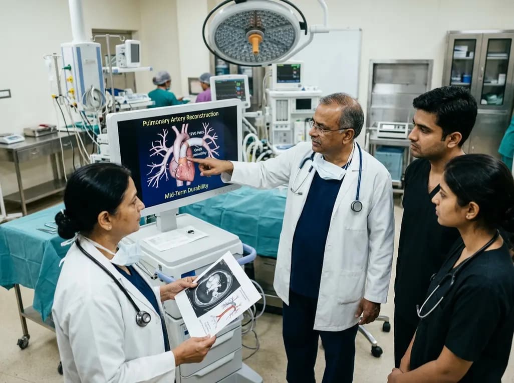
Congenital heart defects frequently require surgical reconstruction of hypoplastic or stenotic pulmonary vessels. Specifically, pulmonary artery reconstruction restores balanced lung perfusion and preserves right ventricular function. Autologous pericardium serves as the traditional patch material for these complex repairs. However, pediatric surgeons frequently exhaust native tissue during prior palliative operations. Synthetic substitutes lack growth capacity and provoke chronic inflammation, thrombosis, and calcification. Consequently, clinicians urgently need viable autologous alternatives. Emerging evidence suggests tissue-engineered vascular graft patches created through in-body tissue architecture provide remarkable durability, presenting an effective surgical option.
Congenital cardiovascular anomalies often exhibit hypoplastic branch vessels and diffuse arborization stenosis. Pathologies such as pulmonary atresia with ventricular septal defect, coarctation, and tetralogy of Fallot require meticulous surgical enlargement. Surgeons routinely perform patch angioplasty to expand narrow segments and decrease right ventricular afterload. Nonetheless, choosing an ideal patch material remains difficult.
Glutaraldehyde-treated bovine pericardium and synthetic polymers like polytetrafluoroethylene remain common clinical substitutes. Unfortunately, these biomaterials lack viable cellular elements and cannot remodel dynamically. Consequently, foreign graft materials trigger chronic immunological reactions, progressive dystrophic calcification, and pseudointimal peel formation. Furthermore, prosthetic patches cannot accommodate somatic growth in young children. As a result, patients face recurrent vessel stenosis, necessitating frequent catheter reinterventions or high-risk reoperations. These secondary procedures compound technical difficulty and elevate surgical morbidity. Therefore, pediatric cardiac surgeons require regenerative, non-immunogenic patch materials that integrate into native vascular walls without excessive neointimal proliferation.
To circumvent cumbersome laboratory cell culture, researchers developed in-body tissue architecture as an autologous biomanufacturing platform. This innovative strategy harnesses the patient's intrinsic foreign body reaction to engineer living collagenous conduits. During an initial staged surgery, clinicians embed silicone or polymeric templates subcutaneously in the patient.
Over four to twelve weeks, native fibroblasts and mesenchymal stem cells migrate into the template matrix. These host cells synthesize dense, highly organized type I collagen and extracellular matrix around the mold. Consequently, the patient's own body functions as an ideal physiological bioreactor, avoiding exogenous scaffolds or foreign cellular lines. During subsequent planned cardiovascular repair, surgeons retrieve the mold and extract a compliant tubular collagen tissue, known as a Biotube. Surgeons then divide this living structure longitudinally to create customized tissue-engineered vascular graft patches. Because the tissue derives exclusively from host cells, it exhibits superb biocompatibility, exceptional mechanical compliance, and strong resistance to bacterial contamination.
A rigorous clinical trial evaluated the mid-term safety and durability of these tissue-engineered vascular graft patches. Between July 2014 and December 2024, surgeons implanted subcutaneous molds in nineteen pediatric candidates. Subsequently, seven patients underwent complex vascular augmentation utilizing fifteen autologous patches. The operative cohort presented with challenging diagnoses, including pulmonary atresia with major aortopulmonary collaterals, aortic coarctation, double outlet right ventricle, and tetralogy of Fallot.
Following surgical reconstruction, investigators monitored the cohort over a median follow-up of 2.8 years, extending up to 7.9 years. Remarkably, no operative mortality or late cardiac-related deaths occurred. In addition, serial post-operative imaging confirmed complete absence of patch-related aneurysms, wall thinning, graft dehiscence, or infectious endocarditis. Importantly, none of the implanted patches exhibited dystrophic calcification, a major flaw of glutaraldehyde-preserved bovine matrices. Freedom from patch-related reintervention reached 85.7% at one year and remained steady at 71.4% at five years. These durable results underscore the clinical safety of autologous in-body tissue engineering.
Although the study established overall durability, anatomical location significantly influenced patch longevity. Three out of fifteen patches developed recurrent stenosis, representing a twenty percent patch restenosis rate. Notably, all stenotic events developed exclusively in the central pulmonary artery near the right ventricle-to-pulmonary artery conduit anastomosis. Local hemodynamic turbulence, high shear stress, and geometric compliance mismatch at the conduit interface likely triggered focal intimal hyperplasia.
Fortunately, interventional cardiologists successfully managed two patients using transcatheter balloon angioplasty, avoiding surgical re-exploration. In contrast, all twelve patches implanted across peripheral pulmonary artery branches retained excellent patency throughout surveillance. Peripheral patches showed no signs of luminal obstruction, structural distortion, or excessive scarring. Peripheral branches experience laminar hemodynamics and lower shearing forces, which promote harmonious endothelialization. Therefore, these autologous patches demonstrate superior efficacy for peripheral vascular augmentation. However, when reconstructing central junctions, surgeons must carefully optimize anastomotic geometry to minimize focal turbulence and prevent late narrowing.
These clinical findings carry profound implications for pediatric cardiovascular centers worldwide. In multi-stage congenital heart operations, surgeons repeatedly encounter pericardial scarcity due to dense adhesions and prior harvests. Commercial xenografts and synthetic polymers provide immediate solutions, but their high expense and complications place substantial burdens on healthcare systems. In-body tissue architecture offers an elegant, cost-effective alternative by utilizing the host's natural regenerative machinery.
Furthermore, histological examinations reveal progressive recellularization of implanted matrices by host endothelial and smooth muscle cells. This biological remodeling fosters vascular elasticity and accommodates somatic growth, which static prosthetic patches cannot deliver. Nevertheless, widespread clinical adoption requires standardized mold designs and multicenter trials with extended follow-up beyond ten years. Further biomechanical refinements may reduce compliance mismatch at high-pressure central anastomoses, mitigating restenosis risk. Ultimately, in-body tissue engineering bridges the gap between laboratory regenerative medicine and routine surgical practice, offering children with complex congenital anomalies a reliable, living autologous reconstructive solution.
In-body tissue-engineered patches provide complete biocompatibility because the host organism synthesizes the collagenous matrix entirely from autologous cells. Consequently, these grafts eliminate foreign body immune reactions, resist systemic infections, and do not trigger dystrophic calcification. Furthermore, the living autologous tissue demonstrates progressive endothelialization and cellular remodeling, which allows the augmented vessel to maintain functional compliance and potentially grow with the pediatric patient over time.
Restenosis predominantly develops at the central reconstruction site due to intense mechanical forces, high turbulence, and altered hemodynamics near the conduit anastomosis. The mechanical mismatch between a rigid prosthetic conduit and pliable patch material concentrates wall stress, triggering localized myofibroblast proliferation and intimal hyperplasia. In contrast, peripheral pulmonary branches experience uniform laminar flow and lower shearing stresses, which foster smooth vascular integration and prevent recurrent stenosis.
Conventional tissue engineering requires harvesting patient cells, expanding them in complex ex vivo cell-culture laboratories, and seeding them onto biodegradable synthetic scaffolds under strict regulatory protocols. Conversely, in-body tissue architecture utilizes a simple subcutaneous mold implanted directly into the patient. The body's natural wound healing response encapsulates the mold with autologous collagen and fibroblasts. This innovative approach substantially lowers production costs, avoids contamination risks, and eliminates complex regulatory logistics.
Disclaimer: This content is for informational and educational purposes only and should not be construed as medical advice. Clinical decisions must be tailored to individual patients based on expert medical evaluation. Refer to the latest local and national guidelines for clinical practice.
References

Read summarized clinical updates, watch expert medical content, and earn CME certifications right from your smartphone.
Open your smartphone camera to scan and install.





Autologous tissue-engineered vascular graft patches created via in-body tissue architecture show excellent mid-term durability and safety for pulmonary artery reconstruction in congenital heart surgery, particularly for peripheral branch augmentation.
Today
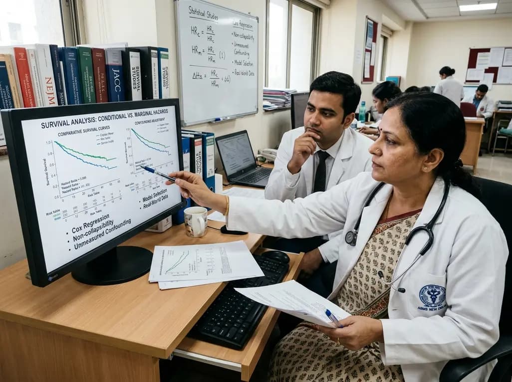
Survival analyses often report hazard ratios, but mathematical non-collapsibility creates crucial differences between conditional and marginal estimates. Clinicians must account for unmeasured confounding and model selection when interpreting trial endpoints and real-world cardiovascular outcomes.
Today
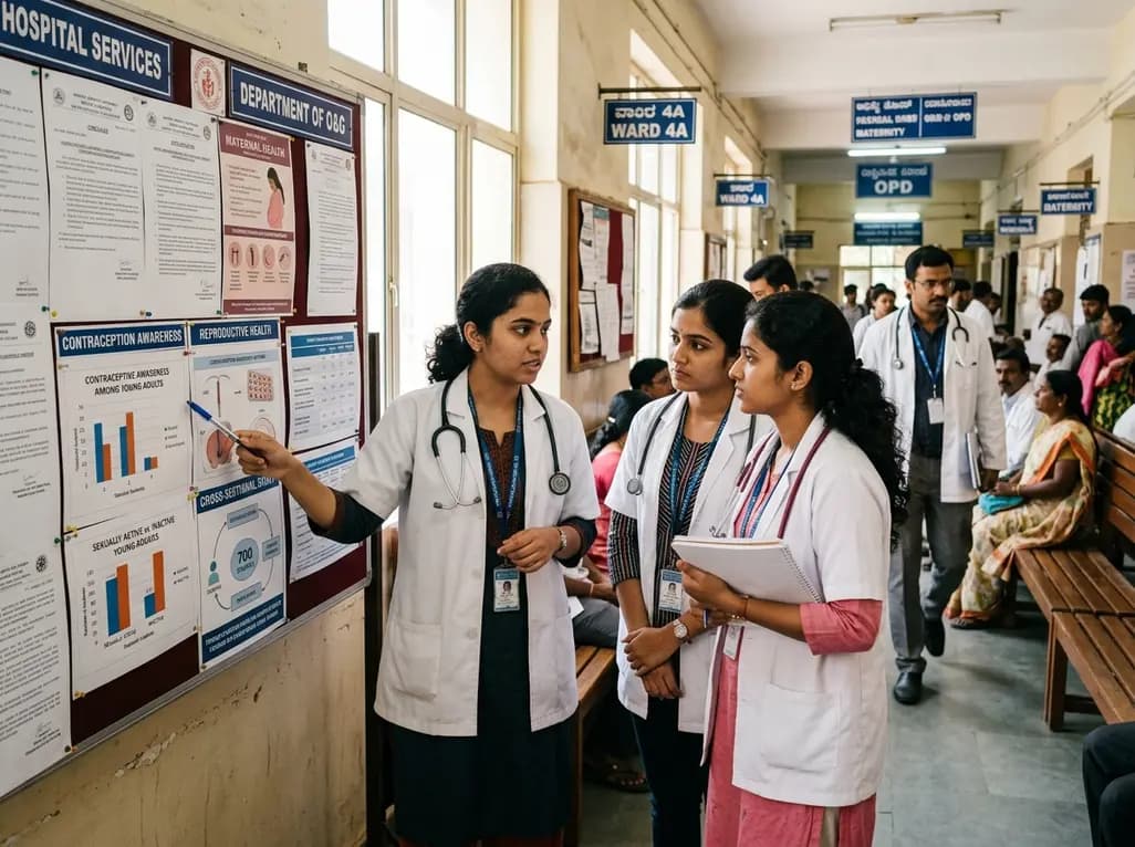
A cross-sectional study of 700 college students highlights significant contraceptive knowledge disparities between sexually active and inactive young adults. The findings demonstrate a critical need for proactive, comprehensive sexual health counseling before sexual debut.
Today

A novel cluster analysis of real-world datasets reveals four distinct CPPD disease phenotypes, validating existing EULAR categories while uncovering previously unrecognized monoarticular and axial presentations to refine patient management.
Today
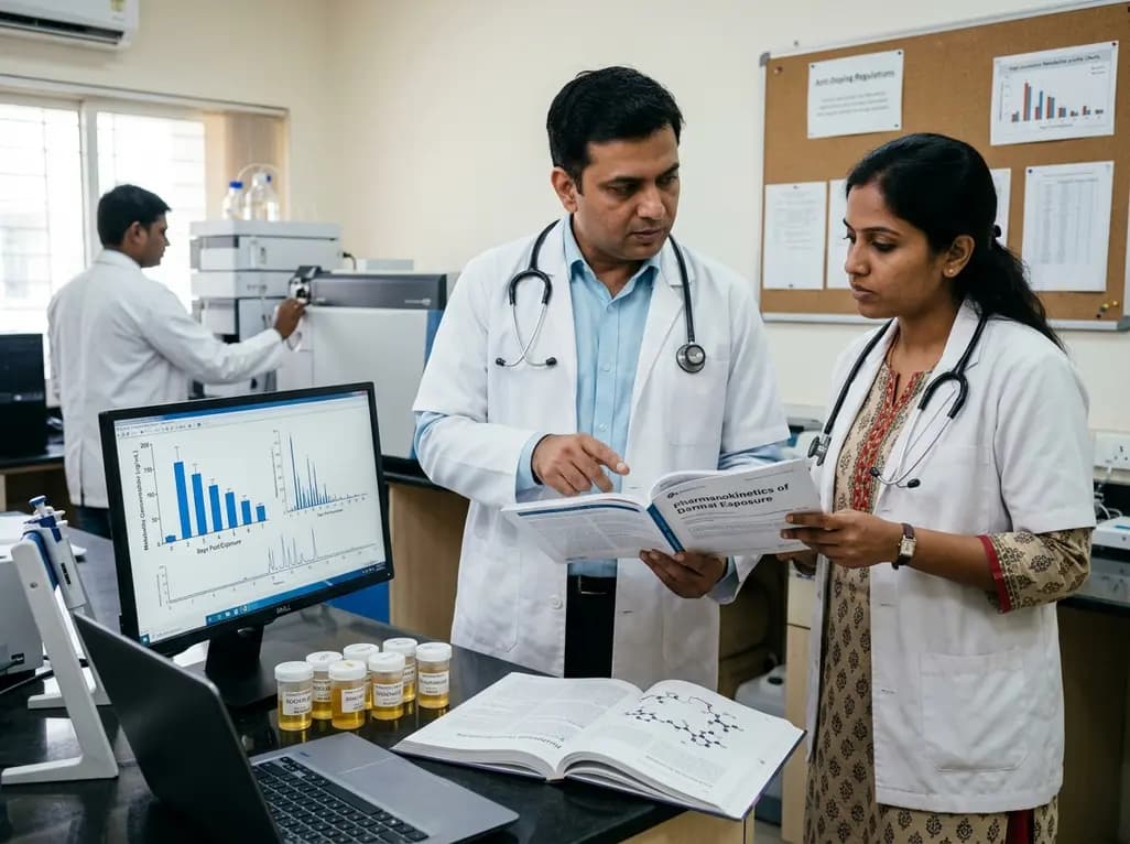
Recent pharmacokinetic research demonstrates that trace dermal exposure to microgram quantities of trenbolone and metenolone produces detectable urinary metabolites for days to over a week, providing critical objective data for evaluating unintentional contamination claims in sports drug testing.
Today
