.webp&w=384&q=75)

Loading, please wait...
Loading, please wait...

Timely identification of neuroinflammatory conditions remains a paramount priority in clinical neurology. The central vein sign has emerged as a groundbreaking imaging biomarker that transforms the diagnostic landscape for demyelinating disorders. Multiple sclerosis often presents with nonspecific neurological symptoms, which creates substantial diagnostic overlap with migraine, vascular leukoencephalopathy, and systemic autoantibody syndromes. Historically, confirming a diagnosis required prolonged observational intervals to establish dissemination in time and space, frequently supplemented by invasive spinal taps. However, the recent formal inclusion of this biomarker into the updated McDonald diagnostic criteria reflects a major paradigm shift toward earlier, biologically definitive disease detection. Clinicians can now leverage high-resolution neuroimaging to identify pathognomonic perivenular inflammatory architecture directly at initial presentation. Consequently, this imaging advance minimizes misdiagnosis risks and empowers care teams to initiate high-efficacy disease-modifying therapies before irreversible axonal damage accumulates.
Historically, establishing an accurate diagnosis of multiple sclerosis demanded a complex synthesis of clinical episodes, radiographic dissemination, and cerebrospinal fluid analysis. While oligoclonal band detection provided essential supportive evidence, lumbar punctures frequently introduced procedural delays, patient anxiety, and post-dural puncture headaches. Moreover, conventional magnetic resonance imaging sequences often struggle to distinguish demyelinating plaques from non-specific white matter hyperintensities common in older populations or patients with migraine. Fortunately, the central vein sign resolves much of this diagnostic ambiguity by targeting the distinct venocentric architecture of demyelinating plaques. Modern susceptibility-weighted imaging and T2-star-weighted fluid-attenuated inversion recovery sequences clearly show tiny venules coursing directly through the center of inflammatory demyelinating lesions. Therefore, incorporating this precise imaging biomarker into frontline neurological workups provides immediate objective confirmation of multiple sclerosis pathology. By prioritizing high-yield neuroimaging at presentation, clinical centers can dramatically streamline patient triaging while maintaining high diagnostic specificity.
Diagnostic latency in neuroimmunology carries profound clinical consequences, as delayed treatment initiation permits ongoing inflammatory attacks and subtle neuroaxonal loss. Standard outpatient referral pathways often subject individuals with suspected demyelinating illness to protracted waiting periods spanning several months between specialist consultations, repeated scans, and cerebrospinal fluid testing. In addition, scheduling invasive lumbar punctures generates operational bottlenecks that overburden day-case procedure units. In contrast, embedding advanced vascular-sensitive sequences directly into initial diagnostic MRI protocols eliminates these multi-step diagnostic hurdles. When radiologists readily verify central venules within suspect cerebral lesions, the diagnostic threshold is reached rapidly without demanding auxiliary invasive procedures. Consequently, patients avoid the physical discomfort and potential complications of spinal taps, such as persistent postural headaches and localized bleeding. Furthermore, eliminating redundant diagnostic steps preserves valuable healthcare resources and relieves clinical backlogs in high-volume neurology services.
A recent quality improvement initiative conducted at an academic multiple sclerosis center in Nottingham, United Kingdom, evaluated the real-world utility of a fast-track diagnostic pathway incorporating routine biomarker analysis. Investigators retrospectively reviewed 59 consecutive individuals referred with brain imaging suggestive of demyelination between March 2024 and July 2025. The clinical team compared diagnostic timelines and procedural utilization between patients managed through this novel protocol and those navigating standard outpatient workflows. Notably, patients enrolled in the fast-track pathway achieved a definitive diagnosis in a mean time of only 2.9 months, compared to 5.6 months in the conventional pathway. This represents a statistically significant mean reduction of 83 days in diagnostic latency. Crucially, zero patients diagnosed within the fast-track cohort required a lumbar puncture, whereas routine pathway patients frequently underwent invasive spinal taps. Additionally, the fast-track pathway yielded substantial economic advantages, reducing the mean cost per evaluated patient from £999 down to £707.
The biological rationale supporting this imaging biomarker lies in the fundamental perivenular anatomy of multiple sclerosis lesions. In demyelinating disease, autoreactive lymphocytes cross post-capillary venules, initiating an inflammatory cascade that drives focal perivascular myelin destruction. Consequently, active and chronic demyelinating plaques develop around a central parenchymal vein, creating a concentric or cylindrical inflammatory zone. In contrast, microvascular ischemic white matter lesions, migraine-related gliosis, and vasculitic injuries arise through arteriolar occlusion, hypoperfusion, or diffuse endothelial injury without central venous orientation. Radiologically, a lesion demonstrates this sign when a thin, hypointense linear structure or dot traverses the exact geometric center of a hyperintense lesion on susceptibility-based imaging. Histopathological correlation studies consistently demonstrate that over forty percent of cerebral plaques in true demyelinating disease harbor this perivenular structure. Conversely, nonspecific vascular lesions rarely exhibit central venous configurations, making this feature extraordinarily specific for inflammatory demyelination.
Widespread adoption of this vascular biomarker in routine clinical workflows requires standardized, accessible neuroimaging acquisition protocols. Dedicated three-dimensional T2-star FLAIR sequences, combining fluid suppression with magnetic susceptibility sensitivity, allow reliable visualization of microvascular anatomy on standard 3-Tesla and optimized 1.5-Tesla MRI systems. Although calculating whole-brain lesion percentages was traditionally considered labor-intensive, simplified scoring rules have drastically reduced radiological interpretation time. Validated frameworks, such as the Select-3 and Select-6 rules, permit radiologists to establish biomarker positivity by identifying as few as three or six distinct perivenular lesions across standard brain regions. Importantly, imaging specialists evaluate discrete, non-confluent lesions larger than three millimeters in diameter to ensure diagnostic precision. Adopting these rapid visual assessment metrics ensures seamless workflow integration for busy radiology departments. As artificial intelligence segmentation algorithms continue to mature, automated identification of perivenular lesions will further accelerate diagnostic reporting across diverse clinical settings.
The successful integration of biomarker-guided diagnostic pathways delivers transformative benefits for global healthcare delivery. Shortening the diagnostic interval from nearly half a year to under three months enables timely discussions regarding highly efficacious disease-modifying therapies. Early suppression of neuroinflammation prevents relapses, halts silent subclinical progression, and protects long-term cognitive and functional reserves. Furthermore, the economic benefits demonstrated in clinical audits highlight a compelling strategy for cost containment within resource-constrained medical systems. Avoiding invasive lumbar punctures significantly reduces disposable equipment usage, day-bed occupancy, and procedural complication management. In developing healthcare environments and emerging economies where access to specialized spinal fluid laboratories or procedural suites may be limited, high-field MRI assessment provides a robust, non-invasive alternative. Therefore, healthcare leaders and radiology administrators should actively modernize baseline neuroimaging protocols to ensure standardized, equitable, and rapid multiple sclerosis diagnosis worldwide.
The central vein sign serves as a highly specific imaging biomarker that confirms perivenular inflammatory demyelination. Its presence reliably differentiates multiple sclerosis plaques from non-specific white matter lesions caused by small vessel ischemia, migraine, or systemic vasculitis, enabling faster and more accurate diagnosis.
Yes, in many straightforward presentations, demonstrating this sign on susceptibility-weighted MRI fulfills modern diagnostic criteria without requiring cerebrospinal fluid analysis. However, clinicians may still perform lumbar punctures during atypical presentations, red flag symptoms, or when imaging remains inconclusive.
Detecting this biomarker requires high-resolution susceptibility-sensitive imaging sequences, particularly three-dimensional T2-star-weighted fluid-attenuated inversion recovery (FLAIR*) or susceptibility-weighted imaging (SWI). These advanced protocols are readily accessible on standard 3-Tesla and optimized 1.5-Tesla MRI platforms.
Disclaimer: This content is for informational and educational purposes only and does not constitute medical advice, diagnosis, or treatment recommendations. Clinical decisions should always be made by qualified healthcare professionals based on individual patient assessment and comprehensive clinical judgment. Refer to the latest local and national guidelines for clinical practice.
References
Gilmartin CGS et al. A Faster Way to Diagnose Multiple Sclerosis: The Central Vein Sign in Routine Clinical Practice. Neurol Clin Pract. 2026 Oct. doi: 10.1212/CPJ.0000000000200647. PMID: 42546228.
Montalban X, et al. Diagnosis of multiple sclerosis: 2024 revisions of the McDonald criteria. Lancet Neurol. 2025;24(10):850–865.
Sinnecker T, Clarke MA, Meier D, et al. Evaluation of the central vein sign as a diagnostic imaging biomarker in multiple sclerosis. JAMA Neurol. 2019;76(12):1446-1456.
Maggi P, Absinta M, Grammatico M, et al. Central vein sign differentiates multiple sclerosis from central nervous system inflammatory vasculopathies. Ann Neurol. 2018;83(2):283-294.

Read summarized clinical updates, watch expert medical content, and earn CME certifications right from your smartphone.
Open your smartphone camera to scan and install.





Integrating the central vein sign into routine clinical pathways significantly accelerates multiple sclerosis diagnosis, eliminates the need for invasive lumbar punctures, and lowers healthcare costs, according to real-world findings from a specialized UK quality improvement study.
last month

Groundbreaking research reveals that naive CD8 T cells are not functionally uniform. Over time, prolonged homeostatic tonic TCR signaling primes long-lived naive cells into superior effectors with enhanced survival and memory potential, providing new opportunities for immunotherapy and vaccine design.
Today

Explore the crucial link between dietary protein and immunocompetence. This review examines how low-protein nutrition shifts metabolic resources away from pathogen defense toward early reproduction, compromising host survival and immunity against severe bacterial infections.
Today
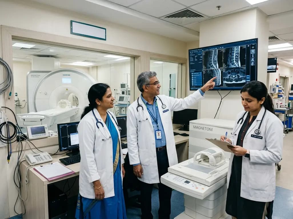
Under India's PLI initiative, domestic production of 57 critical medical devices has commenced, including MRI and CT scanners, cath labs, and heart valves. Global partnerships and local investments are expanding high-end technology, curbing import reliance, and strengthening clinical accessibility nationwide.
Today

A longitudinal study reveals that patients undergoing staged bilateral hip arthroscopy experience progressive contralateral labral deterioration during surgical intervals. However, structural degradation does not compromise 2-year postoperative clinical outcomes or patient-reported satisfaction scores.
Today

New immunological research reveals how PTPN22 forms a delayed inhibitory signaling complex to restrain T cell activation, explaining how the autoimmune-linked R620W variant disrupts feedback control and promotes persistent inflammation in conditions like rheumatoid arthritis and type 1 diabetes.
Today
