

Loading, please wait...
Loading, please wait...

Orthodontic clinicians routinely employ sequential distalization with aligners to address mild-to-moderate Class II malocclusions without permanent premolar extractions. In contemporary digital practice, doctors pair clear aligners with Class II intermaxillary elastics to preserve anterior anchorage during posterior tooth translation. However, the precise nature of molar movement has long remained controversial among dental professionals and clinical researchers. Conventional two-dimensional cephalometrics and plaster study casts obscure the true biomechanical expressions occurring at the crown and root levels. Recent advanced investigations combine cone-beam computed tomography with high-resolution intraoral surface scans. This state-of-the-art methodology finally illuminates the intricate three-dimensional mechanics underlying maxillary first molar displacement.
When orthodontists prescribe sequential distalization with aligners, digital treatment planning software typically projects true bodily translation. Clinicians program the maxillary second molar to move distally first, followed sequentially by the first molar, premolars, and anterior teeth. Nevertheless, clear thermoplastic appliances deliver orthodontic forces coronal to the center of resistance of the dentition. Consequently, aligner forces inevitably generate an uncontrolled tipping moment. In a recent rigorous investigation of thirty adult patients, researchers observed significant distal tipping rather than bodily movement. Specifically, the mesiobuccal cusp moved distally by an average of 1.75 mm, achieving 69.7% of planned displacement. In contrast, the mesiobuccal root apex moved only 0.35 mm, reflecting a modest 27.6% expression of the planned movement. Therefore, the clinical tooth movement manifested primarily as coronal tipping rather than bodily translation. Furthermore, the molar exhibited a mean distal tipping of 7.14 degrees, which significantly exceeded the initial digital projection. These findings highlight that thermoplastic materials alone cannot easily deliver counteractive moments necessary for root translation. As a result, clinicians must anticipate crown tipping and plan overcorrections accordingly.
Evaluating true orthodontic root movement requires imaging modalities that bypass the inherent artifacts of traditional radiography. Historically, clinicians relied on two-dimensional lateral cephalograms to evaluate sagittal dental changes. However, bilateral landmark superimposition and geometric magnification obscure unilateral dental displacement. Furthermore, digital dental casts capture only coronal anatomy, leaving root positions completely unseen beneath the gingiva. To solve these diagnostic dilemmas, investigators utilized advanced image registration software. They fused pre- and post-treatment CBCT images with surface intraoral scans. This technique captures sharp crown morphology from optical scans while accurately recording root boundaries from volumetric tomography. Subsequently, operators superimposed these fused models onto stable maxillary anatomical structures using CAD/CAM software. By establishing a rigid three-dimensional coordinate system, researchers quantified spatial changes across all three planes of space. Consequently, this multi-modal digital framework provides orthodontic specialists with clear objective data. The methodology demonstrates exactly how the maxillary first molar displaces within the alveolus. Moreover, this diagnostic approach highlights where digital software models fail to reflect biological reality. Thus, multimodal superimposition sets a new benchmark for orthodontic clinical research and biomechanical evaluation.
Orthodontic mechanics in the transverse and rotational planes profoundly impact the stability of Class II correction. During maxillary molar distalization, the aligner pushes against the mesiobuccal surface of the crown. Because this force acts buccal to the center of resistance, it frequently induces unwanted mesio-out rotation. In the examined patient cohort, maxillary first molars exhibited an average mesial-out rotation of 8.22 degrees. Interestingly, this rotational change was only slightly greater than planned in the digital setup. This observation indicates that aligners track rotational movements more predictably than pure root translation. However, rotational drift alters the effective arch perimeter and influences intercuspation with opposing mandibular teeth. Additionally, uncontrolled crown tipping commonly causes premature balancing contacts or posterior open bites. As the crown tips distally, the mesial cusps elevate while the distal marginal ridges press gingivally into occlusion. Therefore, practitioners must carefully evaluate transverse width changes, vertical alterations, and occlusal interferences throughout active treatment. When doctors overlook rotational tendencies, tracking errors accumulate rapidly across sequential aligner stages. Consequently, clinicians must incorporate targeted biomechanical compensations directly into the digital setup.
Sequential distalization requires robust anchorage preservation to prevent anterior teeth from flaring during posterior staging. To counteract reciprocal forces, clinicians routinely prescribe Class II intermaxillary elastics. These elastics run from mandibular molars to maxillary canines or premolars. They provide an essential mesial force vector that supports the anterior maxillary segment. However, elastics also introduce complex reciprocal forces that affect the lower dental arch. In clinical practice, elastics can inadvertently cause mandibular molar mesialization and incisor proclination. Moreover, Class II elastics exert an extrusive vertical force on maxillary canines and mandibular molars. While elastics effectively resist reciprocal incisor flaring, they do not prevent posterior molar tipping. In fact, elastics primarily reinforce anterior anchorage rather than delivering root torque to distalizing molars. Consequently, the posterior crown continues to tip distally along the path of least resistance. To overcome this limitation, some practitioners integrate temporary skeletal anchorage devices into the palate or alveolar bone. Skeletal anchors eliminate reliance on patient compliance and offer superior vector control. Therefore, selecting between intermaxillary elastics and skeletal anchorage demands meticulous diagnostic evaluation.
The discrepancy between planned and achieved movements carries crucial implications for clear aligner prescription writing. Because the mesiobuccal root apex achieved only 27.6% of planned translation, clinicians cannot expect full expression of digital setups. To mitigate crown tipping and rotational distortion, practitioners must intentionally engineer overcorrection into the digital staging. First, clinicians can prescribe exaggerated root movement, commonly termed virtual distal root tip. By programming compensatory mesial crown torque, the aligner applies continuous counter-tipping pressure throughout treatment. Second, utilizing optimized rectangular vertical attachments on the buccal surface improves mechanical grip. These attachments help the plastic deliver essential rotational and bodily control. Third, clinicians should avoid excessively fast distal staging rates. Staging movement at 0.15 to 0.2 mm per aligner instead of 0.25 mm reduces aligner deformation and material fatigue. Furthermore, clinicians must closely inspect tracking at every recall appointment. If clear aligners exhibit lag, initiating mid-course refinements prevents severe biological and mechanical derailments. Ultimately, combining intelligent digital overcorrection, strategic attachment design, and diligent clinical monitoring ensures predictable, healthy, and stable orthodontic outcomes.
Recent volumetric research reveals that clear aligners achieve approximately 70% of planned distal crown movement but less than 30% of planned root movement. Consequently, molar distalization occurs largely through distal tipping rather than true bodily translation. Orthodontists must anticipate this biomechanical limitation during treatment planning. By designing virtual overcorrections, applying optimized attachments, and utilizing auxiliary anchorage, practitioners can improve tracking and achieve predictable molar positions.
Maxillary molars tip distally because clear aligners apply mechanical forces coronal to the tooth's center of resistance. This distance generates an unavoidable tipping moment that tilts the crown backward while leaving the root apex behind. Although Class II elastics prevent anterior anchor loss, they cannot generate sufficient counter-moments at the posterior root. Therefore, clinicians must engineer deliberate distal root torque into the digital setup to resist tipping forces.
Merging cone-beam computed tomography with high-definition intraoral surface scans creates a comprehensive three-dimensional digital model. Intraoral scans provide high-resolution surface detail of clinical crowns, while CBCT accurately captures roots and alveolar bone structures. Superimposing these fused models onto stable anatomical reference structures eliminates the magnification errors of two-dimensional radiographs. This robust methodology allows researchers and clinicians to quantify tooth displacement accurately across all spatial planes.
Disclaimer: This content is for informational and educational purposes only and does not constitute medical advice. While we strive to provide accurate and up-to-date information, the content should not be used as a substitute for professional clinical judgment, diagnosis, or treatment. Medical knowledge and practices are constantly evolving. Always seek the advice of a qualified healthcare provider with any questions you may have regarding a medical condition or treatment. Refer to the latest local and national guidelines for clinical practice.
References

Read summarized clinical updates, watch expert medical content, and earn CME certifications right from your smartphone.
Open your smartphone camera to scan and install.





CBCT merged with intraoral scans demonstrates that sequential distalization with aligners produces primarily distal crown tipping and mesial-out rotation rather than bodily translation in Class II patients.
Today
Momelotinib offers unique clinical benefits for myelofibrosis patients with moderate-to-severe anemia via dual JAK and ACVR1 inhibition. While registration trials show positive hematologic outcomes, emerging real-world data underscore the need for vigilant surveillance of renal function and peripheral neuropathy.
Today

Delhi is undergoing a major healthcare overhaul with enhanced Ayushman Bharat insurance up to ₹10 lakh, increased MBBS and PG medical training seats, expanded primary network mandirs, new tertiary neuro-ICUs, and public-private reproductive technology initiatives to build a resilient, preventative healthcare model.
Today
A 78-year-old patient with high-grade aneurysmal subarachnoid hemorrhage complicated by refractory Takotsubo cardiogenic shock achieved full recovery following microaxial flow pump (Impella 5.5) rescue, demonstrating the safety and feasibility of advanced mechanical circulatory support during dual antiplatelet therapy.
Today
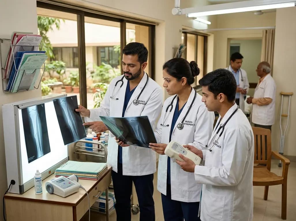
Non-union fractures occur in 2-5% of cases, causing significant disability and cost. Conservative modalities such as LIPUS, PEMF, and focused ESWT offer non-surgical healing options for defects up to 5-10 mm when adequate mechanical stability exists without infection.
Yesterday
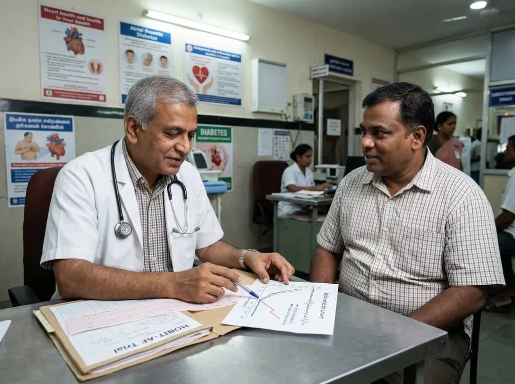
The randomized HOBIT-AF trial reveals that while structured weight loss after catheter ablation significantly improves cardiometabolic health in obese patients with paroxysmal atrial fibrillation, it does not reduce arrhythmia recurrence compared to standard care.
Today
