

Loading, please wait...
Loading, please wait...

Modern radiation oncology constantly seeks strategies to maximize tumor control while minimizing damage to healthy tissues. Consequently, carbon ion minibeam radiotherapy has emerged as an innovative paradigm that integrates spatially fractionated delivery with heavy ion physics. Specifically, carbon ions offer steep Bragg peaks and elevated linear energy transfer. Furthermore, when clinicians combine these physical qualities with submillimetric beam arrays, they achieve remarkable healthy tissue sparing. However, clinical implementation requires addressing complex dosimetric, biological, and motion-related hurdles. Therefore, this review examines the physics, advanced dynamic phantoms, radiobiology, and motion mitigation solutions essential for clinical translation.
Spatially fractionated radiotherapy distributes high radiation doses into narrow peaks while maintaining low doses in adjacent valleys. Therefore, intervening normal tissues tolerate higher overall peak exposures because uninjured stem cells migrate from valley regions to accelerate regeneration. Furthermore, carbon ions display distinct radiobiological properties compared to conventional megavoltage photons or protons. Heavy charged particles deliver elevated linear energy transfer, producing dense clusters of complex, unrepairable DNA double-strand breaks. Consequently, carbon ions yield a higher relative biological effectiveness, which effectively overcomes tumor hypoxia and radioresistance.
In addition, preclinical investigations show that heavy ion minibeams trigger robust systemic anti-tumor immune responses. The dense ionization patterns stimulate the cyclic GMP-AMP synthase pathway and promote tumor antigen cross-presentation. Meanwhile, the submillimetric beam geometry preserves surrounding capillary networks and local immunological microenvironments. For refractory malignancies like unresectable pancreatic adenocarcinoma, conventional therapies usually fail due to normal organ intolerance. Therefore, combining carbon ions with minibeam architecture provides a unique radiobiological advantage. Specifically, tumor cells receive lethal, clustered ionization while surrounding gastrointestinal mucosal barriers remain viable. Ultimately, this cooperative mechanism expands the therapeutic window for radioresistant tumors.
Accurate dose verification represents a critical prerequisite for clinical adoption. However, assessing spatially fractionated carbon ion fields presents extraordinary technical difficulties. Minibeam arrays generate microscopic peaks and valleys with submillimetric widths, creating steep dose gradients. In addition, the radiation field exhibits mixed linear energy transfer spectra across the target volume. As a result, standard ionization chambers suffer from severe volume-averaging artifacts because their sensitive cavities exceed beamlet dimensions.
To overcome these physical limitations, medical physicists utilize ultra-high-resolution detectors. Specifically, radiochromic films, micro-diamond detectors, and fluorescent nuclear track detectors demonstrate superior spatial fidelity. Furthermore, researchers validate experimental measurements using detailed Monte Carlo simulations. These computational frameworks accurately predict nuclear fragmentation, secondary particle generation, and track-structure variations. Clinicians require precise characterization of the peak-to-valley dose ratio across diverse tissue depths. Consequently, dosimetry must capture both physical absorbed dose and variable biological weighting. Recent developments in silicon strip microdosimetry and high-resolution optical tomography provide enhanced readout accuracy. Moreover, international scientific consortia actively collaborate to standardize these specialized dosimetric workflows. Nevertheless, establishing consensus calibration protocols remains essential before initiating multicenter clinical trials.
Translational radiation research depends heavily on realistic experimental validation tools. For example, standard homogeneous water tanks fail to capture the complex anatomical interfaces encountered in human patients. Therefore, biomedical engineers design multi-material anthropomorphic phantoms using high-precision three-dimensional printing technologies. These models accurately reproduce patient-specific tissue heterogeneities, including bony structures, adipose layers, and visceral soft tissues. In addition, researchers calibrate tissue-mimicking polymers to match clinical carbon ion stopping power ratios and Hounsfield units.
Furthermore, abdominal and thoracic tumors experience continuous physiological displacement. For instance, pancreatic lesions move considerably during spontaneous respiration and gastrointestinal peristalsis. Consequently, sophisticated dynamic phantoms integrate computer-controlled robotic actuators to simulate patient-specific respiratory waveforms. Investigators insert radiochromic films and miniature solid-state sensors directly into target volumes and adjacent simulated organs at risk. Subsequently, these dynamic testbeds allow teams to measure real-time spatial fractionated dose distributions during active motion. Recent evaluations confirm that motion-capable phantoms reliably identify dosimetric degradation caused by respiratory excursion. Similarly, these advanced tools facilitate rigorous benchtop testing of novel delivery nozzles. Thus, anthropomorphic dynamic phantoms provide an indispensable quality assurance platform for validating novel delivery schemes prior to patient treatment.
Breathing-induced anatomical movement poses a major obstacle for all spatially fractionated particle modalities. Because minibeam widths measure less than one millimeter, physiological displacements of five to fifteen millimeters can severely degrade spatial modulation. In particular, when target tissues shift during delivery, minibeam peaks bleed into neighboring valley regions. Consequently, the peak-to-valley dose ratio collapses, which diminishes the normal tissue-sparing effect in critical organs at risk. Simultaneously, relative motion can cause unintended tumor under-dosage or irregular dose hot spots.
Therefore, medical physicists develop multi-layered motion mitigation strategies to safeguard spatial fractionation. For instance, respiratory gating represents one practical solution, delivering beamlets only during stable end-expiration phases. In addition, real-time beam tracking offers continuous adjustment of scanned carbon ion spots to match tumor movement. Researchers also investigate abdominal compression devices and breath-hold techniques to restrict internal organ excursion. Furthermore, four-dimensional robust treatment planning incorporates dynamic anatomical deformations directly into dose optimization algorithms. As a result, clinicians can preserve the integrity of submillimetric minibeam peaks. Ultimately, these adaptive methodologies ensure robust dose distributions despite involuntary patient motion, preventing adverse normal tissue toxicity.
Transitioning this novel modality from experimental physics laboratories to routine hospital clinics demands systematic collaboration. First, manufacturers must engineer dedicated, high-efficiency collimation systems or magnetic focusing nozzles for existing carbon ion gantries. While passive multislit collimators provide sharp beamlet profiles, they generate secondary neutron contamination that requires careful shielding. Conversely, magnetically focused spot-scanning minibeams eliminate collimator scatter and optimize beamlet delivery times.
In addition, standardizing treatment planning algorithms remains a paramount priority. Specifically, clinical software must calculate non-linear cell survival, track-structure microdosimetry, and tissue recovery kinetics simultaneously. Regulatory agencies also expect rigorous pre-clinical safety validation in large animal models before approving early-phase human trials. Historically, recalcitrant malignancies such as locally advanced pancreatic carcinoma, recurrent glioblastoma, and chordomas show unsatisfactory outcomes with conventional radiotherapy. Therefore, oncologists view carbon ion minibeams as a potential breakthrough for these lethal conditions. By combining spatial tissue preservation with dense heavy-ion biological destruction, this approach offers new hope for difficult targets. Ultimately, continued multidisciplinary efforts will define standardized operating procedures, paving the way for definitive clinical investigations and broad therapeutic integration.
Conventional carbon ion therapy delivers a uniform dose across the targeted tumor volume to eradicate malignant cells. In contrast, carbon ion minibeam radiotherapy subdivides the radiation field into an array of submillimetric, high-dose beams separated by low-dose valleys. This unique spatial fractionation triggers robust normal tissue-sparing mechanisms. Healthy cells in valley regions migrate to repair localized micro-damage, while tumor cells within peak tracks experience dense, lethal double-strand DNA breaks.
Minibeam patterns rely on strict submillimetric spatial architecture to maintain distinct peak-to-valley dose gradients. When respiratory or peristaltic organ motion occurs during irradiation, tumor and normal tissue structures shift by several millimeters. Consequently, moving tissues blur the alternating dose profiles, causing high peak doses to spill over into protective valley zones. This degradation abolishes the normal tissue tolerance advantage and may cause unacceptable toxicity or tumor under-dosage without active mitigation strategies.
Radio-resistant and anatomically challenging malignancies located adjacent to sensitive, dose-limiting organs stand to gain the greatest clinical benefit. For instance, unresectable pancreatic adenocarcinoma represents a primary target because surrounding duodenal and bowel mucosa restrict standard curative radiation doses. Similarly, recurrent glioblastomas, skull base chordomas, and pediatric brain tumors could achieve superior local control. The technique enables clinicians to escalate tumor core doses safely without precipitating catastrophic normal tissue necrosis.
Disclaimer: This content is for informational and educational purposes only... Refer to the latest local and national guidelines for clinical practice.
References

Read summarized clinical updates, watch expert medical content, and earn CME certifications right from your smartphone.
Open your smartphone camera to scan and install.





Carbon ion minibeam radiotherapy combines high linear energy transfer and relative biological effectiveness with spatial fractionation. This review explores dosimetry, dynamic anthropomorphic phantoms, radiobiology, and organ motion mitigation strategies required for clinical translation in refractory tumors.
Today

A breakthrough study demonstrates that fullerol-primed extracellular vesicles generated in a 3D SECCS bioreactor attenuate ferroptosis and restore myogenic regeneration in diabetes via the FOXO3 pathway, offering a cell-free therapeutic strategy for diabetic musculoskeletal degeneration.
Today

A recent maternal mortality case in Kota has drawn national scrutiny toward operational deficits in emergency transport networks. With low ambulance utilization among high-risk pregnant women, delayed inter-facility transfers, and preventable clinical complications, urgent systemic reforms are vital for patient safety.
Today

A study reveals that the MYLK c.4079G>A, p.(Gly1360Asp) variant is linked to high-penetrance heritable thoracic aortic disease and early-onset aortic dissection, showing a founder effect in Finnish cohorts. Findings emphasize early cascade screening and vigilant surveillance.
Yesterday

A novel sacrificial-template technique combines FDM polymer 3D printing and micro-arc oxidation to fabricate orientation-controlled porous titanium scaffolds. These bioactive constructs offer tailored stiffness, exceptional multidirectional load resistance, and robust osseointegration for bone defect repair.
Today
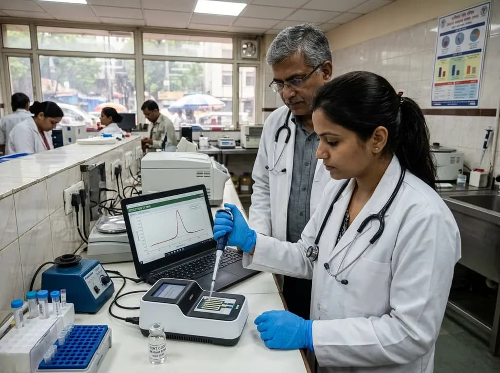
A novel label-free electrochemical biosensor utilizing magnetic nanocomposites enables ultrasensitive TERT C228T mutation detection (LOD: 0.67 pM) for rapid risk stratification in papillary thyroid cancer, offering promising point-of-care utility for endocrine oncology.
Today
