

Loading, please wait...
Loading, please wait...

Cerebrovascular specialists traditionally consider cerebral autosomal dominant arteriopathy with subcortical infarcts and leukoencephalopathy an ischemic disorder characterized by recurring lacunar strokes and progressive cognitive decline. However, recent clinical investigations reveal that CADASIL intracerebral hemorrhage represents a major hemorrhagic threat, particularly across Asian populations. Clinicians frequently encounter severe hemorrhagic complications in patients carrying specific genetic mutations alongside untreated cardiovascular risk factors. Understanding this dual etiology helps physicians optimize clinical surveillance and deliver targeted therapeutic interventions.
CADASIL results from cysteine-altering mutations in the NOTCH3 gene, which encodes a transmembrane receptor predominantly expressed in vascular smooth muscle cells. Typically, these genetic alterations trigger abnormal receptor aggregation, progressive vascular smooth muscle degeneration, and diffuse arteriopathy throughout the cerebral microcirculation. Consequently, the thickened, fibrotic vessel walls lose structural integrity and autoregulatory capacity.
While subcortical white matter ischemia remains the primary clinical manifestation in Western cohorts, hemorrhagic vessel rupture frequently occurs in East Asian demographics. In particular, the NOTCH3 R544C missense mutation impairs vascular elastance without causing classic extensive temporal pole leukoencephalopathy. Therefore, damaged penetrating arterioles become exceptionally vulnerable to mechanical shear stress.
Furthermore, persistent microvascular remodeling degrades pericyte support and extracellular matrix scaffolding. When exposed to fluctuating intravascular pressures, these fragile arterioles rupture rather than occlude. Consequently, clinicians must recognize that the underlying vascular biology differs fundamentally between conventional small vessel disease and genetically driven arteriopathies. Additionally, chronic basement membrane disruption promotes vessel wall permeability, allowing proteinaceous fluid and erythrocyte extravasation into surrounding brain parenchyma. This structural degradation accelerates microvascular fragility and predisposes vulnerable brain areas to catastrophic bleeds.
A multicenter investigation evaluated 552 patients with confirmed CADASIL from French and Taiwanese cohorts to define hemorrhagic risk factors. Notably, 34 individuals developed spontaneous intracranial hemorrhage, reflecting an overall prevalence of 6.2%. The clinical analysis demonstrated that patients with intracerebral bleeding were significantly older and presented distinct vascular risk profiles.
Specifically, multivariate logistic regression demonstrated that the NOTCH3 R544C variant independently increased hemorrhage risk nearly tenfold, with an odds ratio of 9.91. Concurrently, systemic hypertension conferred an independent odds ratio of 7.90. Although investigators observed no multiplicative mathematical interaction between these variables, the two factors demonstrated a powerful additive effect.
Consequently, individuals possessing neither risk factor exhibited a baseline hemorrhage risk of only 0.6%. In contrast, patients harboring solely the R544C variant demonstrated an 8.8% risk, while hypertensive patients lacking the mutation had a 5.4% risk. However, when both hypertension and the R544C mutation coexisted, the risk surged dramatically to 36.9%. Therefore, the convergence of chronic hemodynamic wall tension and genetic vascular fragility creates a devastating synergistic hazard for spontaneous intracranial bleeding.
Neuroimaging plays a pivotal role in detecting early microvascular damage before symptomatic intracranial bleeding manifests. In this binational study, investigators utilized the standardized CADA-MRIT inventory tool to evaluate brain magnetic resonance imaging scans. Consequently, several distinctive radiological hallmarks separated patients with hemorrhagic presentations from those with purely ischemic trajectories.
First, patients with a history of hemorrhage exhibited a significantly higher burden of cerebral microbleeds. These hemosiderin deposits clustered densely within deep brain structures, particularly the basal ganglia and thalamus. Additionally, affected individuals displayed marked brain atrophy and a higher count of dilated perivascular spaces within the basal ganglia.
Furthermore, extensive microbleed burdens signal profound vascular wall compromise and advanced endothelial breakdown. Because dilated perivascular spaces reflect impaired glymphatic clearance and severe arteriolosclerosis, their presence alerts clinicians to fragile deep penetrating vessels. Therefore, regular surveillance with susceptibility-weighted imaging offers indispensable prognostic data. Physicians should actively track these neuroimaging biomarkers to evaluate bleeding risk before deciding on aggressive antithrombotic regimens. Moreover, progressive lobar and deep white matter hyperintensities often parallel these structural disruptions, serving as clear indicators of escalating cerebrovascular vulnerability.
Managing stroke risk in patients carrying the NOTCH3 R544C variant demands a meticulous balance between ischemic prevention and hemorrhage mitigation. Traditionally, neurologists prescribe antiplatelet agents routinely for secondary stroke prevention in CADASIL patients experiencing transient ischemic attacks or lacunar infarcts. However, aggressive antithrombotic therapy in the presence of extensive cerebral microbleeds substantially magnifies the hazard of fatal intracranial hematomas.
Consequently, clinicians must individualize antiplatelet use, especially when patients exhibit multiple deep microbleeds or poorly controlled blood pressure. Furthermore, stringent blood pressure regulation represents the single most actionable modifiable target in mitigating CADASIL intracerebral hemorrhage. Because hypertension amplifies the baseline genetic risk by nearly sevenfold, aggressive blood pressure lowering shields weakened penetrating arteries from excessive hydrostatic shear stress.
Accordingly, medical teams should establish strict target systolic thresholds, often below 130 mmHg, using well-tolerated antihypertensive regimens. Clinicians should also discourage high-risk lifestyle habits, including smoking and excessive alcohol consumption, which further compromise cerebral vascular resistance. Additionally, close home blood pressure monitoring empowers patients to maintain sustained vascular stability. Thus, comprehensive risk-factor modification remains the cornerstone of clinical safety in this vulnerable population.
Effective management of hereditary cerebral small vessel diseases requires integrated genetic and clinical screening frameworks. Given the substantial prevalence of the NOTCH3 R544C variant among East Asian populations and diaspora communities, genetic testing provides vital risk-stratification data. Clinicians encountering atypical stroke presentations or strong family histories of early-onset strokes should maintain high diagnostic suspicion.
Furthermore, when genetic testing confirms an R544C mutation, physicians must immediately screen for coexisting cardiovascular risk factors. Multidisciplinary teams, including stroke neurologists, geneticists, and primary care physicians, should collaborate to construct customized monitoring schedules. Consequently, clinical protocols must incorporate baseline MRI scans with blood-sensitive sequences like susceptibility-weighted imaging to map parenchymal microvascular damage.
Moreover, clinicians should re-evaluate neuroimaging biomarkers periodically to detect rapid accumulation of microbleeds or progressive perivascular space dilation. If microbleeds multiply rapidly, specialists must reassess the safety profile of ongoing antithrombotic drugs. Simultaneously, proactive lifestyle education helps families implement dietary sodium restriction and regular aerobic activity. Therefore, implementing standardized clinical care pathways directly improves long-term neurological outcomes across diverse patient populations. Ultimately, early identification and personalized risk stratification safeguard patients against life-threatening hemorrhagic stroke complications.
The NOTCH3 R544C variant alters vascular smooth muscle cell function without causing the classic severe anterior temporal white matter lesions seen in typical CADASIL. Instead, this specific mutation severely weakens the structural integrity of deep penetrating cerebral arterioles, making them exceptionally vulnerable to mechanical wall stress and spontaneous rupture.
Clinicians should implement stringent blood pressure control, targeting systolic levels below 130 mmHg to protect fragile penetrating arterioles. Because hypertension combines additively with genetic mutations to escalate hemorrhage risk up to 36.9%, routine home monitoring and consistent long-term antihypertensive therapy are essential to prevent life-threatening intracranial bleeding.
Antithrombotic therapies require extreme caution in patients harboring the NOTCH3 R544C variant, especially those with coexisting hypertension. Because these individuals frequently develop extensive cerebral microbleeds, standard antiplatelet therapy may significantly increase intracranial hemorrhage risk. Clinicians should carefully weigh ischemic benefits against bleeding hazards using dedicated MRI screening.
Disclaimer: This content is for informational and educational purposes only... Refer to the latest local and national guidelines for clinical practice.
References

Read summarized clinical updates, watch expert medical content, and earn CME certifications right from your smartphone.
Open your smartphone camera to scan and install.





A binational cohort study reveals that the NOTCH3 R544C variant and hypertension independently and additively elevate intracerebral hemorrhage risk in CADASIL patients. Co-occurrence escalates bleeding risk to nearly 37%, underscoring the urgent need for stringent blood pressure control in genetically vulnerable cohorts.
Today

The DOMINO study protocol presents a multidisciplinary implementation mapping approach to overcome provider, patient, and payer barriers to antiobesity medications, offering vital insights for Indian clinicians managing the growing diabesity burden.
Today

An intracranial EWSR1::CREM fusion sarcoma in a 31-year-old female manifested as a paraneoplastic inflammatory syndrome with anemia and secondary infertility. The dural mass mimicked meningioma, but gross-total resection resolved her systemic inflammation and restored natural fertility within one month.
Today

A real-world cohort study of 6,537 patients from the FOUNTAIN platform demonstrates that initiating finerenone significantly reduces albuminuria by 40.7% with minimal eGFR decline and manageable hyperkalemia risk, confirming its practical effectiveness and safety in diabetic kidney disease.
Yesterday

A comprehensive analysis of MAP therapy resistance in osteosarcoma, examining canonical and non-canonical mechanisms across damage avoidance, enhanced DNA repair, and replication stress tolerance to identify novel vulnerabilities.
Today
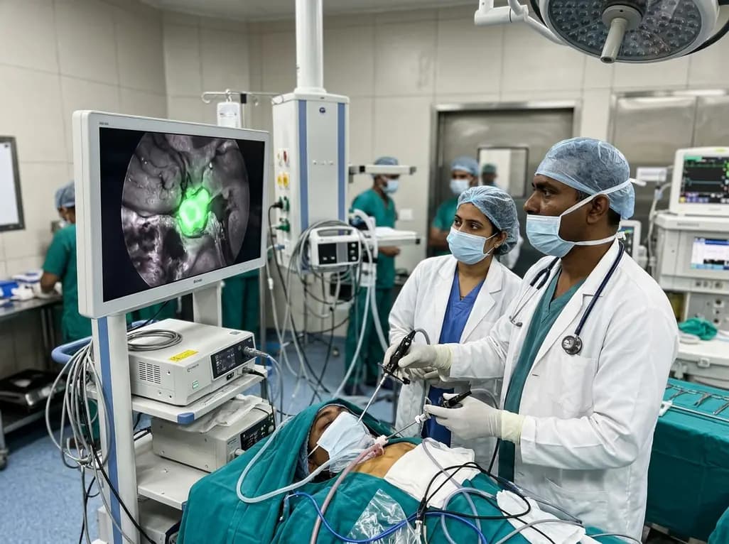
Near-infrared autofluorescence provides real-time label-free optical guidance during endoscopic transsphenoidal pituitary surgery. By highlighting stark signal contrasts between normal pituitary gland and adenomas, this technique aids gross-total tumor resection while safeguarding critical endocrine function.
Today
