.webp&w=384&q=75)

Loading, please wait...
Loading, please wait...

Transverse maxillary deficiency represents a common skeletal discrepancy that complicates malocclusion and compromises upper airway dimensions. Consequently, clinicians frequently utilize C-expander maxillary expansion to correct severe transverse arch collapsibility in adolescent and mature patients. Traditional rapid palatal expanders deliver excessive lateral loads directly to posterior teeth. Therefore, conventional tooth-borne appliances frequently induce undesirable dentoalveolar tipping, gingival recession, and buccal cortical fenestration. In contrast, bone-borne miniscrew-assisted rapid palatal expansion systems deliver orthopedic forces directly to the basal maxillary bone. This targeted approach effectively opens the midpalatal suture while mitigating adverse tooth movement. Moreover, modern digital diagnostic tools allow orthodontists to assess palatal bone morphology with remarkable precision. In this case report, a patient presented with typical skeletal characteristics of severe transverse maxillary deficiency. Specifically, diagnostic records revealed a significant maxillomandibular discrepancy of 6.8 mm. Because skeletal resistance increases as cranial sutures mature, the team selected a bone-anchored appliance. Thus, the clinical team designed a personalized C-expander to optimize orthopedic separation and minimize collateral dental side effects.
Precise anatomical evaluation remains essential before inserting palatal temporary anchorage devices. Consequently, the team merged cone-beam computed tomography datasets with optical intraoral scans using BlueSky Plan 4 software. This digital workflow allowed clinicians to visualize bicortical bone thickness and evaluate adjacent root structures thoroughly. Furthermore, virtual modeling enabled the accurate determination of miniscrew angulation, diameter, and insertion depth. Therefore, practitioners avoided delicate neurovascular bundles along the greater palatine canal and incisive foramen. Additionally, three-dimensional planning ensured that the miniscrews engaged dense cortical plates along the hard palate to provide maximum anchorage. Following virtual verification, the clinicians fabricated a customized surgical guide using additive manufacturing technology. This 3D-printed template transferred the exact digital plan directly to the surgical environment without spatial deviations. As a result, the guided protocol eliminated manual placement errors during chairside installation. Furthermore, the appliance fit securely against the palatal vaults without rocking or mucosal impingement. Ultimately, this integration of CBCT imaging and surgical guides streamlined the placement procedure, improving clinical safety and biomechanical predictability.
Following successful guide seating, the clinician placed the palatal miniscrews and installed the C-expander securely. The operator immediately confirmed stability and verified that the framework avoided excessive pressure on the palatal mucosa. Subsequently, the clinical team initiated a progressive activation protocol alongside weekly clinical monitoring visits. Weekly follow-ups allowed the orthodontist to evaluate tissue health, ensure suture split progression, and detect potential complications early. Furthermore, the controlled expansion schedule generated steady orthopedic stress across the circummaxillary sutures without provoking severe pain. Patients rarely tolerate sudden, aggressive expansion forces well; hence, gradual screw activation preserved patient comfort throughout the active phase. In addition, routine checks confirmed that the miniscrews maintained rigid skeletal anchorage without loosening or peri-implant inflammation. Because clinicians monitored the patient weekly, they could readily identify the clinical signs of suture opening, including an anterior diastema. Therefore, this vigilant monitoring protocol safeguarded periodontal structures while guaranteeing steady skeletal advancement. Once active expansion concluded, the appliance remained in place as a passive rigid retainer to consolidate newly formed bone.
Postexpansion cone-beam computed tomography demonstrated remarkable skeletal orthopedic improvement across the midpalatal suture. Specifically, follow-up axial tomographic views revealed a nearly parallel opening pattern measuring approximately 6.5 mm. Conventional tooth-borne expanders typically create a triangular, V-shaped opening with maximum separation anteriorly and minimal widening posteriorly. In contrast, the C-expander delivered uniform forces along the palate, achieving an efficient and parallel sutural separation. Consequently, this parallel split successfully resolved the initial 6.8 mm maxillomandibular transverse discrepancy. Additionally, the transverse gain provided adequate arch space to accommodate crowded maxillary dentition without extractions. Furthermore, coronal CT reconstructions confirmed robust lateral displacement of both maxillary shelves without significant horizontal rotation. Thus, the direct bone-anchored design bypassed the flexibility of the periodontal ligament, converting mechanical load into genuine skeletal expansion. Moreover, early tomographic verification confirmed adequate suture integrity without structural damage to adjacent bones. These quantitative outcomes confirm that digitally guided skeletal anchorage provides reliable transverse correction even in cases with severe transverse deficiency.
In addition to skeletal changes, the postexpansion analysis revealed favorable dentoalveolar and soft-tissue responses. Dentoalveolarly, the upper intermolar distance increased by 7.1 mm, while the lower intermolar distance increased spontaneously by 1 mm. Meanwhile, intermolar angulation increased by only 2° in the upper arch and 5° in the lower arch. Consequently, the appliance minimized buccal crown tipping, which represents a common complication of traditional expanders. Furthermore, the anterior occlusal relationships shifted favorably; the overjet increased by 1 mm and the overbite decreased by 15%. Beyond dental parameters, 3D frontal facial reconstructions revealed noteworthy soft-tissue midfacial adaptations. Specifically, the intercanthal distance increased minimally by 0.3 mm, indicating negligible negative impact on upper facial esthetics. Conversely, the nasal interalar distance increased by 3.5 mm, and the nasal base width widened by 4.4 mm. These nasal changes reflect skeletal widening of the piriform aperture, which often improves nasal airway patency. Therefore, the digitally guided C-expander successfully achieved true skeletal expansion with minimal dentoalveolar tipping and controlled facial changes.
Digital surgical guides translate three-dimensional cone-beam computed tomography plans into clinical reality with submillimeter precision. Consequently, clinicians accurately orient miniscrews into regions of greatest palatal bone thickness while avoiding dental roots, nerves, and major blood vessels. Furthermore, this guided workflow ensures optimal bicortical bone engagement, which maximizes mechanical stability. Therefore, digitally guided placement substantially reduces surgical complications, prevents mucosal damage, and eliminates the risk of premature implant failure during active orthopedic expansion.
Traditional expanders apply lateral loads directly to premolars and molars, typically generating a triangular opening due to posterior cranial resistance. In contrast, the C-expander anchors directly into dense palatal bone via four bicortical miniscrews. Because the appliance transfers orthopedic forces at the level of the basal maxilla, it overcomes posterior pterygomaxillary resistance more effectively. Consequently, the midpalatal suture separates in a nearly parallel fashion, providing uniform expansion from anterior to posterior segments.
Skeletal expansion of the midpalatal suture directly widens the nasal cavity floor and the piriform aperture. In this clinical protocol, the patient gained 3.5 mm in nasal interalar width and 4.4 mm at the nasal base. Consequently, this structural widening significantly lowers nasal airway resistance and improves airflow dynamics. Moreover, these orthopedic adaptations facilitate daytime and nighttime nasal breathing without causing adverse cosmetic distortion to the intercanthal or upper midfacial soft tissues.
Disclaimer: This content is for informational and educational purposes only and does not constitute medical advice, diagnosis, or treatment. It is not intended to replace the clinical judgment of qualified healthcare professionals. Always consult appropriate healthcare providers or clinical specialists for specific medical guidance. Refer to the latest local and national guidelines for clinical practice.
References
Miranda-Astocondor E et al. Maxillary Expansion With C-Expander Using Digital Planning and CBCT-Based Surgical Guidance: A Case Report. Case Rep Dent. 2026 undefined undefined. doi: 10.1155/crid/3316626. PMID: 42841089.
Cantarella D, Savio G, Grigolato L, et al. A new methodology for the digital planning of micro-implant-supported maxillary skeletal expansion. Med Devices (Evid Res). 2020;13:93-106. doi: 10.2147/MDER.S247751.
Minervino BL, Barriviera M, Curado MM, et al. MARPE Guide: A Case Report. J Contemp Dent Pract. 2019;20(9):1102-1107. doi: 10.5005/jp-journals-10024-2649.

Read summarized clinical updates, watch expert medical content, and earn CME certifications right from your smartphone.
Open your smartphone camera to scan and install.





A case report demonstrates successful C-expander maxillary expansion planned with CBCT and surgical guides. The protocol achieved 6.5 mm parallel midpalatal suture separation with minimal dental tipping and favorable nasal soft-tissue changes.
Today
A longitudinal study reveals that clonal hematopoiesis occurs in 12.5% of chronic idiopathic neutropenia patients, identifying key mutational patterns, variant allele frequency thresholds, and clonal dynamics that predict malignant transformation to myeloid neoplasms.
Today
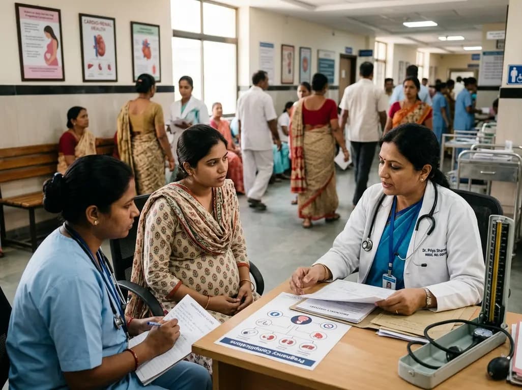
A large UK cohort study of 1.4 million women shows that pregnancy complications, including gestational diabetes, hypertension, and preterm birth, substantially raise long-term risks for hypertension, chronic kidney disease, type 2 diabetes, and cardiovascular events, highlighting early postpartum intervention needs.
Today

Giant cardiac hemangiomas are rare benign vascular tumors that can cause severe hemodynamic compromise and arrhythmias through mass effect. This comprehensive review covers clinical presentation, advanced multimodality imaging, histopathological diagnosis, surgical resection, and long-term surveillance protocols.
Today
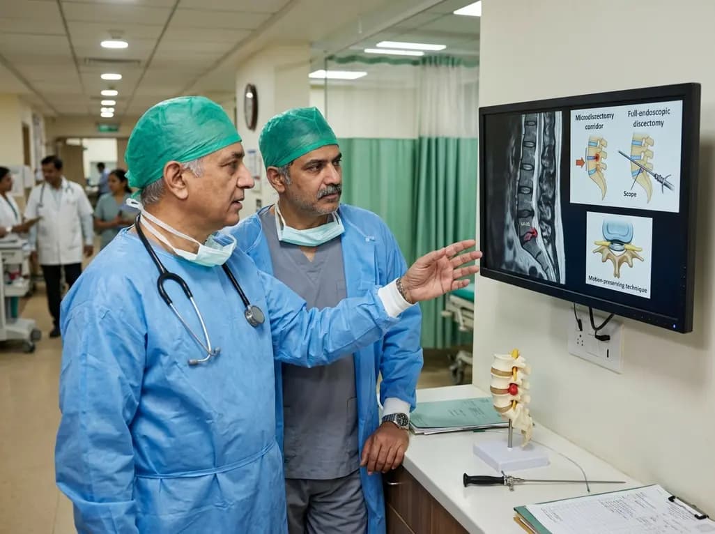
This clinical overview examines recent advancements in lumbar disc herniation surgery, comparing microscopic and full-endoscopic discectomy while highlighting biportal techniques and emerging motion-preserving innovations for spine specialists.
Today

The World Health Organization has issued its first global guidelines for managing child and adolescent obesity. The recommendations strongly endorse family-based dietary interventions, regular physical activity, and behavioral support as frontline therapy, reserving GLP-1 therapies and surgery for select cases.
Today
