

Loading, please wait...
Loading, please wait...

Traumatic disruption of neural pathways drastically alters how the human brain processes somatosensory inputs. Consequently, the dynamic neural representation of one's physical self, termed body schema, undergoes profound reorganization following trauma. Understanding body schema in SCI provides vital insights into why patients develop sensorimotor disconnects and neuropathic pain. When physical trauma severs peripheral connectivity, the brain struggles to maintain accurate internal models. Historically, research primarily emphasized gross motor and sensory loss following spinal cord damage. However, emerging cognitive neuroscience demonstrates that physical deafferentation actively reshapes higher-order sensorimotor representations. Patients do not simply lose peripheral transmission; instead, their central neural circuits reorganize dynamically. Furthermore, central cortical representations depend heavily on continuous reafferent feedback from intact extremities. When severe spinal lesions interrupt these pathways, cortical maps experience degradation and disorientation. Clinicians increasingly recognize that cortical reorganization directly correlates with complications, including phantom sensations and pain. Therefore, evaluating early electrophysiological markers allows clinicians to track cortical integrity in real time. Consequently, understanding these early neural signals uncovers essential targets for therapeutic intervention.
Event-related potentials provide an exceptional window into real-time millisecond-level cortical processing. Specifically, sensory and cognitive evoked potentials capture the immediate neural responses triggered during perceptual tasks. The posterior P100 wave reflects early visual processing and attentional allocation across occipitotemporal networks. In contrast, the anterior N100 represents early discriminative processes, sensory gating, and frontoparietal attention control. When individuals encounter complex body-related imagery, these early electrophysiological signals illustrate how rapidly the brain retrieves motor templates. In healthy individuals, both P100 and N100 exhibit robust modulation when resolving challenging spatial rotations. Nevertheless, individuals with central deafferentation show pronounced disruptions during these temporal windows. An amplitude reduction in P100 signals impaired early perceptual encoding within visual somatosensory association areas. Similarly, an attenuated anterior N100 indicates deficits in recruiting top-down executive resources required for mental body manipulation. Consequently, these electrophysiological alterations serve as objective biomarkers of altered cortical processing. Researchers utilize high-density electroencephalography to track these precise waveforms during cognitive motor testing. By measuring changes in amplitude and latency, clinicians can quantify cortical reorganization with remarkable neurophysiological precision. Thus, these electrophysiological recordings provide an objective window into silent neuroplastic changes.
To assess internal body models objectively, neuroscientists frequently deploy laterality judgment tasks. In these paradigms, participants view images of hands or feet rotated at varying angles. Participants must quickly decide whether the presented image represents a right or left limb. To accomplish this task, the brain implicitly rotates its own internal limb representation to match the visual stimulus. Therefore, this mental rotation process engages the frontoparietal motor network and somatomotor representations. Researchers introduced two distinct rotation angles to evaluate cognitive load: an easy angle of 75 degrees and a difficult angle of 150 degrees. Moreover, investigators contrasted body-related stimuli with non-body control objects, such as geometric shapes or letters. Healthy individuals effortlessly recruit motor execution networks to resolve biomechanically demanding angles like 150 degrees. In contrast, patients with severe spinal cord lesions experience substantial cognitive strain when rotating body-related stimuli. Notably, this difficulty does not occur when observing non-body stimuli. This distinct divergence proves that the deficit specifically involves corporeal representation rather than generalized visuospatial impairment. Consequently, laterality judgment tasks expose subtle disruptions in internal motor simulation that standard neurological examinations miss. Accordingly, this paradigm offers clinicians a sensitive instrument for tracking covert cognitive changes.
The anatomical level of spinal damage plays a decisive role in determining the extent of cortical disruption. Cervical injuries cause tetraparesis, impairing sensorimotor signaling across both upper and lower extremities. Conversely, thoracic injuries preserve full upper limb sensorimotor function while paralyzing the lower limbs. Electrophysiological studies demonstrate that individuals with cervical trauma exhibit significantly reduced posterior P100 and anterior N100 amplitudes compared to thoracic cohorts. Importantly, this electrophysiological suppression emerges selectively when individuals attempt to rotate body-related stimuli at challenging 150-degree angles. Because thoracic patients maintain intact sensorimotor feedback from their arms and hands, their brains preserve robust upper-body schemas. Therefore, thoracic patients recruit preserved cortical sensorimotor circuits to resolve complex mental rotations efficiently. In sharp contrast, individuals with cervical injuries experience profound sensorimotor deprivation across all four limbs. This extensive deafferentation compromises the cortical neural substrates responsible for executing implicit motor imagery. Furthermore, the presence of concurrent chronic neuropathic pain amplifies this cortical dysfunction, corrupting cognitive body schema maintenance. Thus, injury level fundamentally dictates the severity of cognitive body schema distortion.
These electrophysiological discoveries carry immediate implications for modern neuro-rehabilitation protocols. Traditional physical therapy predominantly concentrates on peripheral muscle strengthening, mobility aids, and joint range of motion. However, restoring functional independence requires targeting central neuroplasticity alongside peripheral mechanics. Because individuals with cervical injuries demonstrate specific deficits in mental rotation and early cortical processing, rehabilitation programs must incorporate cognitive-motor paradigms. For instance, graded motor imagery, left-right laterality discrimination training, and virtual reality mirror therapy can directly stimulate dormant somatosensory circuits. Clinicians can utilize structured mental practice to reinforce central body schema representations without requiring overt physical execution. Moreover, pairing non-invasive brain stimulation, such as transcranial direct current stimulation, with mental rotation exercises could restore P100 and N100 wave dynamics. Early intervention appears especially critical to prevent irreversible cortical degradation and attenuate refractory neuropathic pain syndromes. In addition, physical therapists can stratify rehabilitation intensity based on injury level, providing cervical patients with dedicated cognitive-sensory training. Ultimately, integrating neurophysiological assessments into clinical practice will foster personalized, mechanism-driven rehabilitation paradigms that optimize neurological recovery. In conclusion, bridging cognitive neurophysiology and clinical rehabilitation promises improved quality of life for affected patients.
Cervical spinal cord injuries cause widespread sensorimotor deafferentation affecting both upper and lower extremities, significantly diminishing early event-related potentials during complex mental rotation tasks. In contrast, thoracic injuries preserve upper extremity neural pathways and afferent feedback, allowing patients to maintain more robust cortical body schema representations during cognitive motor evaluations.
Posterior P100 and anterior N100 waveforms serve as sensitive electrophysiological biomarkers reflecting early visual processing, spatial orientation, and attentional allocation. Attenuation of these evoked potentials demonstrates impaired early recruitment of sensorimotor networks during mental body manipulation, enabling clinicians to objectively quantify cognitive cortical reorganization following traumatic spinal trauma.
Clinicians can integrate graded motor imagery, laterality training, and virtual reality into conventional rehabilitation paradigms to reactivate dormant sensorimotor pathways. Specifically targeting cortical body representations through structured cognitive exercises helps correct sensorimotor incongruence, potentially reducing intractable neuropathic pain and enhancing functional recovery in patients with high-level spinal cord injuries.
Disclaimer: This content is for informational and educational purposes only. It does not constitute medical advice, diagnosis, or treatment recommendations. Refer to the latest local and national guidelines for clinical practice.
References

Read summarized clinical updates, watch expert medical content, and earn CME certifications right from your smartphone.
Open your smartphone camera to scan and install.





A comparative EEG study reveals that cervical spinal cord injury causes greater impairment in cortical body schema representation than thoracic trauma, shedding light on post-injury sensorimotor loss and neuropathic pain.
Today

A landmark study reveals that congenital DLK1 deficiency in women with a history of central precocious puberty causes visceral adiposity, mitochondrial dysfunction, oxidative stress, and neutrophil activation, highlighting the critical need for lifelong cardiometabolic surveillance.
Today

Estetrol (E4), a native fetal estrogen, offers a unique tissue-selective profile with minimal hepatic impact and lower thrombotic risk. This review highlights its clinical use in oral contraception, menopausal symptom management, and emerging applications across oncology and neurology.
Today
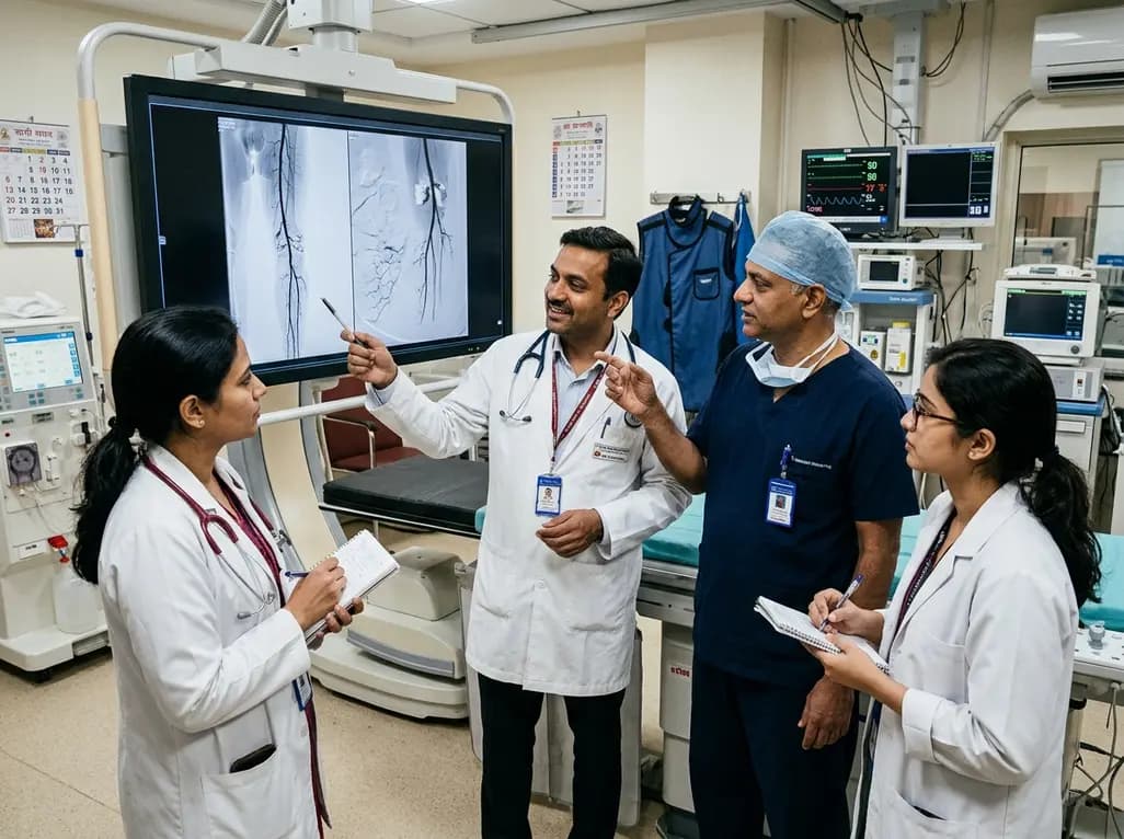
Explore revascularisation strategies in dialysis-dependent chronic limb-threatening ischaemia. This review examines surgical bypass vs endovascular therapy, BEST-CLI trial findings, procedural morbidity, conduit availability, and multidisciplinary decision-making for optimal limb salvage.
Today

A recent cross-sectional analysis reveals that over a quarter of randomized controlled trials for non-specific low back pain face discontinuation or unknown status, while more than 36% of completed trials remain unpublished, exposing notable reporting gaps in spine and musculoskeletal clinical research.
Today
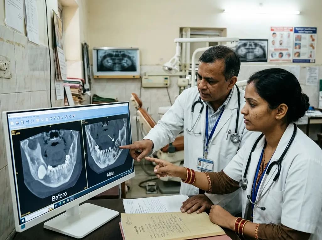
This case report examines the longitudinal CBCT evolution of Stage 2 MRONJ in a 70-year-old woman on denosumab at the site of a pre-existing mandibular dense bone island. It emphasizes the radiographic features, differential diagnosis, and multidisciplinary strategies required for high-risk bone lesions.
Today
