

Loading, please wait...
Loading, please wait...

Vascular access dysfunction remains a persistent threat to hemodialysis delivery worldwide. In routine clinical practice, central venous stenosis and occlusion cause progressive vascular access failure. Prior central venous catheterization frequently triggers thoracic vein intimal hyperplasia, producing dramatic venous hypertension. As a result, patients develop limb swelling, elevated venous dialysis pressures, prolonged cannulation bleeding, and vascular access thrombosis. Nephrologists and vascular surgeons must evaluate thoracic central veins promptly to preserve functional lifelines. Historically, clinicians have relied on catheter-based digital subtraction angiography. Although invasive angiography remains the reference diagnostic gold standard, it requires direct arterial or fistula puncture, expensive catheterization lab infrastructure, and substantial operator time. Duplex ultrasonography offers a noninvasive alternative, but the thoracic cage severely limits sound wave penetration into the innominate veins and superior vena cava. Consequently, clinicians urgently need reliable, noninvasive, and rapid thoracic imaging strategies. Bilateral peripheral CT venography has emerged as a promising cross-sectional modality to address this persistent diagnostic dilemma. This noninvasive examination comprehensively visualizes central thoracic venous architecture while avoiding direct fistula cannulation.
A recent clinical investigation evaluated the real-world utility of bilateral peripheral CT venography in 46 consecutive hemodialysis patients with dysfunctional arteriovenous fistulas. The imaging protocol emphasizes speed, patient comfort, and uniform bilateral vascular enhancement. Radiologic technologists place peripheral intravenous catheters into bilateral upper extremity veins, typically using superficial forearm or hand veins. Subsequently, automated dual-barrel power injectors simultaneously administer iodinated contrast media into both arms alongside normal saline chasers. This simultaneous bilateral injection guarantees symmetric opacification of both the left and right brachiocephalic systems. Therefore, radiologists can distinguish true central venous stenosis from hemodynamic flow artifacts or asymmetric unopacified blood washouts. In addition, the imaging team completes the entire diagnostic acquisition rapidly. The researchers recorded an astonishingly brief mean procedure duration of four minutes, with examination times ranging between three and five minutes. Thus, patients spend minimal time inside the scanner gantry, which significantly reduces procedural distress. Furthermore, multidetector computed tomography provides volumetric datasets that allow isotropic multiplanar reformations and three-dimensional volume-rendered reconstructions. Vascular interventionists can easily evaluate thoracic anatomy before planning surgical or catheter-directed recanalization.
The study demonstrated outstanding technical success and an exceptionally high diagnostic yield. Specifically, the radiologic team achieved a 100% technical success rate across all 46 study participants without procedural abandonment. Bilateral peripheral CT venography successfully identified central venous abnormalities in 44 patients, representing 95.7% of the cohort. Among these findings, isolated or multifocal venous stenosis appeared in 31 patients, accounting for 67.4% of cases. Meanwhile, total central venous occlusion occurred in 11 patients, representing 23.9% of the examined cohort. In addition, radiologists observed extensive venous aneurysmal changes in two patients. To assess clinical reliability, investigators evaluated comparative imaging findings from puncture-based invasive angiography or duplex ultrasonography in 38 patients. Notably, CT venography demonstrated remarkable concordance with comparative modalities in 37 of these 38 patients, yielding an agreement rate of 97.4%. The authors appropriately interpreted this observation as descriptive concordance rather than formal diagnostic accuracy. Nevertheless, such strong concordance highlights the dependable visualization capability of modern cross-sectional venography. Radiologists readily traced tortuous collaterals, collateral decompression pathways, and cephalic arch morphology. Consequently, surgeons can determine definitive endovascular or surgical trajectories with high anatomical confidence.
Evaluating safety parameters and radiation burden remains crucial when deploying computed tomography in vulnerable end-stage renal disease cohorts. Fortunately, the investigators reported an excellent safety profile during the entire evaluation period. Overall, only minor adverse events occurred, and the study documented zero severe procedure-related complications. Specifically, contrast extravasation occurred in only one patient, representing an incidence of 2.2%. In that case, conservative supportive management quickly resolved the localized swelling without tissue compromise. Additionally, mild allergic reactions appeared in three patients, representing 6.5% of the study group. These patients responded promptly to standard oral or antihistamine administration. Regarding radiation parameters, the researchers quantified radiation exposure using standardized computed tomography indices. The mean volume computed tomography dose index was 11.87 mGy, while the mean dose-length product reached 784.15 mGy·cm. These radiation metrics compare favorably with conventional multi-projection fluoroscopic digital subtraction angiography. Moreover, peripheral venography avoids arterial puncture risks, pseudoaneurysm formation, hematoma expansion, and vessel dissection at the fistula cannulation site. Because hemodialysis removes water-soluble iodinated contrast during subsequent scheduled sessions, residual contrast-induced systemic toxicities remain minimal in anuric dialysis populations.
Integrating bilateral peripheral CT venography into multidisciplinary hemodialysis management pathways offers distinct strategic benefits. In busy tertiary care hospitals, catheterization laboratory time represents an expensive, constrained resource. Deploying rapid CT venography permits nephrologists and vascular interventionalists to triage access dysfunction noninvasively before booking catheterization suites. When cross-sectional images confirm severe occlusion or complex extrinsic bony compression, clinicians can prepare specialized recanalization tools, crossing wires, and stents beforehand. Conversely, when CT imaging excludes central pathology and identifies peripheral venous runoff stenosis, teams can avoid unnecessary central venography. Furthermore, bilateral imaging clarifies mediastinal venous collateral pathways that bypass occluded segments. This anatomical insight guides surgeons when they evaluate surgical bypass options versus endovascular balloon angioplasty. Nevertheless, clinicians must maintain judicious contrast stewardship, particularly in patients who retain residual native kidney function. While anuric patients tolerate iodinated contrast well before dialysis, nephrologists should schedule imaging sessions closely prior to regular hemodialysis treatments. Overall, this noninvasive approach substantially streamlines diagnostic workflows, minimizes direct vascular trauma, and supports durable vascular access longevity. Future prospective randomized trials will further clarify long-term access patency outcomes following CT-guided revascularization.
Bilateral peripheral CT venography acquires rapid cross-sectional thoracic images using simultaneous peripheral intravenous contrast injections rather than direct fistula puncture. Unlike invasive catheter angiography, it does not require an operating theater or arterial sheath placement. Instead, multidetector scanners map thoracic venous anatomy, extrinsic compression, and collateral vessels noninvasively within four minutes. Consequently, this technique significantly reduces patient procedural discomfort and eliminates direct access puncture complications while delivering detailed three-dimensional vascular reconstructions.
The reported protocol delivers a mean volume computed tomography dose index of 11.87 mGy and a mean dose-length product of 784.15 mGy·cm. These exposure metrics fall well within standard thoracic diagnostic thresholds and compare favorably with protracted fluoroscopic interventional procedures. Because modern multidetector scanners utilize automated tube current modulation, the examination restricts radiation exposure safely while producing high-resolution cross-sectional images suitable for definitive endovascular intervention planning.
Unilateral contrast injection frequently produces turbulent flow artifacts, incomplete mixing, and asymmetric unopacified blood washouts from the contralateral jugular or subclavian veins. In contrast, simultaneous bilateral peripheral injection uniformly opacifies both brachiocephalic veins and the superior vena cava concurrently. This balanced opacification prevents false-positive interpretations of central filling defects. Furthermore, simultaneous bilateral imaging clearly delineates contralateral venous anatomy, which helps vascular surgeons plan alternative hemodialysis access sites accurately.
Disclaimer: This content is for informational and educational purposes only. It is not intended as medical advice or to substitute for professional clinical judgment. Refer to the latest local and national guidelines for clinical practice.
References

Read summarized clinical updates, watch expert medical content, and earn CME certifications right from your smartphone.
Open your smartphone camera to scan and install.





A recent study highlights bilateral peripheral CT venography as a rapid, noninvasive, and safe technique for assessing central venous lesions in hemodialysis patients with dysfunctional arteriovenous fistulas, demonstrating 100% technical feasibility and high concordance with invasive imaging.
Today

A landmark study evaluates artificial intelligence models and prompting strategies in qualitative lupus pregnancy research, revealing that few-shot prompting in advanced LLMs reliably captures maternal experiences, clinical anxieties, and perinatal outcomes across diverse languages.
Today
A comprehensive computational and clinical analysis evaluates incretin anti-obesity therapies, comparing oral orforglipron with injectable semaglutide and tirzepatide. Findings from head-to-head trials and multi-criteria decision analysis guide personalized clinical management.
Today
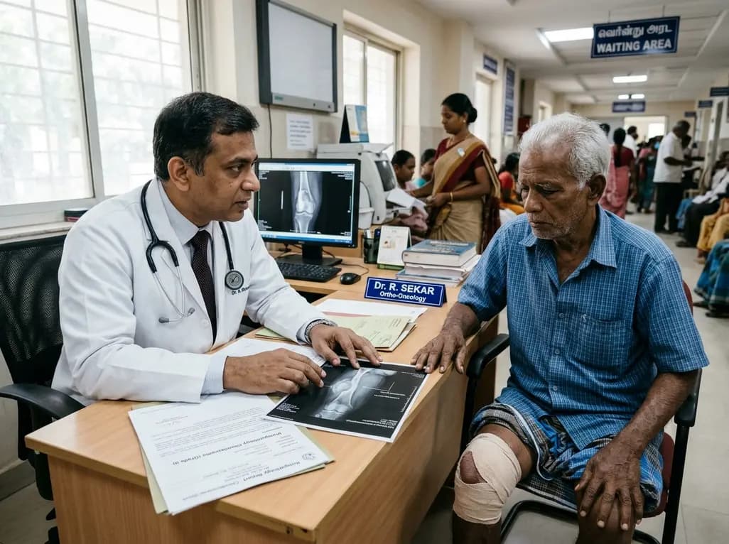
A recent single-institution analysis identifies advanced patient age and progressive systemic disease as primary negative prognostic factors for five-year survival in chondrosarcoma, underscoring the urgent necessity of precise risk stratification, clear surgical margins, and dedicated oncologic follow-up.
Today
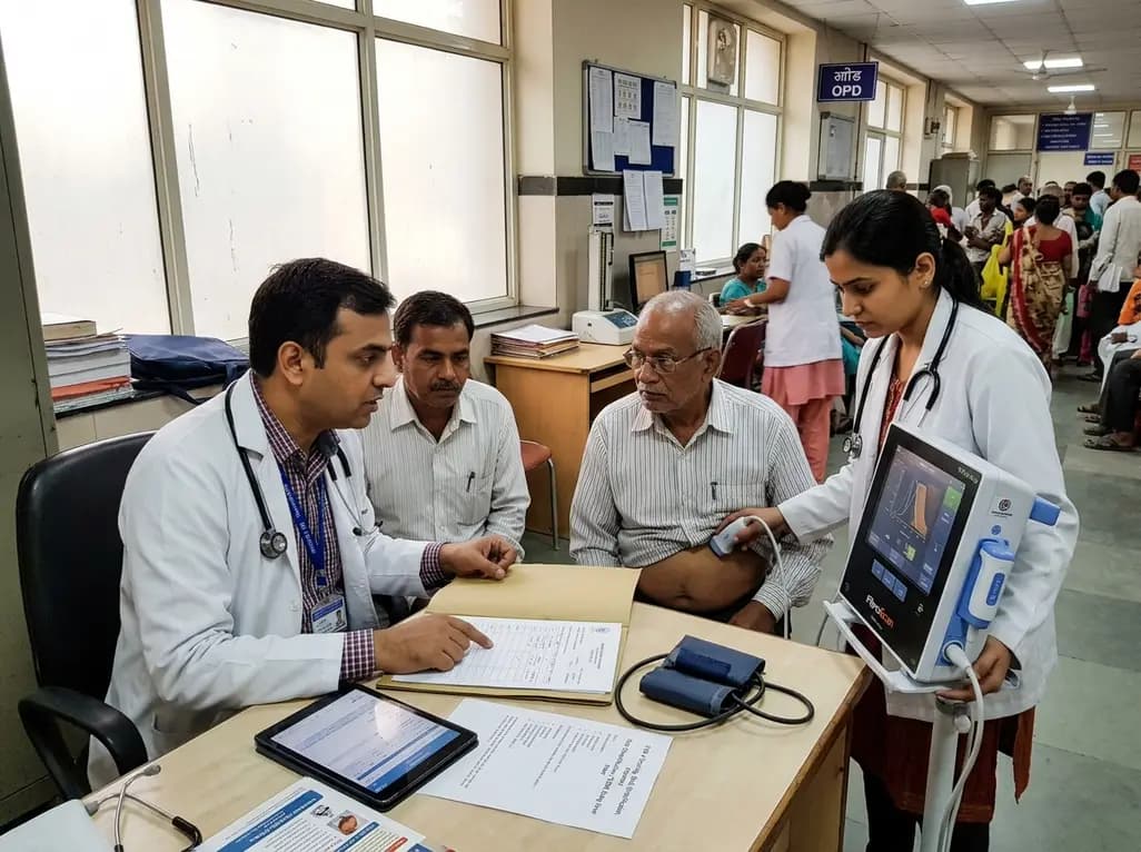
Metabolic dysfunction-associated steatotic liver disease affects nearly 70% of Indian adults with type 2 diabetes. Implementing systematic risk stratification with FIB-4 scoring, transient elastography, and patient-centric digital engagement transforms early detection into sustainable, long-term clinical care.
Today
