.webp&w=384&q=75)

Loading, please wait...
Loading, please wait...
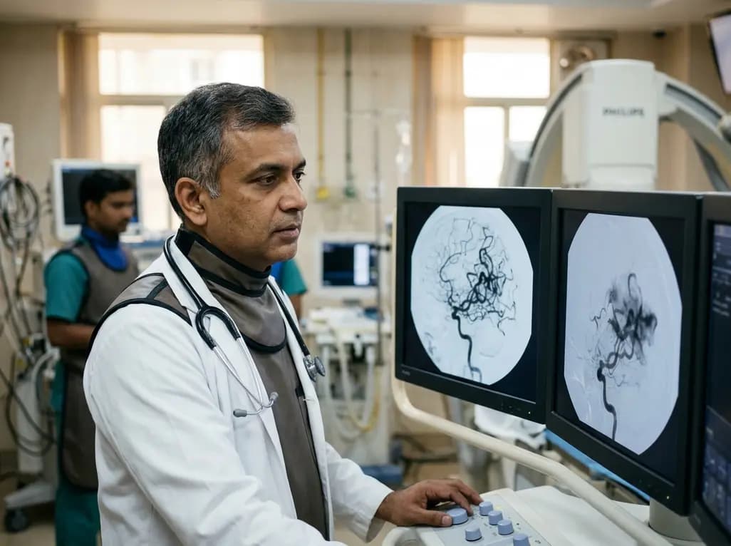
Brain arteriovenous malformations (bAVMs) represent a significant challenge in neurovascular surgery and interventional radiology. These complex lesions consist of a nidus where arteries and veins connect directly without an intervening capillary bed. While endovascular therapy (EVT) has become a mainstay in managing these malformations, it is not without risks. Historically, clinicians attributed bAVM endovascular therapy complications primarily to hemodynamic shifts. However, recent evidence suggests that the physical architecture of the vessel wall itself plays a pivotal role. Understanding how these vessels respond to pressure changes during embolization is crucial for improving patient outcomes. Researchers are now looking beyond flow dynamics to the microscopic structural heterogeneity of the vascular wall. This approach allows for a more nuanced understanding of why some patients experience catastrophic hemorrhage while others remain stable throughout the procedure. By examining the histomorphological features of the nidus, specialists can better predict which vascular structures might fail under procedural stress. Consequently, this shift in focus from macro-hemodynamics to micro-structural integrity offers a fresh perspective on risk stratification in neurointervention.
A recent study utilized advanced digitized hematoxylin-eosin sections to analyze fifty-one patients who underwent surgical resection following endovascular treatment. The research team focused on extracting quantitative vessel-level histomorphological features to categorize the diverse architecture of bAVM vessels. Through principal component analysis and unsupervised clustering, they identified five distinct vascular phenotypes, labeled Types A through E. Specifically, Type B stood out due to its unique structural characteristics. This phenotype exhibited increased textural variability and significant structural heterogeneity within the vessel walls. Furthermore, the distribution of these phenotypes was not uniform across all patient groups. Type B vessels were significantly more prevalent in patients who suffered from bAVM endovascular therapy complications, particularly hemorrhagic events. This finding suggests that the intrinsic makeup of the malformation is a major determinant of procedural safety. Identifying these phenotypes prior to or during treatment could revolutionize how neurosurgeons approach complex nidal architectures. Moreover, the study highlights that vascular homogeneity often correlates with better structural stability during the high-pressure environment of liquid embolic agent injection.
The Type B phenotype presents a particularly dangerous profile for clinicians managing brain malformations. When compared to the more homogeneous and stable Type C phenotype, Type B vessels showed a significantly lower thickness-to-radius ratio. Additionally, these vessels demonstrated a higher coefficient of variation in wall thickness and a much smaller minimum wall thickness. These structural weaknesses mean that Type B vessels are inherently less capable of withstanding the sudden pressure increases associated with embolization. When an embolic agent fills the nidus, it alters the internal pressure gradients. In a heterogeneous wall with thin segments, these focal points of weakness become prime locations for rupture. Therefore, the presence of Type B architecture serves as a morphological marker for fragility. The study confirmed that this enrichment in the hemorrhagic complication group was statistically significant, even after adjusting for false discovery rates. Consequently, understanding the bAVM endovascular therapy complications requires a deep dive into these specific histological markers. Clinical teams must recognize that not all nidal vessels are created equal, and some possess inherent structural flaws that predispose them to failure regardless of the surgical technique employed.
The discovery of vascular phenotypes has profound implications for clinical practice in India and globally. Currently, most neurointerventionists rely on digital subtraction angiography to guide their decisions. While angiography provides excellent temporal and spatial resolution of blood flow, it cannot visualize the microscopic structural heterogeneity of the vessel wall. Consequently, the field is moving toward integrating advanced imaging and histological data to refine risk assessments. If a clinician could identify a Type B-dominant nidus beforehand, they might opt for a more staged embolization approach or prioritize surgical resection. Furthermore, this research underscores the importance of multidisciplinary care involving pathologists and radiologists. By correlating histomorphological data with procedural outcomes, the medical community can develop more accurate predictive models. These models would specifically target the prevention of bAVM endovascular therapy complications by identifying patients who require specialized blood pressure management or specific embolic material choices. Ultimately, the goal is to move toward a precision medicine approach where the structural vulnerabilities of each individual's bAVM are mapped and mitigated before the first catheter is ever placed.
Looking forward, the integration of artificial intelligence and machine learning could further enhance our ability to detect high-risk phenotypes. By training algorithms on digitized histological sections and corresponding imaging, researchers may find non-invasive surrogates for Type B architecture. For instance, certain patterns on high-resolution vessel wall MRI might correspond to the structural heterogeneity observed in the study. Additionally, future research should investigate whether specific genetic markers or molecular signals contribute to the development of Type B vessels. Understanding the biological pathways that lead to wall thinning and textural variability could lead to pharmacologic interventions that stabilize the vessel wall prior to EVT. Such advancements would drastically reduce the incidence of bAVM endovascular therapy complications and improve the long-term safety of neurovascular interventions. Moreover, expanding the study to larger, multi-center cohorts will be essential to validate these phenotypes across diverse populations. As we refine our understanding of nidal morphology, the focus will increasingly shift from simple occlusion to the holistic management of vascular health. This study serves as a critical stepping stone in that journey, providing a clear link between microscopic architecture and macroscopic clinical outcomes.
To implement these findings, hospital systems must consider the value of routine pathological analysis of resected bAVM tissues. While many centers currently focus on the surgical removal of the nidus, detailed histological reporting could provide feedback loops for endovascular teams. Specifically, identifying the prevalence of Type B vessels in a specific patient's specimen can help explain why certain complications occurred. This information is vital for resident training and quality improvement programs within neurosurgery departments. Additionally, the study's use of linear mixed-effects models provides a robust framework for assessing vessel-level differences that clinical factors alone cannot explain. By isolating the vessel wall as an independent risk factor, the research clarifies that bAVM endovascular therapy complications are not always the result of operator error or aggressive hemodynamics. Instead, they are often the result of an unavoidable interaction between therapy and a pre-existing structural vulnerability. Acknowledging this reality allows for a more objective assessment of procedural risks and benefits. It also empowers clinicians to have more transparent discussions with patients regarding the inherent dangers of treating these complex vascular anomalies in the brain.
Vascular wall heterogeneity, particularly in the Type B phenotype, creates localized zones of extreme weakness within the malformation. When endovascular therapy introduces embolic agents, the internal pressure within the nidus shifts. Vessels with high structural variability and thin walls cannot distribute this mechanical stress evenly. Consequently, the weakest points of the wall succumb to the pressure, leading to the hemorrhagic complications often seen during or after the procedure.
The primary markers for high-risk vessels include a low thickness-to-radius ratio and a high coefficient of variation in wall thickness. This means the vessel wall is not only thin but also inconsistently constructed across its circumference. Furthermore, a smaller minimum wall thickness is a critical predictor of failure. These features define the Type B phenotype, which researchers found was significantly enriched in patients experiencing therapy-related hemorrhagic events.
Currently, standard angiography cannot reliably detect microscopic phenotypes like Type B. However, this research paves the way for using advanced techniques such as high-resolution vessel wall MRI or machine learning-enhanced imaging. By identifying specific radiological signatures that correlate with histological heterogeneity, clinicians hope to non-invasively map these high-risk zones. This would allow for better procedural planning and a reduction in bAVM endovascular therapy complications through personalized treatment strategies.
Disclaimer: This content is for informational and educational purposes only and does not constitute medical advice. It is not intended to be a substitute for professional medical advice, diagnosis, or treatment. Always seek the advice of your physician or other qualified health provider with any questions you may have regarding a medical condition. Refer to the latest local and national guidelines for clinical practice.
References
Zeng N et al. Vascular wall structural heterogeneity and hemorrhagic complications during endovascular therapy for brain arteriovenous malformations. J Clin Neurosci. 2026 Jul 17. doi: undefined. PMID: 42468088.
Hou K et al. Intra- and post-operative acute hemorrhagic complications of Onyx embolization of brain arteriovenous malformations: A single-center experience. Frontiers. 2022; 13: 10.3389/fneur.2022.951016.
Chen CJ et al. Risk factors for hemorrhage of brain arteriovenous malformation. PMC. 2020; 11(3): 120-135.

Read summarized clinical updates, watch expert medical content, and earn CME certifications right from your smartphone.
Open your smartphone camera to scan and install.





Recent research highlights that intrinsic vascular wall structural heterogeneity, specifically the Type B phenotype, significantly increases the risk of hemorrhagic complications during endovascular therapy for brain arteriovenous malformations, suggesting a need for better risk stratification.
3 months ago

As monsoon seasons elevate arboviral disease threats, Tamil Nadu has initiated an ICMR-monitored evaluation of the tetravalent dengue vaccine in Tenkasi district. Clinicians emphasize clinical monitoring, public prevention, and high-quality early pediatric care to reduce disease morbidity across endemic regions.
Today

A comparative cohort study evaluates vNOTES vs TLH hysterectomy for benign conditions, demonstrating shorter operative duration, faster return to daily activities, and equivalent female sexual function scores without increased dyspareunia.
Today

Review how tailored exercise programs benefit frail older patients with heart failure. Explore pathophysiological mechanisms, individualized training prescriptions, and strategies to overcome adherence barriers in clinical practice.
Yesterday

This comparative study evaluates posterior labral tears with and without glenohumeral instability, highlighting distinct demographic patterns, MRI underestimation in stable shoulders, and successful arthroscopic repair outcomes.
Yesterday

A clinical study demonstrates that early intervention with tocilizumab, either as monotherapy or bridged with corticosteroids, achieves significant proptosis reduction and resolves active inflammation in treatment-naive thyroid eye disease.
Today
