

Loading, please wait...
Loading, please wait...

Acute basal ganglia ischemic stroke represents a frequent and debilitating presentation encountered in stroke care. Although the primary structural ischemic injury occurs within deep subcortical nuclei, patients predominantly experience severe contralateral motor weakness. Consequently, clinicians recognize that functional deficits extend far beyond the localized anatomical lesion. Instead, focal infarcts interrupt intricate corticostriatal loops and disturb widespread brain networks across both cerebral hemispheres. Recent neuroimaging investigations indicate that functional diaschisis severely impairs communication between remote cortical motor hubs. Therefore, deciphering these distant functional reorganization patterns provides essential mechanistic clarity. Furthermore, elucidating the molecular processes coordinating this network failure bridges macroscopic imaging phenotypes with microscale biological signatures.
The basal ganglia participate extensively in motor planning, execution, and movement modulation through complex loops connecting cortical motor regions. When acute basal ganglia ischemic stroke strikes these subcortical nuclei, it interrupts descending tracts and feedback projections. Consequently, the primary motor cortex within the precentral gyrus experiences immediate physiological perturbation. Clinicians frequently note that similar lesion volumes produce remarkably heterogeneous motor impairments across different patients. However, standard anatomical imaging fails to capture dynamic functional disruptions occurring in remote intact tissue. Therefore, investigators deploy resting-state functional neuroimaging to measure synchronized spontaneous neural fluctuations. These modalities reliably demonstrate that subcortical damage provokes widespread diaschisis. Specifically, the loss of striatal regulatory tone alters the excitability of bilateral primary motor cortices. Thus, motor paralysis reflects both direct subcortical infarction and downstream network breakdown. Understanding this distributed vulnerability helps clinicians appreciate why rehabilitation must engage broader bilateral cerebral networks.
To map functional network changes, researchers utilize complementary metrics including regional homogeneity and seed-based functional connectivity. Regional homogeneity evaluates local neural synchronization within neighbouring voxels in a given cerebral area. In contrast, seed-based analyses interrogate temporal correlations across distant anatomical structures. A recent study investigated 59 patients presenting with acute basal ganglia ischemic stroke alongside 35 matched healthy controls. Notably, investigators uncovered markedly reduced regional homogeneity within the bilateral precentral gyri. This localized decoupling indicates that acute ischemia disrupts coordinated microcircuit firing even within intact cortical motor strips. Furthermore, seed-based assessments revealed a profound reduction in interhemispheric functional connectivity between the left and right precentral gyri. Under normal conditions, transcallosal pathways maintain balanced reciprocal communication between homologous motor regions. However, subcortical ischemic disruption destabilizes this delicate equilibrium, severely impairing interhemispheric cooperation. Importantly, connectivity loss directly correlated with clinical motor deficits. Therefore, quantitative functional metrics capture meaningful neurophysiological disruption that mirrors bedside motor impairment.
Modern clinical neurology increasingly integrates artificial intelligence to identify reliable imaging biomarkers from complex data. Accordingly, researchers developed support vector machine and logistic regression models to differentiate stroke patients from healthy individuals. Both algorithmic models demonstrated outstanding diagnostic performance, achieving area under the curve values of 0.877 and 0.868, respectively. Furthermore, scientists applied SHapley Additive exPlanations to interpret feature contributions transparently. This interpretability analysis pinpointed reduced interhemispheric functional connectivity seeded in the right precentral gyrus as the top discriminative feature. Consequently, this finding confirms that interhemispheric disconnection represents a primary pathophysiological hallmark rather than an incidental epiphenomenon. Moreover, this predictive feature tracked continuous motor performance scores across the patient cohort. In clinical environments, objective neuroimaging biomarkers enhance prognostic accuracy during acute admission. Physicians can potentially identify individuals at elevated risk for persistent motor impairment before functional plateaus occur. Thus, machine learning algorithms extract actionable diagnostic signatures from resting-state scans, bridging computational data science with stroke management.
Uncovering the biological drivers behind functional disconnection requires integrating neuroimaging with human molecular atlases. Therefore, researchers spatially correlated neuroimaging alterations with Allen Human Brain Atlas transcriptomic profiles using partial least squares regression. Subsequently, functional enrichment analyses revealed that genes associated with the disrupted connectivity profile play crucial cellular roles. Specifically, these identified genes showed marked enrichment in synaptic transmission, transmembrane ion transport, and cellular energy metabolism. Neuronal communication across hemispheres demands substantial metabolic energy and precise ion gradient maintenance. When ischemia disturbs corticostriatal loops, remote cortical neurons experience metabolic stress and altered membrane stability. Furthermore, genes governing neurotransmitter receptor trafficking and synaptic plasticity exhibited significant dysregulation within the contralesional motor cortex. These genomic associations illustrate that macroscopic network breakdowns reflect fundamental alterations in synaptic machinery and mitochondrial respiration. Consequently, transcriptomic cross-referencing offers an innovative lens to explore post-stroke diaschisis. Ultimately, these genetic insights illuminate novel molecular pathways for targeted neuroprotective and neurorestorative drug discovery.
These mechanistic discoveries provide profound implications for contemporary neurorehabilitation protocols in stroke care. Historically, rehabilitation therapists focused primarily on repetitive physical exercises for the hemiparetic limb. However, network-level insights underscore the critical need to restore synchronized bilateral motor communication. In clinical settings, non-invasive brain stimulation techniques like repetitive transcranial magnetic stimulation can modulate transcallosal excitability. Specifically, clinicians can apply excitatory stimulation to the affected motor cortex or inhibitory protocols to the unaffected hemisphere. This targeted strategy mitigates maladaptive interhemispheric inhibition and fosters re-synchronization between bilateral precentral gyri. Furthermore, understanding metabolic and synaptic vulnerabilities suggests synergistic pharmacological strategies. Agents supporting mitochondrial bioenergetics or enhancing synaptic neurotransmission might augment connectivity restoration during early therapeutic windows. In diverse clinical environments, identifying precise neuroimaging targets optimizes resource allocation. Accordingly, clinicians can tailor rehabilitation intensity based on baseline interhemispheric functional integrity. Integrating connectomic data into clinical pathways transforms empirical therapy into personalized, mechanism-guided neurorestoration.
The convergence of resting-state functional neuroimaging, machine learning, and transcriptomics heralds an exciting era for precision stroke neurology. Nevertheless, several crucial questions require rigorous investigation in future longitudinal trials. For instance, current evidence captures cross-sectional snapshots during acute presentation. Clinicians must determine how interhemispheric connectivity reorganizes dynamically over weeks and months during spontaneous recovery. Furthermore, researchers must validate whether early restoration of precentral gyrus synchrony directly predicts successful long-term functional independence. Expanding multi-center neuroimaging registries across diverse populations will establish standardized diagnostic thresholds. Additionally, combining functional MRI with bedside functional near-infrared spectroscopy could facilitate real-time monitoring of motor connectivity during active rehabilitation sessions. Similarly, discovering pharmacological agents that upregulate identified metabolic and synaptic pathways could protect fragile interhemispheric circuits. In conclusion, bridging macroscopic functional networks with microscale transcriptomic mechanisms deepens our comprehension of post-stroke motor deficits. Consequently, this multi-scale framework equips clinicians with powerful tools to predict recovery, optimize rehabilitation, and improve functional outcomes.
Basal ganglia ischemic stroke disrupts vital subcortical-cortical loops that coordinate voluntary movement. Consequently, remote cortical motor regions lose regular striatal regulatory signals. This induces functional diaschisis across the motor cortex, reducing local neural synchronization and impairing interhemispheric communication between the bilateral precentral gyri, which directly impairs motor performance.
Resting-state functional MRI measures spontaneous low-frequency blood oxygen level fluctuations without requiring active patient movement. Thus, clinicians and researchers can objectively quantify intrinsic neural synchronization and remote circuit disruption. These functional connectivity metrics accurately reflect motor impairment severity and serve as valuable biomarkers for predicting post-stroke motor recovery trajectories.
Researchers map spatial neuroimaging alterations onto human brain gene expression atlases using multivariable regression techniques. Consequently, this identifies specific gene modules enriched in areas of network disruption. In acute stroke, disrupted motor connectivity strongly associates with genes regulating synaptic transmission, ion transmembrane transport, and cellular energy metabolism in remote cortex.
Disclaimer: This content is for informational and educational purposes only... Refer to the latest local and national guidelines for clinical practice.
References

Read summarized clinical updates, watch expert medical content, and earn CME certifications right from your smartphone.
Open your smartphone camera to scan and install.





A novel neuroimaging study reveals that acute basal ganglia ischemic stroke causes disrupted interhemispheric functional connectivity between bilateral precentral gyri, spatially associated with transcriptomic pathways regulating synaptic function, ion transport, and cellular energy metabolism.
Today

The DOMINO study protocol presents a multidisciplinary implementation mapping approach to overcome provider, patient, and payer barriers to antiobesity medications, offering vital insights for Indian clinicians managing the growing diabesity burden.
Today

An intracranial EWSR1::CREM fusion sarcoma in a 31-year-old female manifested as a paraneoplastic inflammatory syndrome with anemia and secondary infertility. The dural mass mimicked meningioma, but gross-total resection resolved her systemic inflammation and restored natural fertility within one month.
Today

A real-world cohort study of 6,537 patients from the FOUNTAIN platform demonstrates that initiating finerenone significantly reduces albuminuria by 40.7% with minimal eGFR decline and manageable hyperkalemia risk, confirming its practical effectiveness and safety in diabetic kidney disease.
Yesterday

A comprehensive analysis of MAP therapy resistance in osteosarcoma, examining canonical and non-canonical mechanisms across damage avoidance, enhanced DNA repair, and replication stress tolerance to identify novel vulnerabilities.
Today
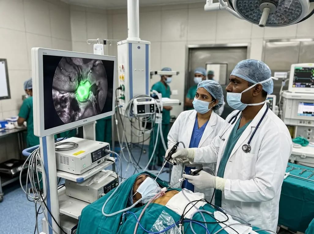
Near-infrared autofluorescence provides real-time label-free optical guidance during endoscopic transsphenoidal pituitary surgery. By highlighting stark signal contrasts between normal pituitary gland and adenomas, this technique aids gross-total tumor resection while safeguarding critical endocrine function.
Today
