

Loading, please wait...
Loading, please wait...
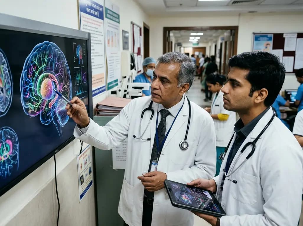
Neuroscientists have achieved a historic milestone by mapping deep brain architecture with single-cell precision. Through the BRAIN Initiative Cell Atlas Network, international researchers unveiled a comprehensive basal ganglia cell atlas across human donors and mammalian models. The basal ganglia coordinate motor initiation, reward learning, habit formation, and executive decision-making. Pathological disruptions in these circuits drive severe motor and neuropsychiatric conditions. Consequently, this multi-omic resource establishes a long-awaited reference for modern neurology.
To construct this unprecedented database, investigators analyzed over twenty-one million cells derived from post-mortem human donors. Additionally, the team integrated comparative cellular profiles from mice, macaques, and marmosets. Historically, anatomists viewed the basal ganglia as a collection of broad structures, including the striatum, globus pallidus, and substantia nigra. However, this high-throughput catalog exposes exquisite cellular diversity within these classic anatomic boundaries. Specifically, researchers categorized sixty-one consensus cell groups, unveiling previously uncharacterized neuronal subtypes and distinct glial states. Furthermore, the investigative team combined single-nucleus transcriptomics with chromatin accessibility assays to capture cellular dynamics in real time. Because cellular identity depends on spatial context, scientists generated spatial transcriptomic maps across anatomical coordinates. Therefore, clinicians can now visualize where specific cell types reside within human subcortical pathways. Moreover, the project creates a unified taxonomic framework, providing researchers worldwide with a common anatomical language. Ultimately, this granular catalog resolves long-standing debates regarding cellular diversity in primate deep gray nuclei. As a result, scientists can track specific neuronal lineages from embryogenesis to adulthood, transforming our fundamental understanding of brain development.
Translational neuroscience has long relied on rodent and non-human primate models to investigate neurological disease mechanisms. Nevertheless, important biological discrepancies frequently impede the successful translation of preclinical findings to human clinical trials. Fortunately, this multi-species cellular roadmap provides an objective framework to assess cross-species evolutionary conservation. By directly comparing homologous cell types across species, researchers determined which neuronal populations remain functionally conserved and which phenotypes diverged uniquely in humans. For example, although direct and indirect pathway striatal spiny projection neurons display remarkable conservation across mammals, primate brains exhibit distinct subpopulations. Specifically, human striatal tissue contains specialized interneurons that regulate complex decision circuits. Consequently, drug candidates that target rodent receptors might fail if human homologous receptors localize to entirely different cellular subsets. Furthermore, understanding primate-specific transcriptional profiles enables biomedical engineers to refine adeno-associated viral vectors for primate gene delivery. Therefore, this cross-species taxonomy significantly elevates the predictive validity of preclinical neuropharmacology and experimental therapeutics. In doing so, future translational pipelines can avoid costly clinical trial failures and optimize therapeutic translation.
Genome-wide association studies have identified hundreds of genetic loci linked to neurological and psychiatric disorders. However, interpreting these findings presents an immense clinical challenge because more than ninety percent of risk variants reside in non-coding DNA regions. These non-coding sequences function as genetic switches or regulatory enhancers that control spatial and temporal gene expression. Because these switches operate in cell-type-specific configurations, bulk tissue homogenates cannot determine where disease-associated variants exert their pathogenic effects. Fortunately, the basal ganglia cellular atlas integrates single-nucleus epigenomics and chromatin accessibility profiling to solve this dilemma. By mapping open chromatin landscapes across individual cell classifications, researchers can align disease-associated single nucleotide polymorphisms directly with functional regulatory elements. For instance, investigators can now trace a Parkinson's disease risk allele directly to an active enhancer in dopaminergic substantia nigra neurons. Similarly, epigenetic modifications associated with normal brain aging can now be isolated within specific glial lineages. Thus, this regulatory map transforms enigmatic genomic statistical signals into precise biological mechanisms. Consequently, gene therapy developers can design expression cassettes that activate exclusively within targeted subcortical neuronal subsets.
The basal ganglia serve as the primary anatomical substrate for several devastating movement disorders, most notably Parkinson's disease and Huntington's disease. In Parkinson's disease, clinicians observe progressive degeneration of dopaminergic neurons in the substantia nigra pars compacta. Although dopamine replacement therapy and deep brain stimulation offer substantial symptomatic relief, they fail to halt ongoing neuronal demise. Importantly, this cellular atlas provides actionable insight into early pathological vulnerability and cellular resilience. For instance, researchers observed that specific subsets of dopaminergic neurons possess distinct transcriptional defenses against metabolic stress. Therefore, deciphering these intrinsic protection signatures may inspire novel neuroprotective therapeutics for early clinical administration. Additionally, Huntington's disease causes profound striatal atrophy, predominantly annihilating medium spiny projection neurons. By establishing a normative baseline of healthy human striatal cell states, the atlas equips researchers to pinpoint initial transcriptional dysregulation before gross clinical neurodegeneration manifests. Furthermore, functional neurosurgeons performing deep brain stimulation can utilize these precise anatomical boundaries to refine electrode targeting within the subthalamic nucleus. Consequently, precision neuromodulation can minimize stimulation-induced adverse effects while optimizing motor recovery.
Beyond motor execution, the basal ganglia govern complex behavioral loops connecting the prefrontal cortex, limbic system, and striatum. Consequently, neurobiological dysfunction within these striatocortical networks precipitates severe psychiatric and developmental illnesses. For example, conditions such as schizophrenia, bipolar affective disorder, Tourette's syndrome, obsessive-compulsive disorder, and chemical addictions directly involve basal ganglia circuitry. Historically, the absence of high-resolution cellular maps hindered the identification of primary cytopathological culprits in psychiatric medicine. Remarkably, the new atlas revealed previously undetected neuronal populations that specifically harbor risk genes for schizophrenia and bipolar illness. Furthermore, pediatric neurodevelopmental studies within the consortium mapped how basal ganglia precursors migrate and mature from fetal development through infancy. During this developmental trajectory, researchers identified key genetic changes in specific striatal cell types that contribute directly to autism spectrum pathophysiology. Thus, neuropsychiatric disorders are increasingly recognized as cell-type-specific channelopathies and synaptopathies. Looking forward, clinicians and neuroscientists can leverage this global database to benchmark genetic studies in diverse populations, accelerating targeted therapeutics for refractory psychiatric illness.
Q1: Why is the creation of a basal ganglia cell atlas considered a major breakthrough?
The basal ganglia cell atlas provides the first comprehensive, single-cell reference of human and non-human primate subcortical structures. Previously, neuroscientists lacked an integrated roadmap describing individual cell types alongside gene expression and chromatin landscapes. By mapping over twenty-one million human brain cells, this resource connects non-coding genetic variants directly to vulnerable neuronal subtypes, establishing a foundational parts list for neurotherapeutic innovation.
Q2: How does this cellular atlas help clinicians and researchers understand Parkinson's disease?
Parkinson's disease primarily destroys dopaminergic neurons residing in the substantia nigra. The new atlas reveals the distinct molecular subtypes of these dopaminergic neurons, highlighting specific transcriptional switches that dictate cellular vulnerability or resilience against cellular stress. Furthermore, researchers can now study where familial Parkinson's genes turn on within individual cells, facilitating the rational design of cell-specific gene therapies and regenerative stem cell grafts.
Q3: How will this cellular data impact the treatment of psychiatric disorders like schizophrenia?
Psychiatric illnesses like schizophrenia and bipolar disorder involve subtle circuit disruptions rather than macroscopic tissue loss. The atlas identified novel brain cell populations in the basal ganglia that selectively express genetic variants linked to psychiatric disorders. By elucidating how developmental gene regulation influences these specific striatal neurons, the data enables neuropsychiatrists to identify precise receptor targets, paving the way for targeted psychiatric medications.
Disclaimer: This content is for informational and educational purposes only. It does not constitute medical advice or replace professional judgment. Refer to the latest local and national guidelines for clinical practice.
References

Read summarized clinical updates, watch expert medical content, and earn CME certifications right from your smartphone.
Open your smartphone camera to scan and install.





Researchers have unveiled an unprecedented cross-species cellular atlas of the basal ganglia, detailing over 21 million human cells. This breakthrough illuminates how non-coding genomic switches govern neuronal function, offering clinicians new roadmaps for Parkinson's disease, schizophrenia, and targeted gene therapies.
Today

The VOCAL-AHF study reveals that most acute heart failure patients across reduced and non-reduced ejection fraction categories are eligible for in-hospital GDMT initiation. Notably, nearly two-thirds of HFrEF patients qualify for quadruple therapy, while non-reduced EF patients show high eligibility for dual therapy.
Today

Ascertainment bias in cancer case-control studies inflates odds ratios and penetrance estimates when cases are drawn from high-risk clinics. A landmark modeling study demonstrates substantial bias ranging up to 23.72, highlighting the critical need for ascertainment adjustment in genetic risk counseling.
Today

Researchers demonstrate that central induction of synthetic torpor in non-hibernating rats provides striking cardioprotection, reducing myocardial infarct size by ~40% during ischaemia-reperfusion injury through endogenous survival signaling, independent of hypothermia.
Today

A meta-analysis shows that movement representation techniques significantly enhance upper limb and wrist function in distal radius fracture rehabilitation. While effects on pain and grip strength remain inconclusive, early low-cost telerehabilitation and mirror therapy provide promising clinical benefits.
Today

A novel fluorine-functionalized near-infrared copper fluorescent probe selectively illuminates lung tissue, revealing critical links between hyperglycemia, ER stress, and pulmonary malignancy for improved early diagnosis.
Today
