

Loading, please wait...
Loading, please wait...

Emergent neurovascular interventions demand rapid diagnostic clarity and tailored mechanical techniques. Managing an acute PComA occlusion presents distinct hemodynamic challenges, particularly when compounded by anomalous posterior circulation architecture and spontaneous arterial dissection. Clinicians must quickly decipher non-embolic etiologies to deploy safe, vessel-preserving endovascular maneuvers.
Acute ischemic stroke involving the posterior cerebral circulation often produces devastating cognitive, sensory, and motor deficits. An acute PComA occlusion is remarkably uncommon in acute neurovascular triage. Consequently, clinicians frequently mistake these presentations for typical anterior circulation infarctions or basilar apex syndromes. When an occlusion occurs within the posterior communicating artery, immediate tissue survival depends directly on collaterals. Patients commonly present with sudden hemiparesis, altered sensorium, or severe visual field disruptions. Furthermore, traditional thrombolytic therapies may fail to provide sustained reperfusion when underlying mechanical wall disruptions exist. Intravenous tissue plasminogen activator can achieve transient recanalization, yet abrupt reocclusion frequently follows. Therefore, neurointerventional teams must remain vigilant. They need to recognize early ischemic signs and identify underlying vessel instability promptly to avoid extensive parenchymal damage.
Anatomical variations in the circle of Willis significantly influence collateral pathways and stroke patterns. A fetal-type posterior cerebral artery represents a common yet critical embryological variant. In this specific configuration, the posterior communicating artery directly supplies the occipital and posterior temporal lobes. The ipsilateral P1 segment remains hypoplastic or entirely absent. Consequently, the posterior communicating artery serves as the primary inflow conduit for the posterior cerebral artery territory. Any abrupt interruption within this slender arterial bridge jeopardizes the entire posterior vascular bed. Moreover, the vessel lacks redundant collateral inflow from the vertebrobasilar junction. Therefore, an acute occlusion in a fetal variant behaves hemodynamically like an anterior large-vessel occlusion. Interventionalists must appreciate this distinct dependency because standard posterior circulation recanalization pathways are entirely unavailable.
Differentiating thromboembolism from spontaneous intracranial dissection remains crucial during emergent digital subtraction angiography. Embolic occlusions typically demonstrate abrupt vessel cutoffs, clean margins, and rapid response to mechanical aspiration or stent retriever deployment. In contrast, arterial dissection produces irregular stenosis, tapered lumen narrowing, or double-lumen configurations. Intravenous thrombolysis may temporarily clear overlying thrombus, but it unmasks severe residual stenosis. When catheter angiography shows immediate vessel reocclusion without significant embolic burden, clinicians should immediately suspect an intimal disruption. In addition, aggressive mechanical thrombectomy passes over a dissected intimal flap can aggravate wall disruption. Such manipulation risks acute vessel perforation or extension of an intramural hematoma. Thus, early angiographic recognition of non-embolic, stenotic profiles shields the fragile vessel wall from unnecessary procedural trauma.
Navigating dissected intracranial branches requires cautious device selection and minimal shear stress. While intracranial stenting effectively restores luminal caliber, it mandates immediate dual antiplatelet therapy. This antiplatelet requirement significantly elevates the risk of secondary hemorrhagic transformation in acute ischemic stroke. Conversely, gentle balloon angioplasty offers an attractive, stent-free alternative. Low-pressure microballoon inflation can effectively tack down a mobile intimal flap. Furthermore, balloon dilation restores sufficient antegrade blood flow without leaving rigid metallic scaffolding within an inflamed artery. Operators must choose appropriately sized, compliant or semi-compliant neurovascular balloons to avoid arterial rupture. By restoring critical perfusion while avoiding heavy antiplatelet loads, balloon angioplasty achieves robust clinical stabilization. It permits natural vascular remodeling as the dissected wall heals over subsequent weeks.
Confirming an intracranial arterial dissection requires sensitive non-invasive imaging modalities during post-procedural convalescence. High-resolution magnetic resonance vessel wall imaging has revolutionized our ability to inspect intracranial architecture. Specifically, volumetric isotropic turbo spin-echo acquisition sequences provide exquisite detail of the arterial wall. These sophisticated scans directly visualize intramural hematomas, localized wall thickening, and intimal flaps. Additionally, T1-weighted hyperintensity within the arterial wall offers definitive evidence of subacute intramural hemorrhage. Longitudinal imaging surveillance frequently reveals gradual spontaneous remodeling and luminal improvement over three to six months. Documenting these structural improvements confirms spontaneous dissection as the definitive etiology. Furthermore, serial vessel wall imaging reassures clinicians that primary balloon angioplasty provides durable long-term stability without late aneurysmal degeneration.
Executing successful angioplasty within delicate communicative channels requires meticulous procedural safeguards. First, interventionalists must maintain gentle microcatheter tracking across the lesion to prevent true-to-false lumen crossing. Second, operators should favor undersized balloon diameters and slow inflation-deflation cycles. This technique minimizes radial tension on weakened arterial tunics. Third, continuous hemodynamic monitoring prevents post-recanalization hyperperfusion injury. After the procedure, clinicians should initiate monotherapy or tailored antiplatelet regimens based on individual hemorrhagic risks. Close neurocritical care monitoring remains vital during the initial forty-eight hours to detect early recurrent ischemia. Ultimately, this constructive approach demonstrates that judicious balloon angioplasty effectively rescues acute ischemic penumbra. Clinicians can achieve favorable 90-day functional independence even in complex, dissection-induced occlusions involving unique anatomical variations.
A fetal-type posterior cerebral artery variant occurs when the embryological posterior communicating artery directly provides primary blood flow to the posterior cerebral artery. In this anatomical configuration, the P1 segment arising from the basilar artery is hypoplastic or absent. Consequently, the occipital and posterior temporal lobes depend entirely on anterior circulation inflow from the internal carotid artery.
Balloon angioplasty is often favored over permanent intracranial stenting because it avoids mandatory acute dual antiplatelet therapy. In acute ischemic stroke, potent antiplatelet agents significantly raise the risk of catastrophic intracranial hemorrhage. Simple balloon dilation can successfully restore antegrade flow and tack down disrupted intimal flaps, allowing the vessel wall to heal naturally over time.
High-resolution magnetic resonance vessel wall imaging confirms intracranial dissection by visualizing structural changes inside the arterial wall itself. Advanced sequences, such as volumetric isotropic turbo spin-echo acquisition, reveal eccentric wall thickening, intimal flaps, and true-versus-false lumens. Furthermore, high-signal T1 methemoglobin accumulation directly identifies the intramural hematoma pathognomonic of spontaneous arterial dissection.
Disclaimer: This content is for informational and educational purposes only. It is not intended as medical advice, diagnosis, or treatment. Always consult a qualified healthcare provider for specific clinical concerns. Opinions expressed are those of the authors and do not necessarily reflect the official stance of any institution. Refer to the latest local and national guidelines for clinical practice.
References
Nakajima S et al. Balloon angioplasty for acute posterior communicating artery occlusion due to dissection: illustrative case. J Neurosurg Case Lessons. 2026 Nov 09. doi: undefined. PMID: 43002772.
Goyal M, Menon BK, van Zwam WH, et al. Endovascular thrombectomy after large-vessel ischaemic stroke: a meta-analysis of individual patient data from five randomised trials. Lancet. 2016;387(10029):1723-1731.
Strambo D, Nannoni S, Sirimarco G, et al. Fetal-type posterior cerebral artery and ischemic stroke: clinical and radiological characteristics. Stroke. 2021;52(10):3232-3240.

Read summarized clinical updates, watch expert medical content, and earn CME certifications right from your smartphone.
Open your smartphone camera to scan and install.





Explore an illustrative case of acute PComA occlusion caused by spontaneous arterial dissection in a fetal-type PCA variant. Discover how primary balloon angioplasty achieved successful vessel recanalization and favorable neurological recovery without the immediate risks of intracranial stenting.
Today
An intracranial EWSR1::CREM fusion sarcoma in a 31-year-old female manifested as a paraneoplastic inflammatory syndrome with anemia and secondary infertility. The dural mass mimicked meningioma, but gross-total resection resolved her systemic inflammation and restored natural fertility within one month.
Today
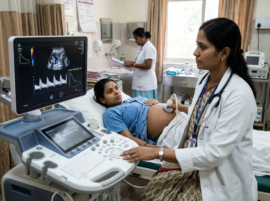
Adaptive spectral analysis of umbilical venous Doppler recordings reveals that an elevated second-trimester maternal-fetal power ratio can distinguish pregnancies at risk of preeclampsia, offering a novel biophysical approach to enhance early vascular risk stratification.
Today

A real-world cohort study of 6,537 patients from the FOUNTAIN platform demonstrates that initiating finerenone significantly reduces albuminuria by 40.7% with minimal eGFR decline and manageable hyperkalemia risk, confirming its practical effectiveness and safety in diabetic kidney disease.
Yesterday

A comprehensive analysis of MAP therapy resistance in osteosarcoma, examining canonical and non-canonical mechanisms across damage avoidance, enhanced DNA repair, and replication stress tolerance to identify novel vulnerabilities.
Today
Near-infrared autofluorescence provides real-time label-free optical guidance during endoscopic transsphenoidal pituitary surgery. By highlighting stark signal contrasts between normal pituitary gland and adenomas, this technique aids gross-total tumor resection while safeguarding critical endocrine function.
Today
