.webp&w=384&q=75)

Loading, please wait...
Loading, please wait...

Enterohemorrhagic Escherichia coli infections cause acute hemorrhagic colitis and severe vascular complications worldwide. Recent mucosal investigations show that bacterial serine protease EspP plays a central role in driving intestinal damage. EspP acts as an extracellular cytotoxin that disrupts the epithelial architecture. Clinicians recognize that enteric pathogens remodel host tissue dynamics, yet the underlying transcriptional alterations have remained obscure. Groundbreaking experimental findings now reveal that acute exposure to bacterial serine protease EspP rapidly halts crypt proliferation and reprograms mucosal differentiation pathways.
Enterohemorrhagic Escherichia coli produces several virulence determinants, including Shiga toxins and plasmid-encoded autotransporters. Among these factors, bacterial serine protease EspP functions as an enzymatic cytotoxin that damages colonic surfaces. Historically, investigators focused primarily on Shiga toxin-mediated microvascular thrombosis. However, non-Shiga toxins exert profound effects on the mucosal barrier. In human colonoid models, acute exposure to purified EspP mimics the tissue destruction observed during natural infection. The protease degrades structural brush border proteins and digests mucosal scaffolding. Furthermore, this enzymatic attack disrupts tight junctions and alters epithelial integrity. Consequently, fluid homeostasis fails and transepithelial electrical resistance collapses. These acute architectural changes illustrate how EspP functions independently of bacterial attachment. Therefore, targeting such secreted proteases offers an important conceptual avenue for mitigating bacterial enterotoxicity.
Human stem cell-derived colonoids provide a robust model to investigate host-pathogen interactions. These organoids recapitulate native architecture without confounding variables from microbiota, stroma, or immune cells. When exposed to EspP, the organoids exhibited immediate transcriptional shifts. Droplet-based single-cell RNA sequencing revealed that the toxin markedly disrupts normal proliferative cycles. Specifically, transit-amplifying cells and stem cells downregulate replication machinery. In contrast, the colonic epithelium redirects its regenerative commitment toward secretory pathways. Rather than generating absorptive colonocytes, the tissue undergoes preferential differentiation into enteroendocrine cells. Furthermore, this lineage shift occurs rapidly following acute toxin exposure. Consequently, the epithelial monolayer modifies its functional state within hours. These observations confirm that bacterial cytotoxins can directly rewrite crypt differentiation cascades in human mucosal tissues.
High-resolution single-cell clustering demonstrated that EspP exposure induces a significant expansion of enteroendocrine progenitors. In addition, the newly differentiated cells predominantly display enterochromaffin characteristics. These enterochromaffin cells serve as the primary sources of mucosal serotonin and regulatory peptides. Under physiologic conditions, enteroendocrine cells constitute less than one percent of the colonic epithelium. However, EspP exposure dramatically multiplies this fraction. The surge in enteroendocrine differentiation alters local neurohumoral signaling within the crypt niche. Moreover, increased hormone production affects mucosal blood flow and smooth muscle motility. This rapid expansion suggests an active host adaptation or a pathogen-induced evasion mechanism. Therefore, the disproportionate generation of neuroendocrine cells could explain the abdominal pain and dysmotility seen in acute enteric colitis.
Surprisingly, the newly formed enteroendocrine cells exhibit an active inflammatory transcriptomic profile. Rather than functioning solely as hormone producers, these cells express elevated levels of chemokines and cytokines. Specifically, single-cell analysis identified transcripts associated with neutrophil and macrophage chemotaxis. Thus, epithelial cells act as frontline immune sensors during bacterial challenge. Consequently, the injured epithelium actively coordinates leukocyte recruitment before professional immune cells arrive. Furthermore, this innate chemokine release explains the severe inflammatory infiltrates characteristic of enterohemorrhagic colitis. By inducing this pro-inflammatory phenotype, EspP accelerates tissue infiltration and local destruction. Clinicians must recognize that epithelial cell reprogramming directly fuels mucosal inflammation. Understanding these secretory signaling events clarifies how enteric pathogens exacerbate gut barrier injury.
These findings reshape our mechanistic understanding of infectious diarrhea and post-infectious intestinal pathology. Enterohemorrhagic Escherichia coli infections frequently trigger protracted mucosal injury and secondary irritable bowel syndrome. Because the bacterial cytotoxin drives enterochromaffin cell expansion, chronic serotonin dysregulation may persist long after bacterial clearance. Furthermore, standard antibiotic therapy remains contraindicated in Shiga toxin-producing infections due to hemolytic uremic syndrome risks. Consequently, targeting protease activity or blocking downstream chemokine cascades could provide viable supportive therapies. In addition, preserving stem cell renewal during acute infectious insults might limit aberrant secretory differentiation. As organoid platforms evolve, translational research will identify targeted therapeutic compounds. Ultimately, delineating toxin-specific epithelial pathways will enhance supportive management strategies for severe enteric infections.
Bacterial serine protease EspP is an extracellular cytotoxin secreted by enterohemorrhagic Escherichia coli. The enzyme belongs to the autotransporter family and degrades host proteins, disrupting epithelial barrier integrity. In addition, EspP stimulates electrogenic ion flux and reprograms intestinal epithelial differentiation independently of whole-bacteria contact, driving secretory cell expansion and severe mucosal inflammation during acute colonic infection.
EspP suppresses proliferative pathways in intestinal stem cells and transit-amplifying progenitors within colonic crypts. Simultaneously, the cytotoxin diverts epithelial lineage commitment toward the secretory trajectory. This selective drive triggers a rapid, de novo expansion of enteroendocrine cells and enterochromaffin subtypes, substantially altering normal mucosal cell ratios and modifying local hormonal and signaling dynamics in the gut.
The newly differentiated enteroendocrine cells express a distinct immune transcriptomic profile in response to EspP cytotoxicity. They transcribe key chemokines and cytokines designed to recruit neutrophils and macrophages to the damaged mucosa. Consequently, these epithelial cells function as early inflammatory sentinels, bridging acute barrier disruption with innate immune cell recruitment during bacterial infection.
Disclaimer: This content is for informational and educational purposes only and should not be considered medical advice or substituted for professional clinical judgment. Medical knowledge evolves rapidly; clinicians must exercise their independent diagnostic and therapeutic decisions. Refer to the latest local and national guidelines for clinical practice.
References
Thompson MN et al. Bacterial serine protease EspP induces differentiation and expansion of enteroendocrine cells in human colonoids. Physiol Genomics. 2026 Sep 09. doi: 10.1152/physiolgenomics.00094.2026. PMID: 42715556.
Tse CM, In JG, Yin J, et al. Enterohemorrhagic E. coli (EHEC)—Secreted Serine Protease EspP Stimulates Electrogenic Ion Transport in Human Colonoid Monolayers. Toxins (Basel). 2018;10(9):351.
In JG, Foulke-Abel J, Zachos NC, et al. Enterohemorrhagic Escherichia coli reduce mucus and intermicrovillar bridges in human stem cell-derived colonoids. Cell Mol Gastroenterol Hepatol. 2016;2(1):48-62.e3.

Read summarized clinical updates, watch expert medical content, and earn CME certifications right from your smartphone.
Open your smartphone camera to scan and install.





A breakthrough study using human colonic organoids demonstrates that bacterial serine protease EspP, an EHEC cytotoxin, halts epithelial proliferation and preferentially triggers enteroendocrine cell differentiation, promoting pro-inflammatory chemokine release and immune cell recruitment.
Today
A pilot randomized controlled trial demonstrates that HPVVaxFacts, a tailored pre-visit mobile web app, significantly increases adolescent HPV vaccine initiation by addressing parental concerns and fostering provider communication.
Today

A landmark AHA study of 206,467 patients reveals marked hospital-level variation in post-ROSC survival (25.0% to 44.8%). Lower community social deprivation was associated with higher risk-standardized survival, highlighting critical targets for post-resuscitation quality improvement.
Today
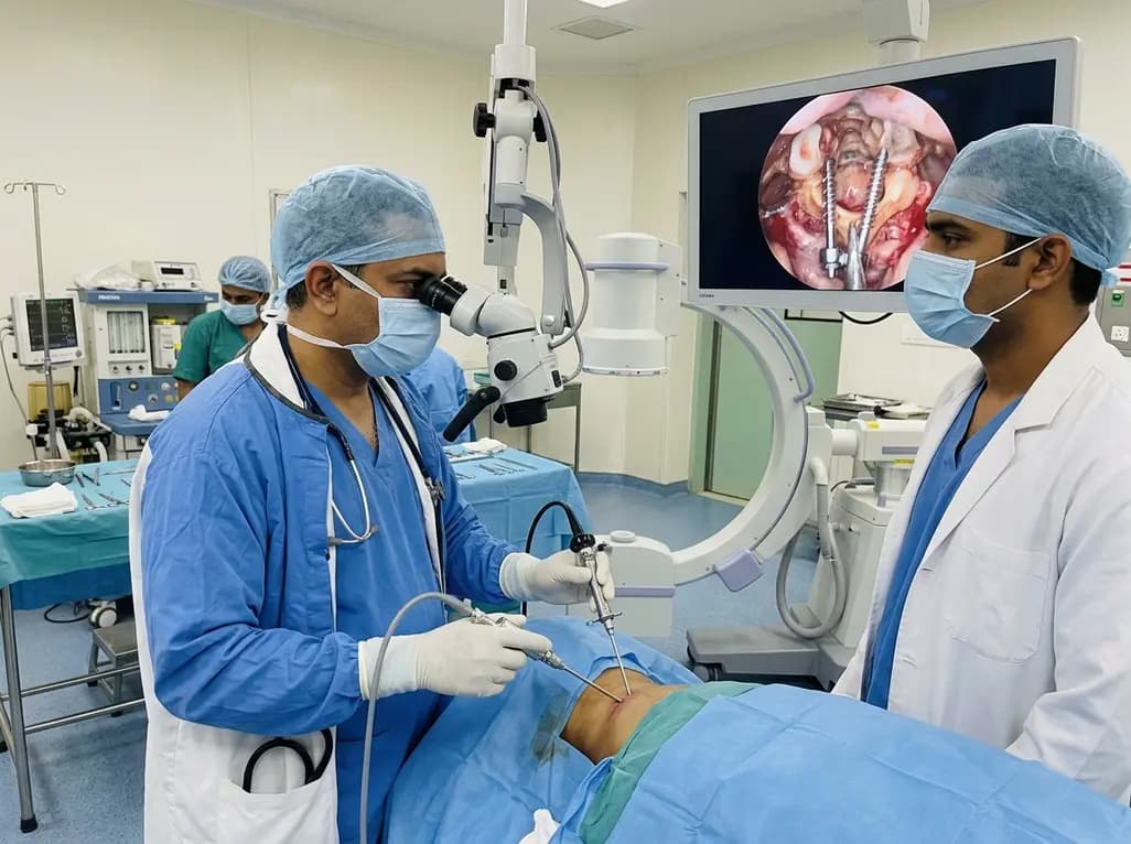
Endoscopic posterior cervical fusion combines minimally invasive decompression, joint preparation, and rigid screw-rod fixation for atlantoaxial pathologies. Early clinical findings demonstrate solid bony union, excellent symptom relief, and minimal soft-tissue morbidity without significant vascular compromise.
Today

Researchers have developed an innovative ultrasound-responsive nano-contrast agent that co-delivers CIITA-siRNA and rapamycin directly to inflamed thyroid tissue, significantly reducing autoimmune injury and thyroid autoantibodies in preclinical models of Hashimoto thyroiditis.
Today
