.webp&w=384&q=75)

Loading, please wait...
Loading, please wait...

Preserving cognitive functions and vocational identity represents a crucial objective in modern neuro-oncology. Historically, neurosurgeons focused primarily on safeguarding basic sensorimotor and speech pathways. However, patients presenting with World Health Organization grade 2 gliomas are often young and professionally active, making the preservation of specialized vocational talents essential. Recent neurosurgical investigations demonstrate that awake connectome-based resection offers an exceptional approach to protect complex neural networks without sacrificing cytoreductive efficacy. In particular, professional and semiprofessional artists present unique surgical challenges because artistic creation integrates multi-level cognitive, emotional, and sensorimotor processes. When managing these cases, traditional localizationist models often prove inadequate. Consequently, neurosurgeons have adopted a dynamic connectomal framework that monitors distributed brain networks. This clinical review explores groundbreaking evidence demonstrating the preservation of artistic creativity following connectome-guided glioma resections.
Low-grade gliomas predominantly affect young adults who maintain active professional careers. Therefore, balancing maximal oncological resection with long-term functional preservation remains essential. Awake craniotomy with direct electrical stimulation mapping serves as the clinical gold standard for identifying critical cortical and subcortical pathways. Traditionally, surgeons adhered to a modular model of cerebral localization, assuming distinct anatomical zones govern isolated faculties. However, contemporary cognitive neuroscience indicates that human cognition arises from dynamic, interconnected neural meta-networks.
The connectome-based surgical philosophy replaces rigid localization with network-level mapping. During awake connectome-based resection, the surgical team navigates cortical boundaries and subcortical white matter tracts while actively monitoring distributed cognitive networks. By evaluating motor, visual, semantic, and executive pathways in real time, the surgeon identifies individual functional boundaries. Consequently, resection continues safely until functional limits appear, rather than halting at arbitrary anatomic landmarks. Furthermore, this dynamic approach respects individual neuroplasticity induced by the indolent neoplasm. Thus, surgical teams achieve significant tumor removal while safeguarding the neural connections necessary for complex human creativity.
A landmark clinical study evaluated nineteen consecutive artists who underwent awake surgical resection for IDH-mutated grade 2 gliomas. Specifically, the cohort comprised fifteen professional artists and four semiprofessional artists. The professional subgroup included five architects, two comedians, two musicians, two dancers, a sculptor, a plastic artist, a writer, and an art professor. Meanwhile, the semiprofessional artists included two musicians, a poet, and a painter, who also worked as a chemistry professor, a computer scientist, a theater administrator, and a social worker.
Demographically, the study group included ten men and nine women with a mean age of 36.8 years. Sixteen patients were right-handed, reflecting typical cerebral dominance. Preoperatively, patients maintained remarkable baseline functional independence, demonstrated by a mean Karnofsky Performance Scale score of 94.7. Tumors involved both hemispheres, with eleven left-sided and eight right-sided lesions distributed across all five cerebral lobes. The mean preoperative lesion volume was 52.8 cubic centimeters. Except for one architect who suffered from intractable epilepsy, all individuals maintained active creative practice prior to surgical intervention.
Clinicians frequently debate whether preserving artistic abilities requires specialized intraoperative tasks during tumor resection. For instance, some practitioners have advocated having musicians play instruments or having painters sketch during craniotomy. However, the Montpellier neurosurgical team implemented a standardized continuous multitasking protocol without introducing any specific artistic tasks. Throughout tumor removal, patients performed real-time tasks assessing basic motor execution, visual processing, semantic cognition, and executive control.
The surgical team systematically employed dual-task paradigms to challenge cognitive flexibility and working memory. Specifically, patients performed motor coordination tasks while simultaneously naming items or processing visual stimuli. Direct electrical stimulation mapped critical cortical areas and essential subcortical tracts, including the arcuate fasciculus, inferior fronto-occipital fasciculus, and superior longitudinal fasciculus. By continuously monitoring these structural foundations, the surgical team preserved the distributed meta-networks that sustain artistic cognition. Consequently, this protocol avoided the logistical complexities of intraoperative artistic instruments while ensuring comprehensive protection of higher-order cognitive functions.
The surgical results established outstanding oncological and neurological outcomes. Specifically, the mean extent of resection reached 91.2 percent, leaving a mean residual tumor volume of only 5.0 cubic centimeters. Histopathological evaluation confirmed twelve diffuse astrocytomas and seven oligodendrogliomas, all displaying IDH mutations. Notably, no patient suffered permanent motor or linguistic deficits. Only one patient experienced an intentional, voluntary-induced hemianopia required to secure an oncologically sound margin. At three months postoperatively, the mean Karnofsky Performance Scale score reached 95.7, confirming rapid recovery.
Long-term follow-up confirmed the durability of these surgical results over a mean observation period of 7.6 years. Overall, 84.2 percent of patients remained alive at their latest evaluation. Among three deaths, one resulted from an unrelated medical condition. Because low-grade gliomas naturally progress, five patients eventually underwent repeat awake surgery. Interestingly, semiprofessional artists showed a significantly higher rate of repeat surgery than professional artists. However, other oncological metrics remained comparable across groups. Ultimately, these findings verify that aggressive cytoreduction does not require compromising functional integrity.
The central finding of this investigation concerned the preservation of artistic vocational performance. Remarkably, all nineteen patients returned to their artistic practice at their pre-surgical professional or semiprofessional level. Furthermore, comprehensive qualitative and neuropsychological evaluations showed that no patient reported a subjective loss of creativity. Instead, artists maintained intact creative drive, technical precision, and imaginative depth throughout long-term follow-up.
These observations illuminate the complex neurobiology of human creative cognition. Creativity does not emanate from a single isolated brain center. Instead, creative performance emerges from coordinated interactions among large-scale networks, including the default mode network, executive control network, and salience network. Because awake connectome-based resection systematically monitors and spares the subcortical white matter pathways interconnecting these systems, it inherently shields creative faculties. Therefore, patients preserve their artistic identity even following extensive tumor resection in eloquent cerebral regions. This observation provides immense reassurance to artists facing neurosurgical procedures.
These empirical findings provide valuable lessons for neurosurgeons, neuro-oncologists, and rehabilitation teams. First, the data challenge the assumption that patients must perform bespoke artistic tasks during awake craniotomy. Bringing instruments or painting materials into the operating theater introduces substantial logistical, ergonomic, and anesthetic difficulties. Consequently, proving that standardized continuous multitasking preserves artistic capabilities simplifies surgical procedures and broadens clinical applicability.
Second, the study expands traditional benchmarks of neurosurgical success. Historically, teams focused primarily on preventing overt paresis or severe aphasia. However, contemporary neurosurgery must safeguard higher-order cognition, personality, and vocational identity. Preserving the ability to paint, design, or perform directly influences postoperative quality of life and psychological well-being. Therefore, clinicians must incorporate thorough vocational assessments into preoperative surgical planning. Furthermore, connectome-based mapping benefits not only professional artists but every patient with low-grade glioma, as all individuals rely on complex cognitive networks. Ultimately, this approach harmonizes aggressive tumor control with the preservation of personal identity.
No, neurosurgeons do not need to test specific artistic activities during awake craniotomy. Standardized intraoperative multitasking protocols sufficiently protect complex artistic talents. By monitoring core sensorimotor, language, visual, and executive networks, surgeons safeguard the underlying neural connectivity required for creative expression and professional artistic performance.
Awake connectome-based resection utilizes direct electrostimulation to identify functional cortical and subcortical boundaries in real time. Rather than relying on localized anatomical landmarks, this technique preserves distributed neural networks and critical white matter pathways. Consequently, surgeons maximize tumor removal while safeguarding the neural foundations of higher-order cognition.
The cohort achieved a mean tumor resection extent of 91.2 percent with minimal residual volume. Across a mean 7.6-year follow-up, 84.2 percent of patients remained alive, and Karnofsky Performance Scale scores improved postoperatively. Five patients safely underwent repeat awake surgery without suffering permanent functional deficits.
Disclaimer: This content is for informational and educational purposes only... Refer to the latest local and national guidelines for clinical practice.
References

Read summarized clinical updates, watch expert medical content, and earn CME certifications right from your smartphone.
Open your smartphone camera to scan and install.





A landmark study in Journal of Neurosurgery reveals that awake connectome-based resection preserves artistic abilities and creativity in 100% of professional and semiprofessional artists undergoing surgery for WHO grade 2 glioma, without requiring specialized intraoperative artistic tasks.
Today

Discover how advanced clinical trial designs, including treat-through, randomized withdrawal, and continuous sequential parallel comparison designs (SPCD), evaluate induction and maintenance efficacy in chronic disease drug development.
Today

Meghalaya recorded a historic 55% reduction in maternal deaths, dropping from 244 to 107 over five years. Through the MOTHER app, dedicated transit homes, and expanded infrastructure like the 150-bed Tura Maternity Hospital with its first public NICU, the state demonstrates an effective public health model.
Today
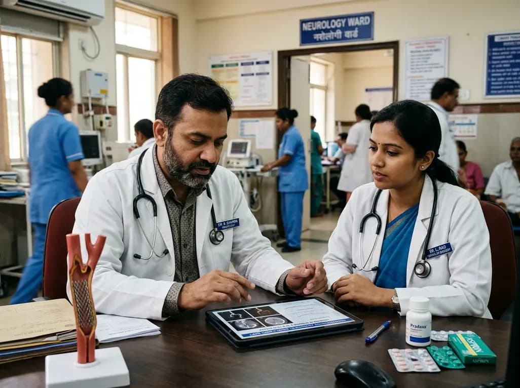
A multicenter propensity-matched study evaluated DOAC plus single versus dual antiplatelet therapy after carotid artery stenting. Findings revealed comparable intracranial hemorrhage and ischemic stroke rates, supporting DOAC plus SAPT as a viable antithrombotic strategy in high-risk patients.
Today

A cross-sectional study reveals that sarcopenic obesity independently increases functional impairment and severe disc degeneration in patients with degenerative lumbar spinal stenosis, highlighting the need for body composition assessment.
Today

At Homa CME 2026 at Hyderabad's T-Hub, healthcare leaders emphasized moving beyond routine metrics through digital calculators and precision stratification. Discover why characterizing individual insulin secretion, resistance patterns, and vascular risks transforms chronic metabolic care in India.
Yesterday
