

Loading, please wait...
Loading, please wait...
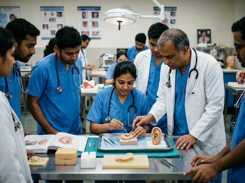
Congenital microtia presents one of the most intricate challenges in modern reconstructive surgery. Successful repair demands exceptional anatomical knowledge, artistic spatial visualization, and precise manual dexterity. Historically, surgical trainees acquired these complex abilities through direct operating room apprenticeships. However, case rarity and limited operative opportunities frequently hinder resident competence. To solve this instructional gap, surgical educators developed structured simulation environments. A groundbreaking study evaluates the educational efficacy of a structured simulation workshop designed for autologous auricular reconstruction. By offering tiered hands-on practice, this innovative curriculum provides a safe, reproducible pathway for surgeons to master critical framework fabrication techniques.
Reconstructing a congenitally absent external ear requires extraordinary technical execution. The native auricle features intricate elevations and hollows, including the helix, antihelix, concha, and tragus. Therefore, recreating these delicate contours from autologous rib cartilage demands both surgical accuracy and three-dimensional sculpting ability. Surgeons commonly utilize the Brent or Nagata techniques, which involve harvesting cartilage from the sixth through ninth ribs. The operator must assemble these harvested segments into a cohesive, structurally stable construct capable of projecting outward under taut cutaneous tissue.
Consequently, the procedure presents a formidable learning curve for novice trainees. When surgeons carve cartilage incorrectly, they cannot easily replace the precious autologous donor tissue. Donor cartilage remains strictly finite, so operative errors directly compromise patient outcomes. Furthermore, most plastic surgery and otolaryngology training programs encounter limited microtia cases annually. As a result, residents rarely gain sufficient primary operator experience before graduation. Simulation workshops offer an essential solution to this educational dilemma, allowing surgeons to practice demanding cartilage carving without jeopardizing patient safety or tissue integrity.
The highlighted educational curriculum utilized a graduated, multi-material training strategy to optimize skill acquisition. Rather than exposing learners immediately to expensive models, educators implemented a sequential hierarchy of physical media. First, participants attended comprehensive didactic lectures detailing normal auricular anatomy, landmark ratios, and foundational assembly principles. Subsequently, attendees initiated tactile exercises using everyday low-fidelity materials, specifically modeling compound and dense raw potatoes. These accessible media allowed learners to grasp basic carving depths, proportion relationships, and outline tracing without performance pressure.
After refining basic spatial awareness on simple media, participants transitioned to high-fidelity synthetic chondral cartilage. These specialized models accurately mirrored the sixth, seventh, eighth, and ninth human costal ribs. Trainees harvested simulated baseplates, shaped outer helical rims, and carved intricate antihelical complexes using specialized gouges and scalpels. Next, they secured the distinct cartilage elements using stainless steel wire and micro-sutures. This structured progression allowed participants to internalize the complex multi-step fabrication sequence methodically. Consequently, the tiered methodology transformed an overwhelming surgical task into manageable, step-by-step technical objectives.
The objective findings from the simulation workshop demonstrate significant educational advancement across all participant cohorts. Across two distinct sessions, thirty surgical learners participated, including integrated plastic surgery residents, independent residents, craniofacial fellows, and senior medical students. Notably, ninety-three percent of these attendees reported zero prior operative experience with ear framework construction. This demographic highlights the widespread scarcity of real-world microtia exposure in standard surgical residency programs.
Pre-workshop and post-workshop evaluations revealed profound subjective and objective benefits. Trainees completed a standardized microtia repair confidence scale measuring their self-assessed understanding of cartilage harvesting, subunit carving, and framework assembly. The mean total confidence score surged by fifty-four percent, increasing from 23.5 at baseline to 36.1 following workshop completion. Furthermore, statistical analysis confirmed that this improvement was highly significant across every measured technical domain. Participants universally praised the hands-on instruction, confirming that realistic synthetic cartilage models effectively simulated the resistance, tactile feedback, and structural behavior of living human cartilage.
These remarkable outcomes underscore the vital necessity of integrating simulation models into modern surgical residency training. In an era governed by work-hour restrictions and heightened operating room efficiency demands, traditional apprenticeship models cannot reliably foster mastery of ultra-specialized procedures. Therefore, residency programs must establish dedicated simulation laboratories that support deliberate practice outside clinical settings. Ear framework carving represents a prime candidate for simulation because errors on synthetic ribs carry no biological consequences.
Moreover, modern three-dimensional printing technologies make high-fidelity rib models increasingly accessible and cost-effective. By standardizing synthetic costal cartilage fabrication, academic departments can organize recurring annual workshops for junior and senior trainees alike. Repeated simulation sessions enable residents to develop fine motor coordination, instrument muscle memory, and spatial problem-solving skills over time. Consequently, when trainees finally encounter an autologous ear reconstruction in the operating theater, they approach framework assembly with grounded confidence rather than hesitation. This educational paradigm shift ultimately elevates resident training quality while safeguarding patient care standards.
In developing nations like India, congenital microtia represents a substantial clinical burden across both urban and rural healthcare settings. Indian plastic surgeons and otolaryngologists frequently encounter adolescent and pediatric patients presenting for delayed auricular reconstruction. However, formal hands-on training opportunities in microtia surgery remain concentrated in a small handful of specialized tertiary centers. Consequently, many reconstructive surgeons across regional teaching hospitals enter practice without substantial experience in autologous cartilage carving.
Adopting scalable simulation models could transform microtia training across Indian surgical institutes. Low-cost training materials, including dental wax, potatoes, and locally cast silicone or polyurethane rib constructs, provide highly affordable alternatives to expensive imported commercial models. Furthermore, national associations such as the Association of Plastic Surgeons of India and the Foundation for Head and Neck Oncology can integrate these tiered carving workshops into national conferences and residency boot camps. By systematically expanding simulation access, surgical training programs can build widespread technical capacity. Ultimately, empowering regional Indian surgeons with refined carving competence ensures that patients nationwide receive high-caliber, cosmetically superior auricular reconstructions.
Surgeons consider this procedure exceptionally challenging because the human ear possesses a delicate three-dimensional topography with subtle convolutions. In addition, harvesting costal cartilage carries donor-site morbidity risks, including pneumothorax and contour deformities. Because clinicians rarely perform microtia repairs in standard training, trainees lack repeated exposure. Consequently, mastering the delicate assembly of structural cartilage frameworks requires intensive spatial appreciation and manual dexterity that traditional observation cannot adequately cultivate.
Low-fidelity materials like modeling compound and raw potatoes offer accessible mediums for deliberate practice. Specifically, they allow novice carvers to understand proportional anatomy and delicate three-dimensional depth without squandering costly synthetic models. Trainees can sculpt basic contours, practice scapha hollows, and refine helix creation repeatedly. Therefore, transitioning through these forgiving materials builds fundamental muscle memory and instrument handling before participants carve high-fidelity costal cartilage analogs.
High-fidelity simulation provides an authentic tactile environment where trainees can manipulate realistic synthetic rib cartilage templates safely. Furthermore, this controlled exposure bridges the steep learning curve inherent to congenital microtia reconstruction. By standardizing framework assembly techniques before entering operating rooms, simulation workshops lower intraoperative error rates, shorten surgical duration, and protect patient safety. Consequently, academic institutions increasingly incorporate these hands-on modules into official craniofacial and reconstructive training curricula.
Disclaimer: This content is for informational and educational purposes only and should not be considered medical advice. Refer to the latest local and national guidelines for clinical practice.
References

Read summarized clinical updates, watch expert medical content, and earn CME certifications right from your smartphone.
Open your smartphone camera to scan and install.





A landmark study evaluates a high-fidelity simulation workshop for autologous auricular reconstruction. By utilizing low-fidelity shaping followed by synthetic rib cartilage carving, surgical trainees significantly improved technical confidence and anatomical mastery for microtia repair.
Today
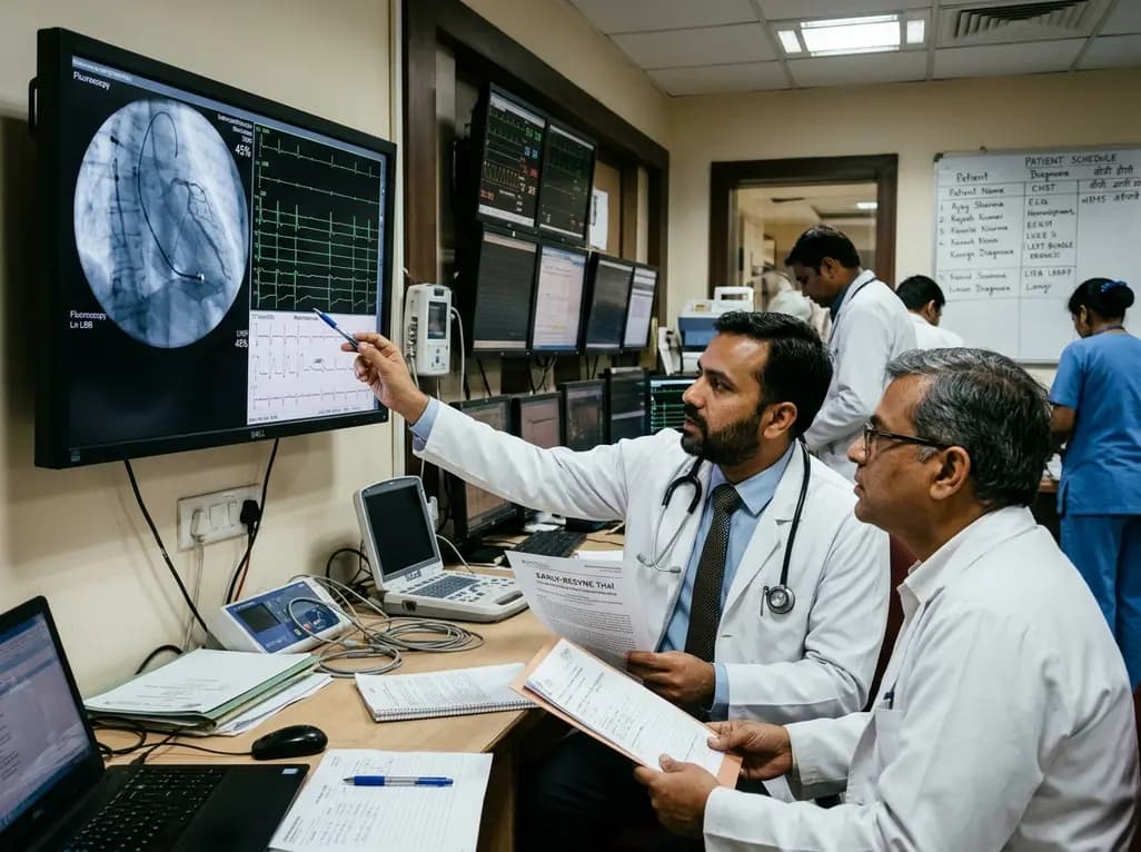
The randomized EARLY-RESYNC trial reveals that early left bundle branch pacing combined with guideline-directed medical therapy significantly improves LVEF and reduces LV end-systolic volume compared to medical therapy alone in patients with complete LBBB and mildly reduced ejection fraction (36%-50%).
Today
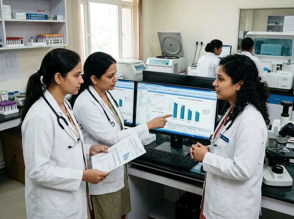
A comparative evaluation of INNO-LiPA, Cobas 4800, and Amplicor highlights significant assay-dependent variation in high-risk HPV detection, emphasizing the clinical distinction between analytical sensitivity and screening specificity.
Today
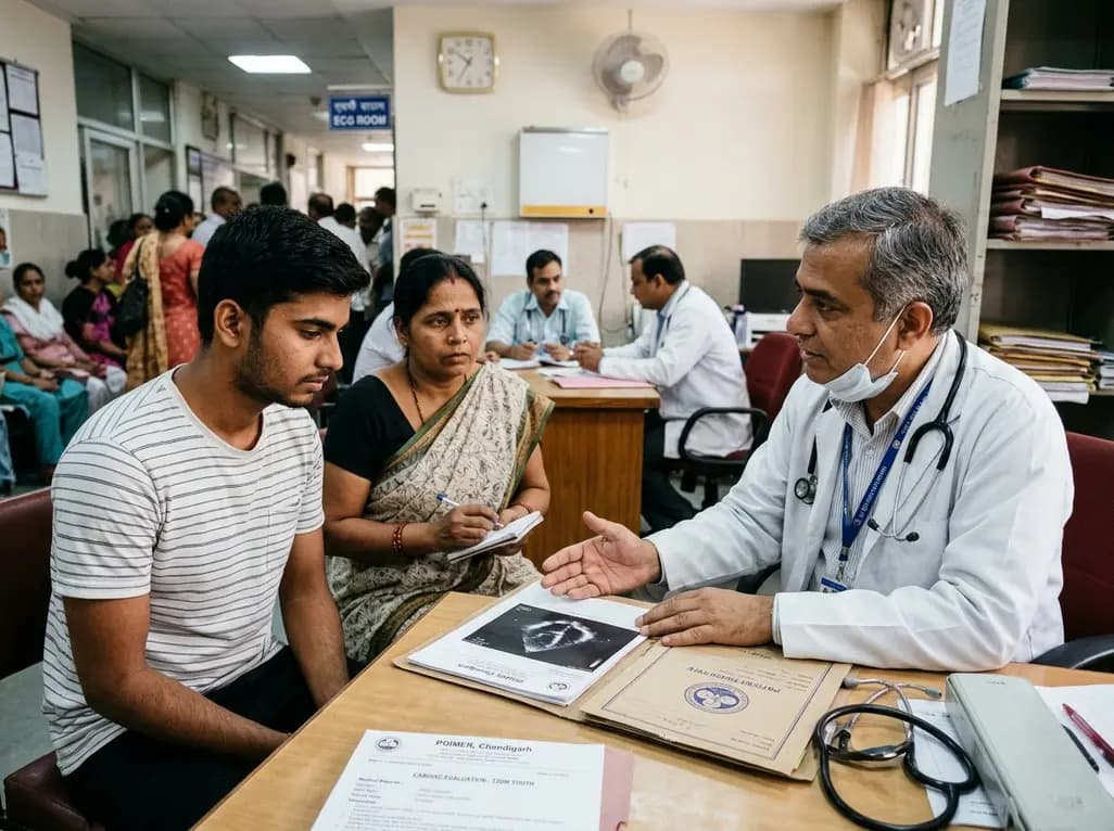
Recent data from PGIMER reveals that young adults diagnosed with type 2 diabetes face an aggressive disease trajectory. They encounter severe cardiovascular and microvascular damage within five years, occurring at higher rates than in type 1 diabetes despite lower mean blood glucose levels.
Today
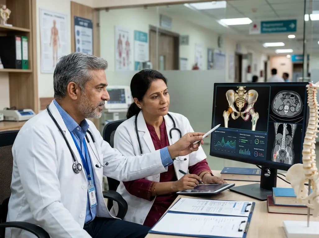
Discover the six core principles of responsible AI in orthopaedics, emphasizing algorithmic fairness, transparency, clinical reliability, and specialist oversight to safeguard patient trust and outcomes.
Today
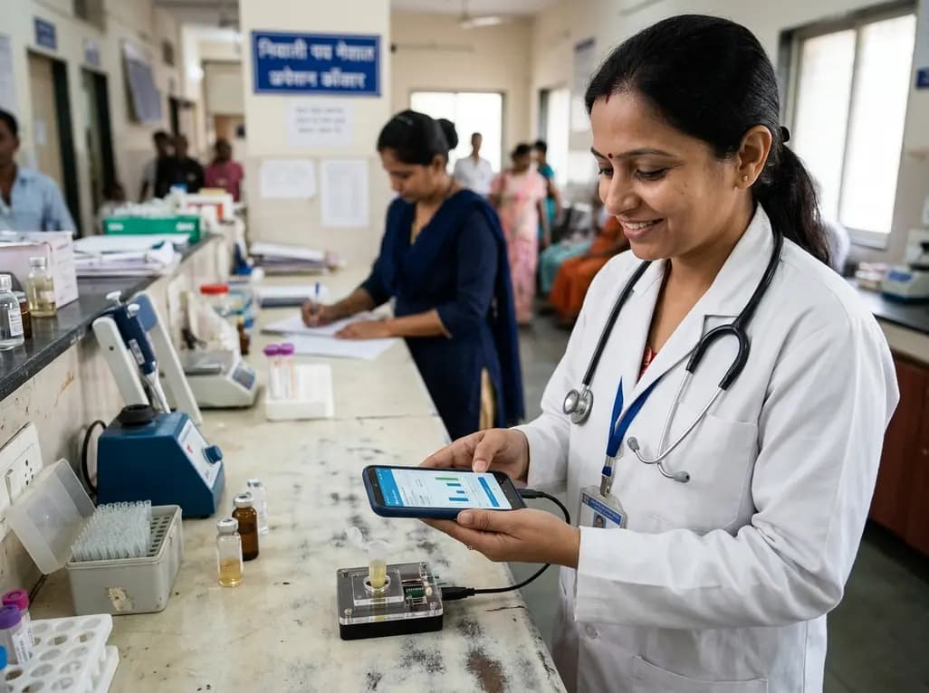
A novel smartphone-assisted biosensor utilizing molecularly imprinted polymers and catalytic nanozymes offers antibody-free, enzyme-free urinary microalbumin testing, enabling rapid, low-cost point-of-care screening for early chronic kidney disease.
Today
