

Loading, please wait...
Loading, please wait...

Neurosurgical interventions for deep intracranial lesions require exceptional anatomical precision and surgical confidence. For decades, traditional frame-based stereotactic brain biopsy has served as the diagnostic benchmark for evaluating deep or eloquent brain pathology. However, mechanical frames inherently lack direct, real-time visual feedback once needle insertion begins. Operating surgeons rely strictly on mechanical scales and pre-calculated stereotactic coordinates. Consequently, this workflow creates a blind trajectory where surgeons cannot visualize underlying vital structures. Specifically, recent clinical investigations demonstrate that wearable augmented reality headsets can effectively bridge this visualization gap. By projecting patient-specific digital holograms directly onto the operative field, augmented reality delivers intuitive trajectory monitoring while preserving mechanical accuracy.
Surgeons historically depend on rigid stereotactic apparatuses to sample obscure intracranial tumors. Systems such as the Cosman-Roberts-Wells frame provide submillimeter mechanical reliability across varied operative trajectories. Nevertheless, traditional frame-based interventions present distinct operational challenges during daily neurosurgical routines. Surgeons face considerable cognitive burdens because they must mentally translate two-dimensional cross-sectional slices into three-dimensional surgical reality. Furthermore, standard navigation screens require clinicians to shift their gaze repeatedly away from the surgical target toward an external monitor. This visual disconnection disrupts concentration and lengthens operative times. Although frameless optical navigation systems offer greater freedom, they often sacrifice rigidity and struggle with mechanical drift. Therefore, integrating mixed-reality headsets into traditional stereotactic setups represents an evolutionary milestone in neurosurgery. Head-mounted displays overlay multimodal imaging datasets directly onto the sterile field. Consequently, the surgical team maintains an uninterrupted field of view while manipulating instruments. This hybrid strategy combines the structural stability of classic stereotactic frames with dynamic three-dimensional computer vision. As a result, neurosurgeons gain immediate spatial awareness without abandoning proven stereotactic principles. This technological convergence ensures both anatomical precision and surgical ergonomics throughout complex intracranial procedures.
In a groundbreaking prospective feasibility study, neurosurgeons evaluated a novel holographic navigation workflow during ten frame-based stereotactic brain biopsy procedures. The operative protocol coupled the Cosman-Roberts-Wells stereotactic frame with the Microsoft HoloLens 2 headset and dedicated Medgital software. Preoperatively, the clinical team acquired high-resolution magnetic resonance images and co-registered them with intraoperative computed tomography data in NeuroSight Arc. Subsequently, the surgical software generated comprehensive holographic models displaying the patient's craniofacial anatomy, target lesion, and planned needle trajectory. A lightweight passive optical tracker attached directly to the biopsy cannula enabled continuous holographic instrument tracking. Thus, the operating surgeon observed a dynamic virtual replica of the advancing needle superimposed precisely over the patient's head. Moreover, this setup eliminated the need for external monitor checks during the critical insertion phase. Additionally, the operative team seamlessly integrated the wearable display into the standard sterile environment without altering traditional draping practices. By overlaying critical anatomical boundaries onto the physical frame, the platform provided continuous visual guidance from skin incision to specimen collection. This seamless digital integration demonstrated remarkable practical compatibility with existing operating theater protocols.
Speed and spatial fidelity remain crucial benchmarks for adopting new intraoperative technologies. Notably, the pilot study achieved a flawless technical success rate, confirming seamless augmented reality navigation in all ten surgical patients. Spatial registration occurred remarkably fast, recording a mean registration time of only 79 ± 27 seconds. Consequently, the holographic setup introduced negligible operational delay into the standard surgical timeline. Furthermore, quantitative assessments demonstrated outstanding spatial alignment between holographic projections and conventional stereotactic trajectory planning. The mean augmented reality trajectory error measured 2.21 ± 0.67 millimeters relative to standard frame calculations. In addition, this sub-three-millimeter margin aligns closely with established acceptable thresholds for cranial biopsy guidance. The tracking platform maintained consistent holographic stability even during minor surgeon head movements across diverse viewing angles. Because the passive marker provided reliable position recognition, the system prevented spatial drift throughout needle deployment. Therefore, the holographic overlay faithfully represented real-time needle depth and axial orientation at every surgical stage. Consequently, clinicians can rely on holographic trajectories without compromising the meticulous precision demanded in deep neurosurgical corridors.
The fundamental objective of any biopsy procedure centers on obtaining adequate diagnostic tissue without provoking neurological harm. In this pilot investigation, the surgical team secured a definitive histopathological diagnosis in all ten patients, achieving a 100% diagnostic yield. On average, the operating neurosurgeon required 2.4 needle passes per patient to acquire sufficient diagnostic tissue samples. Importantly, the real-time visualization of needle depth prevented unnecessary multiple punctures into eloquent brain parenchyma. Furthermore, no patient experienced postoperative intracranial hemorrhage, regional neurological deficits, or mortality. The enhanced visual orientation enabled surgeons to confirm target engagement before opening the biopsy window. Additionally, holographic trajectory visualization allowed clinicians to avoid critical sulcal vessels along the immediate needle path. Standard frame-based systems provide rigid stability, but augmented reality introduces the sensory reassurance that surgeons historically lacked. Thus, the clinical findings confirm that holographic visualization preserves patient safety while maintaining the high diagnostic efficacy of frame stereotaxy. Furthermore, achieving such reliable tissue yields without hemorrhagic morbidity underscores the profound clinical value of augmented navigation.
Beyond technical accuracy, wearable augmented reality markedly improves intraoperative ergonomics and surgeon comfort. Traditional stereotaxy imposes heavy cognitive strain, forcing clinicians to correlate mental reconstructions with physical scales. In contrast, head-mounted displays merge target anatomy, trajectory pathways, and instrument tips into a unified field of view. Neurosurgeons maintain direct visual contact with the patient, eliminating repetitive neck twisting toward peripheral equipment consoles. Moreover, this natural visual alignment flattens the technical learning curve for junior surgical trainees. In academic training centers, supervising faculty and residents can share the identical holographic perspective, enhancing procedural instruction and intraoperative communication. However, several engineering challenges warrant further investigation before widespread clinical implementation. Current headsets present battery life constraints, occasional optical display calibration shifts, and weight considerations during prolonged surgeries. Future technological refinements will likely integrate artificial intelligence for automated trajectory optimization and instant vascular segmentation. As mixed-reality devices become lighter and more intuitive, holographic guidance will inevitably become routine across cranial neurosurgery. Consequently, these developments will empower surgical teams to deliver safer, more efficient patient care worldwide.
Augmented reality provides real-time, three-dimensional visual feedback during needle advancement in stereotactic brain biopsy. By superimposing the planned trajectory, patient anatomy, and tracked biopsy cannula onto the surgical field, it eliminates the traditional blind corridor. Consequently, surgeons verify needle trajectory directly without looking away toward external monitors.
Recent clinical data demonstrates that augmented reality achieves high spatial fidelity, showing a mean trajectory error of 2.21 ± 0.67 millimeters relative to standard frame calculations. Furthermore, holographic registration requires only 79 seconds. This sub-three-millimeter accuracy meets established clinical safety standards for deep cranial biopsies without compromising mechanical precision.
Augmented reality currently complements rather than replaces traditional stereotactic frames. The mechanical frame ensures rigid skull fixation and stable needle alignment, while the holographic headset provides intuitive visual guidance. This hybrid combination maintains the physical stability necessary for precise intracranial sampling while overcoming the visual limitations of traditional frame stereotaxy.
Disclaimer: This content is for informational and educational purposes only... Refer to the latest local and national guidelines for clinical practice.
References
1. Konovalov AN et al. Application of augmented reality in frame-based stereotactic brain biopsy: a case series feasibility study. J Neurooncol. 2026 Nov 20. doi: 10.1007/s11060-026-05886-9. PMID: 43062733.
2. Satoh D, et al. Application of Augmented Reality to Stereotactic Biopsy. NMC Case Rep J. 2019;7(1):1-6. doi: 10.2176/nmccrj.cr.2019-0082.
3. Skyrman S, et al. Augmented Reality Navigation with Intraoperative Imaging in Stereotactic Neurosurgery. Acta Neurochir (Wien). 2023;165(4):1011-1020. doi: 10.1007/s00701-023-05512-y.

Read summarized clinical updates, watch expert medical content, and earn CME certifications right from your smartphone.
Open your smartphone camera to scan and install.





A clinical feasibility study reveals that integrating augmented reality via Microsoft HoloLens 2 into frame-based stereotactic brain biopsy provides real-time needle visualization, a 79-second registration speed, 2.21 mm accuracy, and a 100% diagnostic yield without complications.
Today

A landmark study reveals that congenital DLK1 deficiency in women with a history of central precocious puberty causes visceral adiposity, mitochondrial dysfunction, oxidative stress, and neutrophil activation, highlighting the critical need for lifelong cardiometabolic surveillance.
Today

Estetrol (E4), a native fetal estrogen, offers a unique tissue-selective profile with minimal hepatic impact and lower thrombotic risk. This review highlights its clinical use in oral contraception, menopausal symptom management, and emerging applications across oncology and neurology.
Today

Explore revascularisation strategies in dialysis-dependent chronic limb-threatening ischaemia. This review examines surgical bypass vs endovascular therapy, BEST-CLI trial findings, procedural morbidity, conduit availability, and multidisciplinary decision-making for optimal limb salvage.
Today

A recent cross-sectional analysis reveals that over a quarter of randomized controlled trials for non-specific low back pain face discontinuation or unknown status, while more than 36% of completed trials remain unpublished, exposing notable reporting gaps in spine and musculoskeletal clinical research.
Today
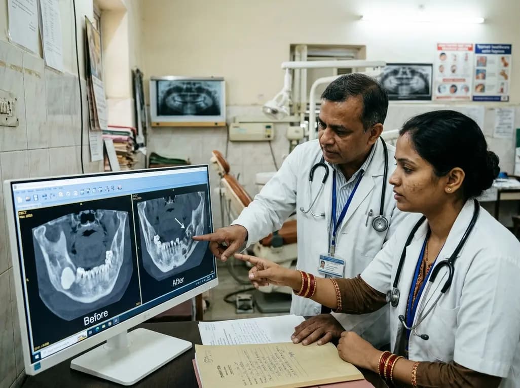
This case report examines the longitudinal CBCT evolution of Stage 2 MRONJ in a 70-year-old woman on denosumab at the site of a pre-existing mandibular dense bone island. It emphasizes the radiographic features, differential diagnosis, and multidisciplinary strategies required for high-risk bone lesions.
Today
