

Loading, please wait...
Loading, please wait...
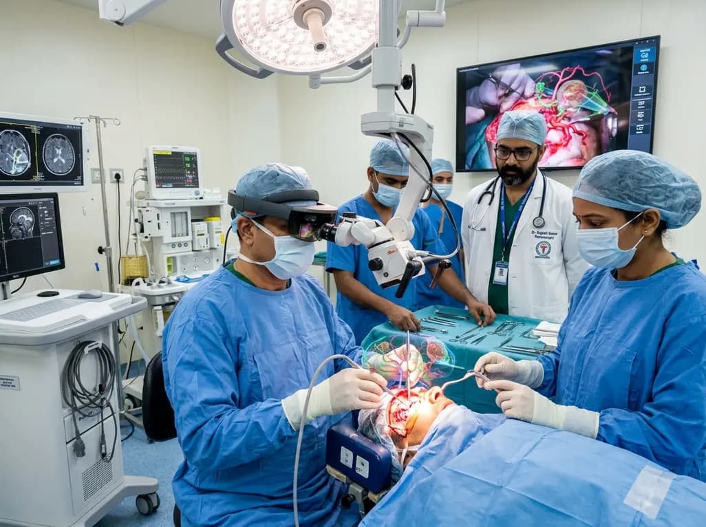
The integration of head-mounted displays has introduced augmented reality neurosurgery into daily clinical practice. Traditionally, surgical navigation required operators to shift their visual attention between the surgical site and external monitors. Consequently, this fragmented visualization disrupted surgical rhythm and hand-eye coordination. Head-mounted systems eliminate this challenge by projecting patient-specific three-dimensional holograms directly onto the operative field. Therefore, surgeons maintain an uninterrupted line of sight while viewing vital structures beneath the anatomical surface. Recent clinical investigations confirm that holographic overlays substantially improve spatial orientation during intricate cranial and spinal procedures. Moreover, wearable navigation platforms help surgical teams align skin incisions and bone flaps with remarkable accuracy. As operating rooms adopt advanced digital visualization, understanding this wearable technology becomes essential for neurosurgeons. However, deploying holographic guidance in real-world suites requires strict assessment of registration accuracy and team workflow.
A single-center investigation evaluated the Medivis head-mounted platform across 105 consecutive neurosurgical procedures performed by eight attending neurosurgeons. The cohort included 60 female and 45 male patients with an average age of 53.5 years. Cranial surgeries formed the vast majority of operations, representing 87.6 percent of the total series. Within this group, neurosurgeons treated intracranial tumors in 69 patients. Meningiomas accounted for 22.9 percent of all cases, while glioblastomas comprised 19.0 percent. Additionally, teams used holographic navigation during cerebrovascular reconstructions, stereotactic punctures, and functional surgeries. Spinal interventions constituted another important subgroup, where operators utilized dedicated spine modules for precision instrumented stabilization. Systematically recording every operation with integrated cameras allowed researchers to evaluate procedural nuances thoroughly. Consequently, this diverse real-world series demonstrated that wearable holographic systems adapt readily to diverse anatomical targets and complex pathologies across cranial and spinal surgery.
Achieving stereotactic accuracy requires robust patient registration and stable reference frame placement. In cranial cases, clinicians utilized custom reference frames in 57 procedures to support dependable spatial tracking. Furthermore, surface matching registration served as the primary alignment method across 93 cranial operations. During this sequence, the headset cameras captured facial surface topography, matching it directly to preoperative volumetric scans. Consequently, this contact-free registration eliminated bulky invasive head-pins during initial setup and reduced anesthesia preparation time. However, neurosurgeons recognized that tissue shift following craniotomy could alter coordinate alignment. Therefore, surgical teams continuously validated holographic projections against palpable bony landmarks and superficial cortical vessels. Whenever mechanical drift occurred, surgeons recalibrated the tracking coordinates before continuing dissection. Thus, combining rapid surface registration with rigorous anatomical verification preserved reliable navigation throughout intricate intradural stages.
Cranial oncological surgery demands maximal tumor resection while protecting eloquent functional brain areas. Notably, multimodal holographic planning guided 69 cranial tumor resections in this cohort. By fusing structural magnetic resonance imaging with diffusion tensor tractography, the software rendered comprehensive three-dimensional projections. Neurosurgeons viewed these structural models directly over the cranium, which facilitated targeted scalp incisions and tailored craniotomies. Consequently, surgical teams achieved gross-total resection in 62 of 69 tumor operations, achieving an impressive 89.8 percent resection success rate. Specifically, operators clearly appreciated the margins of high-grade glioblastomas and skull-base meningiomas before making their initial cortical incision. Furthermore, holographic tractography overlays helped surgeons visualize adjacent motor and language tracts throughout subcortical debulking. Accordingly, multimodal planning offered enhanced surgical confidence and helped clinicians minimize postoperative neurological deficits.
Spine surgery requires absolute navigational precision to prevent devastating spinal cord or vascular injuries. In this real-world case series, surgeons utilized dedicated spine navigation software to guide 34 pedicle screw placements. Remarkably, postoperative imaging confirmed that all 34 pedicle screws achieved a Gertzbein-Robbins grade of A. This perfect accuracy demonstrated zero pedicle breaches, highlighting the reliability of wearable trajectory overlays. Furthermore, holographic navigation enabled operators to place screws smoothly without continually looking away toward remote workstations. Beyond spinal instrumentation, teams successfully used augmented visualization during complex cerebrovascular procedures and percutaneous punctures. Specifically, holographic models identified feeder vessels and defined aneurysmal neck orientations during vascular dissections. Therefore, wearable augmented guidance provides tangible clinical advantages across spinal stabilization, vascular exposure, and deep percutaneous access corridors.
Despite notable clinical success, augmented reality cannot entirely replace conventional neuronavigation systems at present. For instance, head-mounted displays introduce physical ergonomic challenges, including headset weight, visual fatigue, and restricted peripheral vision. Furthermore, significant brain shift occurring after cerebrospinal fluid drainage can degrade hologram alignment during surgery. When registration accuracy declined, neurosurgeons promptly reverted to conventional navigation or trusted anatomical landmarks to ensure patient safety. Consequently, successful integration depends on careful case selection and structured surgical workflows. Centers adopting this technology must establish formal simulation training to flatten the learning curve for operating room staff. In addition, future technological iterations should integrate real-time intraoperative imaging to adjust virtual models dynamically as brain geometry shifts. Ultimately, wearable holographic systems represent a transformative surgical adjunct when clinicians understand and respect their technical boundaries.
Augmented reality neurosurgery projects three-dimensional patient anatomy directly onto the operative field, eliminating the constant distraction of looking away at external monitors. Consequently, this seamless in situ visualization preserves natural hand-eye coordination, accelerates spatial orientation, and supports precise incision planning. Furthermore, multimodal holographic overlays allow neurosurgeons to visualize critical subcortical tracts, deep tumor boundaries, and vascular structures simultaneously, thereby improving resection margins while protecting adjacent eloquent brain tissue during complex procedures.
Intraoperative brain shift poses an ongoing challenge because cerebrospinal fluid loss and tumor debulking alter parenchymal position relative to preoperative scans. To mitigate this issue, surgeons actively verify holographic overlays against fixed anatomical landmarks, such as the skull base and major vessels. Moreover, if registration discrepancies arise, clinicians revert to conventional pointer navigation or intraoperative ultrasound. Future platforms aim to incorporate dynamic intraoperative image updates to correct virtual alignments automatically during surgery.
Flawless pedicle screw placement relies heavily on precise patient registration and stable reference frame fixation to avoid tracking drift. By anchoring rigid markers securely to adjacent spinous processes, the head-mounted system projects virtual trajectories directly onto the bony anatomy. Additionally, surgeons observe real-time alignment angles and entry points through the headset display. This continuous guidance prevents cortical breaches, reduces radiation exposure from repetitive fluoroscopy, and streamlines operative workflow during complex spinal stabilization procedures.
Disclaimer: This content is for informational and educational purposes only and should not be considered medical advice. It does not establish a doctor-patient relationship nor substitute for professional clinical judgment. Always consult relevant clinical guidelines and specialists for specific patient scenarios. Refer to the latest local and national guidelines for clinical practice.
References

Read summarized clinical updates, watch expert medical content, and earn CME certifications right from your smartphone.
Open your smartphone camera to scan and install.





A landmark real-world study evaluates head-mounted augmented reality navigation across 105 neurosurgical procedures, demonstrating high resection rates in cranial tumors and flawless spinal pedicle screw accuracy while highlighting workflow nuances.
Today
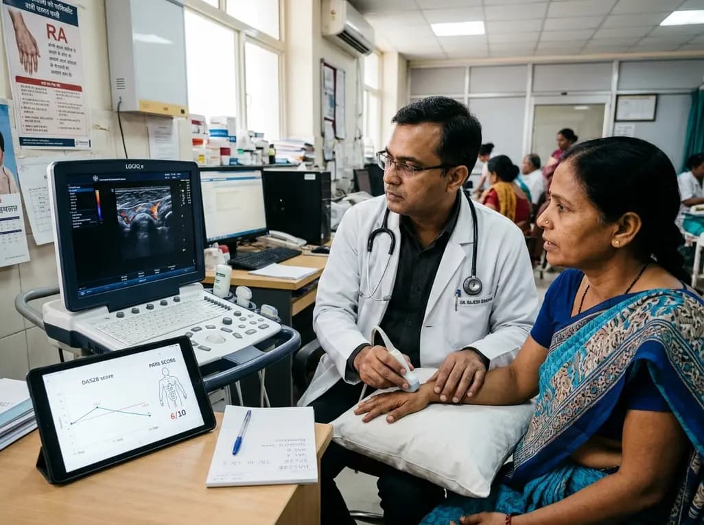
Subjective components in DAS28 often inflate disease activity scores due to central sensitization and emotional distress. Integrating the DAS28-P index, musculoskeletal ultrasound, and emerging digital biomarkers allows clinicians to differentiate true synovitis from non-inflammatory pain and prevent futile escalation.
Today

AIIMS researchers suggest yoga may alleviate gut dysbiosis in Polyendocrine Metabolic Ovarian Syndrome (PMOS) by modulating the gut-brain axis, lowering chronic inflammation, and enhancing parasympathetic tone to support metabolic and hormonal equilibrium.
Today
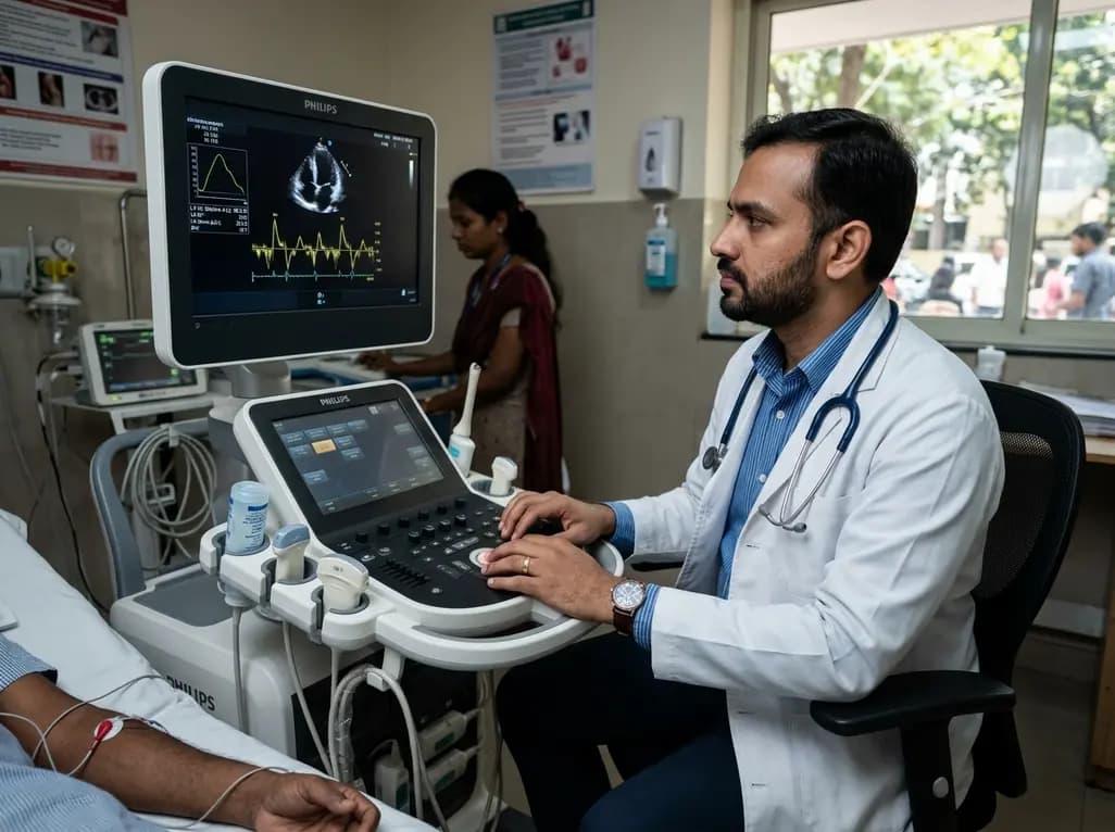
A structured practical guide to left atrial echocardiographic assessment covering biplane volumetry, phasic physiology, speckle-tracking strain, and hemodynamic filling pressure estimation for routine cardiovascular practice.
Today

Age-related spinal kyphosis is driven by vertebral fragility, disc degeneration, and paraspinal muscle dysfunction. Coexisting with locomotive syndrome and frailty, its management requires combined anti-osteoporotic therapy, spinal extensor rehabilitation, and comprehensive nutritional optimization.
Today

A longitudinal feasibility pilot study highlights that combining polygenic risk score disclosure with in-person lifestyle counseling significantly enhances genomic literacy in individuals at risk for type 2 diabetes, overcoming educational disparities and reinforcing patient motivation.
Today
