

Loading, please wait...
Loading, please wait...

Modern cancer management relies heavily on contrast-enhanced computed tomography for staging, response assessment, and surveillance. However, clinicians often worry about contrast-associated acute kidney injury when patients undergo active systemic chemotherapy. Because many antineoplastic agents carry nephrotoxic potential, oncologists and radiologists frequently balance diagnostic urgency against renal safety. Fortunately, emerging evidence indicates that patients with preserved baseline kidney function experience a relatively low incidence of post-contrast renal impairment during systemic therapy.
Contrast-associated acute kidney injury represents an acute decline in renal function occurring within 48 to 72 hours following intravascular iodinated contrast administration. Historically, clinical teams applied the term contrast-induced nephropathy to all post-procedural creatinine elevations. However, contemporary guidelines distinguish between correlative contrast-associated acute kidney injury and true contrast-induced damage caused solely by the agent. Consequently, modern nomenclature reflects the multifactorial origin of renal impairment in hospitalized or complex medical populations.
Cancer patients face multiple concurrent insults that compromise renal hemodynamics. Specifically, nausea, vomiting, decreased oral intake, and tumor-related metabolic disturbances frequently cause subclinical volume depletion. Furthermore, exposure to antimicrobial prophylaxis, nonsteroidal anti-inflammatory agents, and potentially nephrotoxic antineoplastic regimens increases tubular vulnerability. Therefore, identifying whether iodinated contrast independently precipitates acute kidney injury remains critical for rational clinical decision-making. Physicians should avoid withholding essential diagnostic imaging when true renal risk remains negligible.
A recent observational study evaluated 146 adult oncology patients receiving active chemotherapy at a tertiary cancer center. Notably, all enrolled individuals maintained an estimated glomerular filtration rate greater than 50 mL/min/1.73 m² at baseline. The cohort presented a mean age of 51.5 years and included both solid malignancies and hematological cancers. All participants received intravenous contrast-enhanced computed tomography within 45 days of their chemotherapy cycles.
Researchers observed contrast-associated acute kidney injury in only 2 of the 146 evaluated patients, establishing an incidence rate of 1.4%. Furthermore, both instances demonstrated mild, transient creatinine elevations without requiring renal replacement therapy. These reassuring results align closely with international real-world registries. Previous trials demonstrated that intravenous iodinated contrast rarely causes meaningful nephrotoxicity in individuals with an estimated glomerular filtration rate above 45 to 50 mL/min/1.73 m². Therefore, oncologists can confidently utilize contrast-enhanced computed tomography in stable patients without excessive fear of irreversible renal injury.
Interestingly, the study identified a significant disparity regarding the timing between chemotherapy infusion and contrast administration. Specifically, patients who developed renal impairment had a mean interval of only 2 days between therapy and computed tomography. In contrast, patients who maintained stable renal function had an average interval of 23.3 days. Although this finding is exploratory due to the small number of events, it highlights biological plausibility.
Chemotherapeutic drugs such as cisplatin, carboplatin, methotrexate, and ifosfamide frequently produce acute tubular stress. When iodinated contrast enters the renal vasculature shortly after cytotoxic exposure, additive physiological stress may occur. In addition, hyperosmolar contrast agents stimulate transient renal vasoconstriction and direct medullary hypoxia. Consequently, overlapping tubular toxicity and microvascular ischemia can precipitate acute functional decline. Therefore, clinicians should consider spacing elective diagnostic imaging when possible to permit tubular recovery between intensive cytotoxic infusions.
Preventing renal complications in cancer patients requires a proactive, multidisciplinary strategy involving oncologists, nephrologists, and radiologists. First, clinicians must assess volume status thoroughly before every contrast-enhanced scan. Intravenous isotonic saline represents the cornerstone of prevention for dehydrated or marginal-risk individuals. However, routine intravenous hydration is generally unnecessary for euvolemic outpatients with robust baseline renal function.
Second, medical teams must exercise strict nephrotoxin stewardship throughout the peri-imaging window. For instance, temporary discontinuation of nonsteroidal anti-inflammatory drugs, aminoglycosides, and renin-angiotensin-aldosterone system inhibitors can safeguard renal hemodynamics. Moreover, selecting low-osmolar or iso-osmolar contrast media at the lowest diagnostic volume mitigates direct epithelial exposure. Additionally, careful communication between oncology day units and radiology departments prevents accidental contrast administration during periods of severe dehydration. Ultimately, these practical steps ensure patient safety while avoiding unnecessary interruptions in cancer surveillance.
While current findings offer valuable reassurance, substantial knowledge gaps remain regarding post-contrast nephrotoxicity in oncology. Because most existing studies rely on retrospective cohorts, unmeasured confounding variables such as subclinical dehydration often blur results. Therefore, larger prospective studies must evaluate specific antineoplastic drug classes, targeted agents, and immune checkpoint inhibitors. For instance, immunotherapy-mediated interstitial nephritis presents distinct pathophysiology requiring entirely different preventive approaches.
Furthermore, novel structural urinary biomarkers such as neutrophil gelatinase-associated lipocalin and kidney injury molecule-1 may detect subclinical damage earlier than serum creatinine. Integrating these modern biomarkers could provide clearer insights into the true cellular impact of iodinated media. Meanwhile, oncologists and radiologists should continue collaborative risk stratification. Clinicians can deliver high-quality oncologic imaging while safeguarding essential kidney health by maintaining individualized patient assessment and adherence to evidence-based protocols.
Contrast-associated acute kidney injury represents an abrupt decrease in kidney function occurring within 48 to 72 hours after intravascular iodinated contrast administration. Clinicians define it by an absolute serum creatinine rise of at least 0.3 mg/dL or a relative increase exceeding 50 percent from baseline. It reflects a correlative temporal relationship rather than definitive contrast-induced toxicity.
Yes, contrast-enhanced computed tomography is remarkably safe for cancer patients who maintain preserved renal function. Studies consistently demonstrate that the incidence of significant renal impairment remains below two percent in patients with an estimated glomerular filtration rate above 50 mL/min/1.73 m². Diagnostic imaging should not be delayed when clinically indicated for staging or monitoring.
Clinicians do not routinely need to delay urgent diagnostic imaging. However, for elective scans, spacing imaging a few days after intensive, nephrotoxic cytotoxic therapy may minimize cumulative tubular stress. Medical teams should evaluate hydration status, avoid nephrotoxic drug combinations, and maintain open communication between oncology and radiology departments before scheduling non-urgent procedures.
Disclaimer: This content is for informational and educational purposes only. It is not intended to provide medical advice, diagnosis, or treatment. Always seek the advice of qualified healthcare professionals for medical conditions. Refer to the latest local and national guidelines for clinical practice.
References

Read summarized clinical updates, watch expert medical content, and earn CME certifications right from your smartphone.
Open your smartphone camera to scan and install.





A retrospective study evaluates contrast-associated acute kidney injury (CA-AKI) incidence in oncology patients receiving active chemotherapy with preserved renal function, highlighting low overall risk and timing considerations.
Today

A landmark study evaluates artificial intelligence models and prompting strategies in qualitative lupus pregnancy research, revealing that few-shot prompting in advanced LLMs reliably captures maternal experiences, clinical anxieties, and perinatal outcomes across diverse languages.
Today
A cross-sectional study in Zanzibar reveals that 46.5% of adults with type 2 diabetes experience multidomain autonomic dysfunction on the COMPASS-31 tool, primarily orthostatic intolerance, while 45.9% exhibit cardiovascular autonomic neuropathy, highlighting the urgent need for structured clinical screening.
Today
A pilot case-control study reveals that severe preoperative functional impairment, rather than tear size, predicts graft retear following arthroscopic superior capsule reconstruction using a biceps autograft. Despite structural failures, patients achieved equivalent functional gains exceeding clinical thresholds.
Today
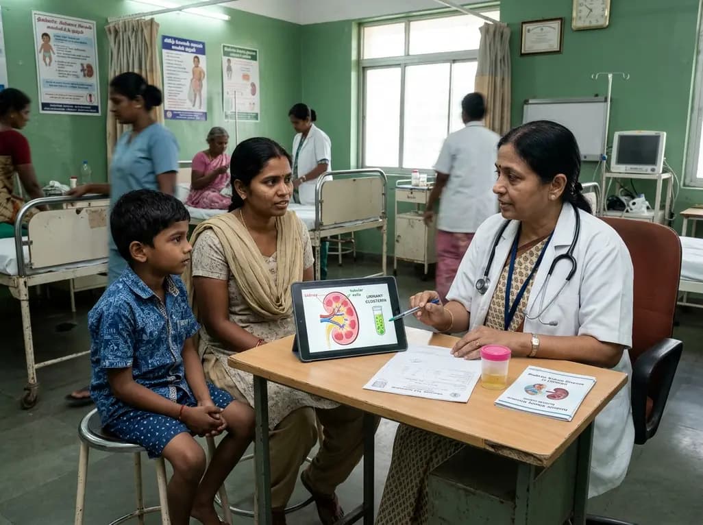
A recent case-control study reveals that urinary clusterin provides superior diagnostic performance for early diabetic kidney disease detection in pediatric type 1 diabetes mellitus, identifying tubular damage before conventional microalbuminuria occurs.
Today
